AutoFUSE or First-Trimester Ultrasound Scan

Conditions: Fetus Disorder

Interventions: Diagnostic Test: AutoFUSE system; Diagnostic Test: Traditional ultrasound scan

Sponsors: Union Hospital, Tongji Medical College, Huazhong University of Science and Technology

Not yet recruiting

Precancerous Pancreatic Lesions May Not Always Lead to Cancer

Pancreatic cancer is an often deadly disease that, even if successfully treated, has a relative survival rate of about 13%, according to the NIH. When detected and treated early, that survival rate increases substantially. Developing strategies to diagnose cancer early or identify precancerous tissues is vital to success and increased survival.

A common precursor to pancreatic cancer is the presence of lesions called pancreatic intraepithelial neoplasia (PanIN). Researchers from the University of Michigan and the University of Maryland have been studying these precursor lesions and previously identified metabolic biomarkers involved with the development of PanIN to cancer. In their new study, published in Cancer Discovery, they expand their work into the PanIN microenvironments—the cells and tissues directly surrounding the lesions—to understand the cellular changes in an effort to find markers to use in diagnostics.

To examine the microenvironments, the team utilized single-cell RNA sequencing and spatial transcriptomics, isolating single cells and mapping their gene expression.

“These lesions are like needles in a haystack,” said co-senior author Timothy Frankel, MD, professor of surgical oncology and co-director of the Rogel and Blondy Center for Pancreatic Cancer at the University of Michigan. “The prior way of looking at this was to look at the entire haystack. You get a lot of information about hay and very little information about the needle.” He explained that using these precise techniques to examine cells individually is both more efficient and allows the researchers “to just focus in on the needle so we can look at multiple needles using the same amount of computing power and resources.”

The researchers used whole donated human pancreases to map the progression from PanIN to cancer, both for the precancerous and cancerous cells themselves, and the microenvironments surrounding them.

The pancreatic cancer microenvironment contains a highly diverse group of interactive cells, including fibroblasts and immune cells. The researchers found that while the epithelial tissue gene expression changes on a continuum from PanIN to cancer, the cells in the microenvironment are much more dynamic.

“Progression to cancer is accompanied by profound geographical reorganization of myeloid cells and lymphocytes and the formation of a cancer-specific fibroblast population characterized by high levels of Smooth Muscle Actin, LRRC15, and the WNT signaling component LEF1,” they wrote.

“It turns out, the microenvironment of these precursor lesions is the same as the microenvironment of the normal pancreas,” explained co-senior study author Marina Pasca di Magliano, PhD, professor of surgical immunology and co-director of the Rogel and Blondy Center for Pancreatic Cancer at the University of Michigan.

“The lesions have not convinced any of the cells around them to change. That’s not what we were expecting. We were expecting the two components, the cells and the microenvironment, to evolve in lockstep. They did not.”

These unexpected results indicate that there are other factors or stressors impacting the microenvironment. This is also in line with prior data showing that while pancreatic cancer is preempted by PanIN, not all, and in fact most, PanIN occurrences do not lead to a cancer diagnosis.

“It is incredible to see how we can uncover the fundamental cellular mechanisms of disease etiology by blending new computational methods and cutting-edge spatial transcriptomics technologies,” said co-corresponding author Elana J. Fertig, PhD, director of the Institute for Genome Sciences at the University of Maryland School of Medicine and associate director of quantitative sciences at the University of Maryland Marlene and Stewart Greenebaum Comprehensive Cancer Center. “Through careful study design, we can use the spatial information to start delving into the unknown dynamics of pancreatic tumor evolution.”

Moving forward, more research is needed to identify those stressors that may impact the microenvironment surrounding PanINs, which would allow the lesions to turn into cancers. Understanding those switches may open the door for developing earlier diagnostics or therapies to help prevent cancer development in the first place.

The post Precancerous Pancreatic Lesions May Not Always Lead to Cancer appeared first on Inside Precision Medicine.


AI Designs Miniprotein Switches for GPCR Targeting

Many scientists first encountered G protein–coupled receptors (GPCRs) as a looping sketch across the cell membrane in an early biology textbook. That simple diagram belied the complexity of a receptor family now known to govern vision, smell, hormone sensing, and the actions of countless medicines. Yet despite their centrality, designing molecules that can precisely switch GPCRs on or off has remained one of drug discovery’s most persistent challenges.

A new study led by the UW Medicine Institute for Protein Design and Skape Bio demonstrates that AI‑driven de novo protein design can finally meet that challenge. The work, published recently in Nature, shows that computationally designed miniproteins—compact proteins under 100 amino acids—can be engineered to either activate or block GPCRs with high affinity, potency, and selectivity. The paper is titled “De novo design of miniproteins targeting G protein-coupled receptors.”

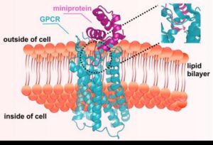
The research team developed a suite of design strategies to create miniproteins capable of slipping into the deep, flexible pockets that govern GPCR signaling. These pockets shift shape depending on whether the receptor is active or inactive, making them difficult to target with conventional biologics. By designing molecules that recognize specific receptor states, the team generated agonists for receptors involved in itch and pain, and antagonists for receptors implicated in cancer, metabolic disease such as diabetes and obesity, and migraine.

GPCR microprotein
A tiny protein (pink) designed on a computer fits into a deep pocket (inset) of a cell surface receptor called a GPCR (blue), allowing scientists to switch cell signaling on or off. [Edin Muratspahić/UW Medicine Institute for Protein Design]

“Protein design takes our understanding of how proteins fold and reverses it—asking if we can envision, with the aid of AI computing, a new protein that sticks to a target in a purpose-built way,” said senior author David Baker, PhD, director of the Institute for Protein Design, professor of biochemistry at the University of Washington School of Medicine, and a Howard Hughes Medical Institute Investigator. “This paper showcases how we can do this repeatedly for different GPCRs in ways that capitalize on their dynamic motion to either activate or inactivate them.”

Cryo‑EM structures of five designed miniproteins closely matched their computational models, underscoring the accuracy of the design pipeline. In one mouse study, a designed chemokine‑receptor antagonist mobilized hematopoietic stem and progenitor cells at levels comparable to a clinically used drug—but with fewer side effects, according to the authors.

For first author Edin Muratspahić, PhD, the moment of validation came when the designed molecules did more than bind. “Seeing computationally designed miniproteins not only bind but actually control GPCR signaling in living cells was a defining moment for me,” he said.

A second major advance reported in the study is a high‑throughput “receptor diversion” screening system that evaluates tens of thousands of designed proteins directly in living human cells. Traditional GPCR screens often require purifying or stabilizing receptors—steps that can distort their natural signaling behavior. By keeping receptors in their native membrane environment, the new system accelerates discovery while preserving biological relevance.

According to corresponding author Christoffer Norn, PhD, co‑founder of Skape Bio, the study lays out a roadmap for all‑computational design of GPCR ligands.

The methods described in the paper are already being adapted at Skape Bio to explore GPCR targets involved in metabolic, inflammatory, and neurologic pathways—areas where conventional discovery efforts have often struggled.

The post AI Designs Miniprotein Switches for GPCR Targeting appeared first on GEN – Genetic Engineering and Biotechnology News.

STAT+: Closely watched experimental Parkinson’s drug fails key clinical trial

Biogen and Denali Therapeutics said Thursday that their experimental therapy for Parkinson’s disease failed to slow the degenerative brain disorder in a randomized trial, dealing a substantial blow to a scientific approach that stoked excitement among advocates and academics. 

In the study, 648 adults with Parkinson’s were randomized to receive either a placebo or a pill targeting a protein called LRRK2. In 2004, researchers discovered that mutations in the LRRK2 gene can cause a rare, inherited form of Parkinson’s. And in 2018, another group of scientists showed that blocking the protein might actually benefit all patients with the disease. 

Thursday’s results are a significant setback to the latter idea. 

Continue to STAT+ to read the full story…