Interventions: Diagnostic Test: Soma Body-Composition Segmentation Software
Sponsors: Nucleo Research, Inc.
Not yet recruiting
Many scientists first encountered G protein–coupled receptors (GPCRs) as a looping sketch across the cell membrane in an early biology textbook. That simple diagram belied the complexity of a receptor family now known to govern vision, smell, hormone sensing, and the actions of countless medicines. Yet despite their centrality, designing molecules that can precisely switch GPCRs on or off has remained one of drug discovery’s most persistent challenges.
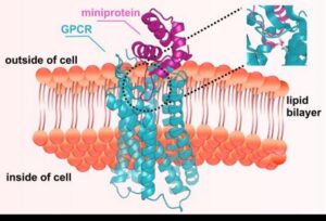
A new study led by the UW Medicine Institute for Protein Design and Skape Bio demonstrates that AI‑driven de novo protein design can finally meet that challenge. The work, published recently in Nature, shows that computationally designed miniproteins—compact proteins under 100 amino acids—can be engineered to either activate or block GPCRs with high affinity, potency, and selectivity. The paper is titled “De novo design of miniproteins targeting G protein-coupled receptors.”
The research team developed a suite of design strategies to create miniproteins capable of slipping into the deep, flexible pockets that govern GPCR signaling. These pockets shift shape depending on whether the receptor is active or inactive, making them difficult to target with conventional biologics. By designing molecules that recognize specific receptor states, the team generated agonists for receptors involved in itch and pain, and antagonists for receptors implicated in cancer, metabolic disease such as diabetes and obesity, and migraine.

“Protein design takes our understanding of how proteins fold and reverses it—asking if we can envision, with the aid of AI computing, a new protein that sticks to a target in a purpose-built way,” said senior author David Baker, PhD, director of the Institute for Protein Design, professor of biochemistry at the University of Washington School of Medicine, and a Howard Hughes Medical Institute Investigator. “This paper showcases how we can do this repeatedly for different GPCRs in ways that capitalize on their dynamic motion to either activate or inactivate them.”
Cryo‑EM structures of five designed miniproteins closely matched their computational models, underscoring the accuracy of the design pipeline. In one mouse study, a designed chemokine‑receptor antagonist mobilized hematopoietic stem and progenitor cells at levels comparable to a clinically used drug—but with fewer side effects, according to the authors.
For first author Edin Muratspahić, PhD, the moment of validation came when the designed molecules did more than bind. “Seeing computationally designed miniproteins not only bind but actually control GPCR signaling in living cells was a defining moment for me,” he said.
A second major advance reported in the study is a high‑throughput “receptor diversion” screening system that evaluates tens of thousands of designed proteins directly in living human cells. Traditional GPCR screens often require purifying or stabilizing receptors—steps that can distort their natural signaling behavior. By keeping receptors in their native membrane environment, the new system accelerates discovery while preserving biological relevance.
According to corresponding author Christoffer Norn, PhD, co‑founder of Skape Bio, the study lays out a roadmap for all‑computational design of GPCR ligands.
The methods described in the paper are already being adapted at Skape Bio to explore GPCR targets involved in metabolic, inflammatory, and neurologic pathways—areas where conventional discovery efforts have often struggled.
The post AI Designs Miniprotein Switches for GPCR Targeting appeared first on GEN – Genetic Engineering and Biotechnology News.
The story of type 1 diabetes begins in the pancreas, long seen as a battleground between insulin-producing beta cells and misdirected immune defenders. Scientists have been searching for ways to spot this internal warfare early enough to prevent a lifelong disease that depletes the body’s source of insulin.
Two new papers published Wednesday in Science Translational Medicine offer new clues to what happens in these beta cells before type 1 diabetes emerges. Experiments in human cells and in mouse models used biosensors and genetic analyses to illuminate this pathway and detect possible ways to halt beta-cell destruction.
In the first study, a team from the Indiana University School of Medicine explored how certain immune cells involved in inflammation, known as signaling cytokine interferon-alpha, normally trigger beta cells to produce other molecules that play a role in inflammation, cell proliferation, and cell death. These reactive oxygen species, ROS for short, sometimes cause collateral damage. But cells from patients with type 1 diabetes did not have ROS-producing beta cells, suggesting they lacked the cytokines that stimulate their ROS production. That dearth might be useful in flagging the decline of beta cells early on in type 1 diabetes, the study authors surmised.
A new study in Science Signaling identifies a previously overlooked control point in receptor tyrosine kinase (RTK) signaling, one that operates not at the plasma membrane, but at the Golgi. The research, published as “Oncogenic receptor tyrosine kinase signaling is driven by the Golgi protein GOLPH3 and its interaction with MYO18A,” reveals that the Golgi‑localized proteins GOLPH3 and MYO18A act together to route RTKs to the cell surface, thereby setting the strength of growth‑factor signaling across multiple pathways.
The work was led by Kyle Starost and colleagues at Case Western Reserve University School of Medicine and the University of California, San Diego. Their findings help explain why GOLPH3 is frequently amplified in human cancers and why its overexpression correlates with poor prognosis across tumor types.
RTKs such as EGFR, insulin receptor, and PDGFR are central drivers of proliferation and survival in many cancers. Although RTK inhibitors are widely used clinically, resistance often emerges, underscoring the need for alternative strategies that modulate signaling upstream of the receptor. The new study identifies one such upstream node: the delivery of RTKs from the Golgi to the plasma membrane.
Using an unbiased signaling analysis, the team found that siRNA knockdown of GOLPH3 or MYO18A impaired phosphorylation of EGFR at Tyr1068 and Tyr1086, as well as downstream AKT and ERK signaling. These defects persisted even when PI3K/AKT/mTOR signaling was pharmacologically blocked, demonstrating that GOLPH3 acts directly at the receptor level rather than through mTOR modulation.
To pinpoint the mechanism, the researchers turned to trafficking assays. Imaging of endogenous EGFR showed that loss of GOLPH3 or MYO18A caused the receptor to accumulate in intracellular puncta rather than at the plasma membrane. A quantitative PDGFR‑GFP surface‑delivery assay confirmed that both proteins are required for Golgi‑to‑surface transport. Treatment with brefeldin A or golgicide A, which disrupt Golgi structure, produced similar reductions in surface receptor levels, reinforcing the conclusion that the GOLPH3–MYO18A complex is essential for RTK delivery.
Overexpression experiments completed the mechanistic picture. Increasing GOLPH3 or MYO18A levels enhanced EGF‑stimulated phosphorylation of EGFR and AKT, while a GOLPH3 mutant unable to bind PI4P failed to do so. These results position the GOLPH3–MYO18A complex as a central determinant of RTK availability at the cell surface.
The authors wrote, “The GOLPH3-MYO18A complex at the Golgi apparatus was required and rate-limiting for RTK signaling across the cell types and receptors assessed.” The findings suggest that targeting Golgi‑based trafficking machinery could offer a new therapeutic angle for tumors that rely on hyperactive RTK signaling or have developed resistance to RTK inhibitors.
The post Oncogenic Signaling Shaped by a Golgi Trafficking Protein Pair appeared first on GEN – Genetic Engineering and Biotechnology News.
The idea of drinking during pregnancy sounds like a generational punchline: Someone’s grandmother drank beer to fatten her fetus, another had a nightly martini to get a healthy amount of sleep — presumably unthinkable behavior in today’s America.
Yet after precipitous declines in the last 50 years, rates of alcohol use in pregnancy in the U.S. started climbing upward a decade ago. More than 1 in 8 pregnant adults reported drinking in the past month, according to STAT’s analysis of 2024 government data, making alcohol use a more common national phenomenon than gestational diabetes. Of those who drank, a quarter reported having four or more drinks in one sitting — binge drinking — in the prior month.
While rates of alcohol use in pregnancy are lower in the U.S. than those of several peer nations, the effects are all around Americans. Alcohol is the key driver of what are, by some estimates, the nation’s top neurodevelopmental conditions: fetal alcohol spectrum disorders, or FASDs.
The exact prevalence of FASDs is difficult to measure, but the most recent federally funded community studies have found as many as 1 in 20 school-aged children may have a disorder caused by prenatal alcohol exposure. By comparison, about 1 in 31 American children has autism, per recent estimates from the Centers for Disease Control and Prevention.
Not all fetuses exposed to alcohol have birth defects or go on to develop intellectual disabilities, researchers say. But every person born with an FASD was harmed by alcohol specifically. They worry this point is being glossed over as Americans question the conventional medical advice of avoiding all alcohol while pregnant.
A University of Bath-led research effort received £500,000 to develop an organ-on-chip device that replicates connections between the brain, gut, and pancreas. The GlucoBrain project is designed to allow researchers to track how signals move between the organs and uncover why diabetes may lead to changes in memory and cognition.
Collaborators include investigators from the University of Oxford and Johns Hopkins. Their findings could pave the way for new treatments to improve the lives of millions of people affected by diabetes, dementia, or both, notes the team.
Diabetes and Alzheimer’s disease are two of the world’s most pressing health problems, especially in aging societies. While diabetes is widely known to affect the heart, kidneys, and eyes, growing evidence suggests it is also linked with problems in memory, learning, and brain function. However, the biological mechanisms behind this link remain poorly understood.
“Our gut, pancreas, and brain are constantly communicating via a network of signals, helping us regulate hunger and blood sugar,” says Despina Moschou, PhD, project lead. “But we still don’t fully understand how these signals interact at a cellular level and why glucose levels are linked to cognitive decline. “By creating a connected system on a chip, we can study in real time how signals travel between organs, how diabetes may impair brain function, and how new drugs could help.”
Most current knowledge on the link between diabetes and dementia comes from animal studies, simple cell cultures, and patient studies. While these are useful, they don’t fully and accurately capture all the complex interactions between our organs, hormones, and cells, points out Moschou.
Organ-on-chip technology uses living human cells in miniature devices that mimic how organs work in the body. Unlike cell cultures grown in a petri dish, these devices allow cells to grow in three dimensions, receive a controlled supply of nutrients and interact more naturally. Researchers will also be able to isolate these individual organs and cell types to understand exactly how they communicate at a molecular level.
The three-year project starts in October, bringing together engineers, clinicians, biologists and computer scientists to model the complex disease interactions. The team will first develop individual chip models for the gut, pancreas, and brain, before connecting them into a multi-organ system. They will gradually increase complexity and measure how each organ responds to glucose, hormones and different drug treatments.
Researchers from the University of Oxford will provide core clinical expertise in diabetes and metabolic disease, ensuring models are physiologically accurate. The team from Johns Hopkins University brings specialist expertise in Alzheimer’s disease and brain organoids.
GlucoBrain is a pilot project established to help researchers understand exactly how diseases like diabetes and dementia work at a deeper, biological level. This early-stage research will build the foundations for even more advanced and realistic models, bringing together more organs and cell types, explain team members. By harnessing the power of artificial intelligence, the devices have the potential to reveal new insights into how diseases emerge and develop.
“Not only would these devices give us an unprecedented way to study diseases, but they could help speed up drug discovery and testing, reducing reliance on animal models and making results more relevant to humans,” continues Moschou. “In the long term, they could pave the way for personalized medicine, using a patient’s own cells to identify the most effective treatment.”
The project is funded by the Engineering and Physical Sciences Research Council (EPSRC) Health Technologies Connectivity Awards.
The post Organ-on-Chip Method Designed to Zero In on Connection Between Diabetes and Dementia appeared first on GEN – Genetic Engineering and Biotechnology News.

Psychostimulant use disorder (PSUD) and nicotine use disorder (NUD) affect tens of millions of people worldwide, yet no clinically-approved medications exist for PSUD and existing NUD treatments have limited long-term efficacy. Recent preclinical evidence suggests that glucagon-like peptide-1 receptor (GLP-1R) agonists could be repurposed for treating PSUD and NUD. This review summarizes the current literature investigating the efficacy of GLP-1R agonists to reduce psychostimulant and nicotine-mediated behaviors in animal models of PSUD and NUD, respectively.