

Single-Cell Atlas Simultaneously Maps 3D Genome Architecture and DNA Methylation

Scientists at the Salk Institute and the Arc Institute, along with their collaborators, unveiled the first body-wide single-cell atlas of two major epigenetic systems: three-dimensional genome folding and DNA methylation, measured simultaneously in the same cells.

The atlas spans 86,689 cells from 16 human tissues, revealing 35 major cell types and 206 subtypes, and is freely available online. The work is part of the National Institutes of Health’s 4D Nucleome (NIH 4DN) program, which aims to understand how the genome is organized in space and time to regulate gene expression in health and disease.

Because the two epigenetic layers were measured together, the researchers could compare what each layer says about a cell’s identity. And while often the two pictures agree, they found that sometimes they do not.

The Salk paper “Human body single-cell atlas of 3D genome organization and DNA methylation”  was published alongside five other NIH 4DN papers in Science, and three others in Science Advances.

The Human Genome Project, completed in 2003, produced a linear read of the three billion DNA letters in the human body. But the letters alone don’t explain how a single genome produces hundreds of different cell types. That information lives in the epigenome in the form of chemical modifications and structural folds layered on top of the DNA sequence, where they can switch genes “on” and “off” in patterns specific to each cell type.
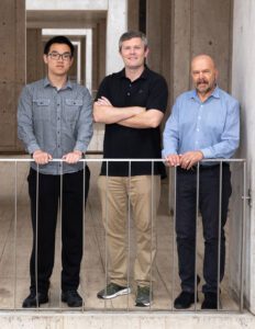
Caption: Salk scientists Jingtian Zhou (left), Jesse Dixon (center), and Joseph Ecker (right) profiled 86,689 cells across 16 human tissues, linking cell-type-specific epigenetic features to disease risk and revealing that a cell’s 3D genome and DNA methylation don’t always tell the same story. [Salk Institute]

Two of the most consequential epigenetic features are 1) DNA methylation, where small chemical groups called methyl groups are attached to specific DNA bases, and 2) 3D genome organization, where intricate loops, folds, and compartments bring distant stretches of DNA into contact. Both influence gene expression, but they had never been measured together in single cells across the human body.

“There has been an appreciation for trying to understand, at the individual cell level, how the genome is organized, so that we can get a better idea of how genetic variants impact disease,” said co-corresponding author Joseph Ecker, PhD, a professor and Salk International Council Chair in Genetics at Salk and a Howard Hughes Medical Institute investigator. “Some cell types may be more vulnerable than others to genetic variants, because the genome is organized differently in different cell types—and whether a variant matters can depend on that organization.”

Why is noncoding DNA relevant in disease?

Most disease-associated genetic variants fall in the noncoding regions of the genome. That has made it difficult to figure out how a variant contributes to disease, which cell type it acts in, and what gene it ultimately affects.

The new atlas identifies more than 1.36 million differentially methylated regions and 283,606 differential chromatin loops across the human body’s cell types, using tissues from the heart, brain, lungs, stomach, skin, and more. When the researchers overlaid genetic variants known to raise disease risk, specific pairings emerged like variants for blood-glucose regulation concentrated in endocrine cells, atrial fibrillation variants in heart muscle cells, balding variants in skin fibroblasts, and bipolar disorder and schizophrenia variants in excitatory and inhibitory neurons.

“A lot of the genetic variation that predisposes someone to disease is in noncoding parts of the genome,” said co-corresponding author Jesse Dixon, MD, PhD, associate professor and Helen McLoraine Developmental Chair at Salk. “By adding in the 3D genome aspect, we can potentially bridge that gap—connecting noncoding variations with the genes they affect in specific cells and tissues.”
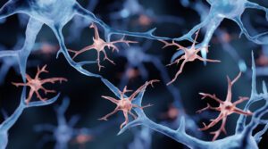
glial cells
Microglia, illustration. Researchers from the New York Genome Center and Columbia University used the atlas’ cross-tissue methylation data to show that a substantial fraction of the brain’s resident immune cells (microglia) are replaced by cells resembling blood monocytes between roughly ages 50 and 75. The finding challenges the long-held view that microglia persist from embryonic development throughout the life span. [Artur Plawgo/Getty Images]

What happens when two epigenetic lenses disagree?

One of the study’s most surprising findings is that DNA methylation and 3D genome structure don’t always tell the same story about a cell. In skeletal muscle, the team found fibers that look like mature, differentiated muscle cells by their 3D genome folding, but still carry the methylation signature of muscle stem cells. The reverse almost never happens. The most plausible explanation, they explained, is that these cells are caught mid-differentiation, with 3D architecture updating first and methylation catching up.

Similar mismatches appeared in Schwann cells of the peripheral nervous system and in placental trophoblasts. The pattern suggests that different epigenetic features update on different time scales during cell state transitions—a finding that could reshape how researchers define “cell type” in adult tissues and how they track cells moving between states in disease.

The atlas also revises a long-standing assumption about “non-CG methylation,” an unusual form of methylation previously thought to be largely confined to brain cells and stem cells. The study shows that it carries cell-identity information across many human tissues, including muscle, pancreas, and immune cell types, at lower but biologically meaningful levels.

“The inconsistency between modalities may be further used to determine what cell populations are switching between each other in adult tissues and diseases, which could, for example, expand our understanding of cancer cell dynamics,” said co-first and co-corresponding author Jingtian Zhou, PhD, a former graduate researcher in Ecker’s lab who now leads his own lab at the Arc Institute.

A public resource for scientists and artificial intelligence

To make the atlas broadly usable, the team built an interactive web browser that lets researchers visualize DNA methylation and 3D chromatin contacts across every tissue, cell type, and subtype in the study. The underlying data, including 195 billion methylation measurements and 18 billion chromatin contacts, are freely available.

The resource arrives as artificial intelligence tools are increasingly used to predict the functional impact of genetic variants. Atlases like this one can provide the labeled, cell-type-resolved training data that models need to make accurate predictions—a bottleneck that has historically limited the field.

For example, in a companion paper in the same issue of Science, a study led by Bing Ren, PhD, from the New York Genome Center and Columbia University used the atlas’ cross-tissue methylation data to show that a substantial fraction of the brain’s resident immune cells, called microglia, are replaced by cells resembling blood monocytes between roughly ages 50 and 75. The finding challenges the long-held view that microglia persist from embryonic development throughout the life span.

“DNA methylation patterns are specific to each cell type and analogous to a cellular barcode,” said Ren, who also co-authored the Salk-led study. “The comprehensive cross-tissue DNA methylation atlases show that the aging microglia in the human hippocampus more closely match the monocytes from peripheral blood than microglia from young adults, providing a crucial clue for the biological identity of these cells.”

The NIH 4D Nucleome consortium, of which this study is a part, aims to extend this kind of mapping into the fourth dimension: time. A 4D understanding of the genome—how its structure and chemistry change as cells develop, age, and respond to disease—remains a major goal, and the cross-tissue atlas provides reference scaffolding that future time-course studies will build on.

Along with scientists from the Salk Institute and Arc Institute,  investigators from the University of California, San Diego, Columbia University, New York Genome Center, University of California, Los Angeles, Harvard, Henan University in China, Vanderbilt University, Stanford University, Broad Institute, University of Sheffield in the U.K., Yale, University of Florida, University of Freiburg in Germany, University of Graz in Austria, and Nanchang University in China; and Chongyuan Luo also contributed to the Science paper.

The post Single-Cell Atlas Simultaneously Maps 3D Genome Architecture and DNA Methylation appeared first on GEN – Genetic Engineering and Biotechnology News.
<![CDATA[Top 2026 psychiatry conference takeaways: mixed-feature bipolar care, GLP-1 addiction trials, schizophrenia adherence, TD gaps, AI prodrome screening.]]>

Optimizing Treatment Strategies in the Bipolar Disorder Spectrum With Classical AI Approaches: Systematic Review of Performance, Bias, and Clinical Applicability

Background: Bipolar disorder (BD) is a complex and heterogeneous psychiatric condition, characterized by fluctuating clinical courses that affect approximately 1%‐2% of the global population in their lifetime. Despite pharmacological advances, treatment response varies significantly among patients, making the identification of individualized treatment strategies a major challenge. Artificial Intelligence (AI), through its classical approaches, has emerged as a powerful tool in precision psychiatry to identify subtle patterns in complex data and inform personalized clinical decisions. Objective: The present systematic review aimed to examine the current evidence on classical AI-supported treatment optimization in the BD spectrum. Methods: The review was conducted in accordance with the PRISMA (Preferred Reporting Items for Systematic Reviews and Meta-Analyses) 2020 guidelines. Four databases (PubMed, Web of Science, Scopus, and Embase) were searched for original studies published after 2015 on the application of classical AI in the treatment of BD in adult patients. Publication bias was evaluated by visual inspection of a funnel plot. The methodological quality, risk of bias, and clinical applicability of the predictive models were assessed using the Prediction Model Risk Of Bias Assessment Tool for prediction models using regression or AI methods (PROBAST+AI; PROBAST+AI Working Group) tool. Results: A total of 35 studies were included and classified into 5 outcome-based categories, including acute symptomatic response, long-term maintenance response, relapse and readmission risk, safety and dose optimization, and brain aging and phenotyping. Acute symptomatic response models performed modestly (pooled area under the curve [AUC] 0.68), while imaging improved accuracy (74%‐77%). Long-term maintenance response models showed moderate-to-high performance (pooled AUC 0.80), with biomarker- and cellular-based models reaching 96%‐99% accuracy. Relapse and readmission prediction achieved a pooled AUC of 0.71, with digital phenotyping and rule-based methods performing best (AUC 0.85‐0.88). Safety and dose optimization models achieved 85%‐97% accuracy. Brain aging and phenotyping studies highlighted accelerated brain aging in BD, partially mitigated by lithium, and revealed novel data-driven subgroups. However, 3 studies were considered at high risk of bias due to small sample sizes associated with disproportionately high-performance estimates. An additional study was identified as potentially biased because it lay markedly distant from the funnel plot’s confidence line. Finally, the PROBAST+AI assessment revealed a high risk of bias in most studies, primarily due to data analysis limitations, small sample sizes, and lack of external validation. Conclusions: The adoption of classical AI tools in BD serves as a driver for therapeutic optimization, although current AI tools in BD should still be considered exploratory rather than ready for clinical use. Effective implementation in real-world clinical scenarios requires more robust, transparent, and externally validated models to ensure reliability and generalizability.
<img src="https://jmir-production.s3.us-east-2.amazonaws.com/thumbs/df358c951a2c44fbd20077fdda306baf" />

Transition Care for FEP

Conditions: Schizoaffective Disorders; Bipolar Disorder With Psychotic Features; Schizophrenia Disorders; Major Depressive Disorder With Psychotic Features; Psychosis NOS

Interventions: Behavioral: Transition Care Team

Sponsors: University of Chicago; Sidney R. Baer, Jr. Foundation

Recruiting
<![CDATA[Learn how clinicians manage bipolar disorder in women who may become pregnant—safer meds, contraception counseling, and relapse prevention.]]>
<![CDATA[Learn how clinicians tackle mood and substance use disorders together, boost patient engagement, and expand evidence-based addiction medications for bipolar and alcohol use.]]>

See, Blind Mice: Consortium’s Drugs Restore Sight

A consortium led by scientists at the Institute for Bioengineering of Catalonia (IBEC) has developed a series of light-activated small molecule drugs that in preclinicial tests restored sight in blind mice. The team’s approach is based on photopharmacology, a technique for reversibly control drug activity using light.

The newly developed compounds, called prosthe6, mimic the function of light sensing photoreceptor cells, which degenerate in blinding diseases such as age-related macular degeneration (AMD) and retinitis pigmentosa (RP).

The prosthe6 compounds target ON-bipolar neurons and in tests were found to successfully restore saccadic eye movements (optokinetic reflex) in blinded zebrafish larvae, a widely used model for studying visual acuity. Even more strikingly, the researchers demonstrated recovery of innate light-avoidance behavior in mouse models of age-related macular degeneration and retinitis pigmentosa.

Test results suggest that the prosthe6 compounds may be administered by injecting them in the eye, or administered as eye drops. In animal studies the photoswitchable molecules also showed promising preliminary safety profiles, pointing to the development of potential drug candidates for restoring vision in patients with degenerative retinal diseases, without the need for genetic manipulation or implanted devices. Importantly, these compounds are designed to work under normal lighting conditions and do not require light-enhancing devices as optogenetics. They are small, water-soluble molecules that respond to ordinary visible or white light, such as indoor lighting or daylight, without requiring intense or specialized light sources.

“These molecules do not cure blindness, because they do not address the cause of photoreceptor degeneration,” said study co-lead Pau Gorostiza, PhD, ICREA Research Professor at IBEC, leader of the Nanoprobes and Nanoswitches group, member of CIBER-BBN. “But they are remarkably effective at restoring sight, and they do so using a very simple and potentially patient-friendly approach.”

Rosalba Sortino, former PhD student at the University de Barcelona, and currently post-doctoral researcher at Gorostiza’s group at IBEC, added, “Our goal was to restore vision using a molecular mechanism that is as close as possible to how the healthy retina works … Instead of bypassing retinal processing, we aimed to reactivate it right at the same level of the retinal circuit than the lost photoreceptor cells.”

Sortino is co-first author of the team’s published paper in Journal of the American Chemical Society, titled “Restoration of saccadic eye movements and visually guided behavior in ambient white light with photoswitchable small molecules.”

Diseases such as age-related macular degeneration and retinitis pigmentosa affect 200 million people worldwide and are the leading causes of visual impairment and blindness. Beyond the personal impact on quality of life and independence, vision loss places a global economic burden estimated at over US$400 billion per year in healthcare costs and lost productivity.

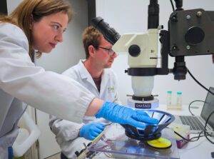
Researchers Rosalba Sortino (left) and Joaquin Martinez Tambella (right) working in the laboratories of the Institute for Bioengineering of Catalonia (IBEC). Sortino is a post-doctoral researcher at the Nanoprobes and Nanoswitches group at IBEC and co-first author of the study. Martinez is a PhD student at the Nanoprobes and Nanoswitches group at IBEC and co-first author of the study. [Institute for Bioengineering of Catalonia (IBEC).]
Researchers Rosalba Sortino (left) and Joaquin Martinez Tambella (right) working in the laboratories of the Institute for Bioengineering of Catalonia (IBEC). Sortino is a post-doctoral researcher at the Nanoprobes and Nanoswitches group at IBEC and co-first author of the study. Martinez is a PhD student at the Nanoprobes and Nanoswitches group at IBEC and co-first author of the study. [Institute for Bioengineering of Catalonia (IBEC)]

In many of these conditions, photoreceptor (PhR) cells—the retina’s light detectors—progressively degenerate and die. Although the downstream retinal neuronal circuitry remains largely intact and functionally viable, it no longer receives the light signals needed to drive visual processing towards the brain. This opportunity has fueled intense research efforts to develop treatments capable of restoring light sensitivity to the eye. Current strategies include gene therapy—effective only for a very small subset of patients with specific mutations—and electronic retinal prostheses, which are invasive, expensive, and require extensive training for effective use.

More recently, optogenetics and light-responsive drugs have entered clinical testing, the latter with encouraging safety results. “Photopharmacology can develop photoswitchable small molecules to restore vision impairment by conferring light sensitivity to ion channels that are widely expressed in the remaining inner retinal neurons, and a first-in-human clinical trial is ongoing,” the team noted. However, achieving high-quality vision at ambient illumination levels remains a major challenge.

The (IBEC)-led consortium has now developed a new class of photoswitchable small-molecule drugs that are capable of restoring key visual functions in animal models of blindness. The team’s photopharmacology-based technique involves modifying a drug’s chemical structure by adding a light-activated molecular switch, enabling control of the pharmacological action using light. “Unlike (opto)genetic manipulation and surgically implanted retinal electronic prostheses, pharmacotherapy is noninvasive, readily reversible, and can be upgraded when new drugs are approved,” the authors noted. “Medicines are preferred by patients, clinicians, and public healthcare systems, they  can be developed and manufactured at lower costs than other approaches and assessed by conventional regulatory procedures and clinical assays.”

The reported work builds on more than a decade of research and was carried out in collaboration with the team co-led by Pedro de la Villa at the University of Alcalá (UAH), as well as researchers from the Institut de Química Avançada de Catalunya (IQAC-CSIC), the University of Barcelona (UB), the Institute Ramón y Cajal of Health Research (IRYCIS), the Autonomous University of Barcelona (UAB), and the Fundació Eduard Soler.

Researcher Joaquin Martinez Tambella working in the laboratories of the Institute for Bioengineering of Catalonia (IBEC). Martinez is a PhD student at the Nanoprobes and Nanoswitches group at IBEC and co-first author of the study. [Institute for Bioengineering of Catalonia (IBEC).]
Researcher Joaquin Martinez Tambella working in the laboratories of the Institute for Bioengineering of Catalonia (IBEC). Martinez is a PhD student at the Nanoprobes and Nanoswitches group at IBEC and co-first author of the study. [Institute for Bioengineering of Catalonia (IBEC)]

The prosthe6 compounds work by acting on a specific type of retinal cells called ON bipolar cells, which normally receive signals from the photoreceptors. “In healthy vision, ON bipolar cells play a key role in passing on information about the presence of light to the rest of the visual circuit,” explained study co-lead de la Villa. “In degenerative eye diseases, although the photoreceptors are lost, much of this underlying circuitry remains intact but inactive. This creates a major therapeutic opportunity.”

By targeting a protein (mGlu6) in this preserved part of the retina, prosthe6 compounds can take over the role of the missing photoreceptors. “… we have targeted metabotropic glutamate 6 (mGlu6) receptors, which are exclusively expressed in ON bipolar cells (OBCs) and localized postsynaptic to PhR cells, thereby leveraging a privileged position to drive physiological visual circuit,” the investigators explained. When light enters the eye, the molecules respond by changing their shape, triggering signals inside the retina in a way that closely resembles natural vision. In this way, the drugs effectively act as “molecular prostheses,” helping the eye process light again without the need for implants or genetic modifications.

Healthy mice naturally prefer to remain in dark environments and instinctively avoid brightly lit areas, a behavior that relies entirely on a functional visual system. Blind mice, by contrast, lose this preference and move indistinctly between light and dark spaces, as they are unable to perceive light. The team showed that after treatment with prosthe6, blind mice once again showed a clear and spontaneous preference for dark areas, indicating that they could perceive light and use this information to guide their behavior.

This recovery occurred without any training and under light levels comparable to those found indoors or on an overcast day, demonstrating that the treatment restores functional light perception capable of driving natural, visually guided behavior.

Two lead compounds, prosthe6-12 and prosthe6-15, showed particularly promising results. The restored behaviors were observed not only after intraocular injection, but also after topical administration as eye drops. “… at least two compounds (prosthe6-12 and -15) appear to be devoid of adverse effects and restore sight by topical administration, which is linked to higher overall clinical success rate than systemic routes for neurological drugs, and to stronger patient adherence,” the investigators pointed out.

The prosthe6 technology is protected by patent and the researchers are now evaluating its safety and formulation to extend the duration of visual rehabilitation. The team is working with Eyelumina, a spin-off company in formation to secure investments that support translational development and future clinical trials.

“Turning this into a therapy is a long and laborious process,” says Gorostiza. “But the results show that there is a realistic possibility of restoring high-quality vision with drugs, non-invasively, reversibly and with a mechanism that is independent of the specific retinal disorder or genetic mutation to reach a majority of patients.”

If successful in humans, the drug-based approach would offer a widely accessible and affordable alternative to existing vision restoration technologies, especially relevant for patients with advanced retinal degeneration for whom no effective treatments currently exist.

In their paper the team further stated, “From a fundamental perspective, prosthe6 constitute new tools for ophthalmology to study the physiopathology of mGlu6 receptors and retinal circuits in vitro and in vivo and contribute to the medicinal chemistry of allosteric modulators. They also achieve the prediction that upstream targeted photopharmacology can deliver nearly native output signals, taking full advantage of the retinal circuit for high-quality vision restoration.”

The post See, Blind Mice: Consortium’s Drugs Restore Sight appeared first on GEN – Genetic Engineering and Biotechnology News.

Fragile self, mechanical world: mechanistic delusions and ego fragility in schizotypal–affective spectrum disorder—a CARE case report

Categorical nosological systems frequently fall short when confronted with patients whose presentations cross established diagnostic boundaries. We report M.S., a 44-year-old Brazilian man involuntarily admitted to a psychiatric inpatient unit who presented with systematized persecutory ideation of mechanistic–technological content (chip implantation, satellite-based surveillance), structural ego fragility, absent insight, and progressive social and occupational deterioration. While prior diagnoses of bipolar disorder and provisional schizophrenia had been considered, neither fully captured the clinical complexity. Psychopathological-dimensional analysis, grounded in phenomenological observation and contemporary psychopathological theory, suggests three potentially interacting axes: (1) structural ego fragility (Ich-Schwäche), potentially arising from impaired early attachment and deficient relational learning; (2) a relational causality deficit replaced by concrete–mechanistic reasoning; and (3) limbic hyperactivation that appears to sustain an anxiety–perplexity–paranoia feedback loop. These converge on a schizotypal–affective spectrum formulation. Laboratory investigations identified severe dyslipidemia and marked hyperandrogenism (total testosterone 1,367 ng/dL), the latter potentially associated with limbic hyperactivation, though causality cannot be established from a single cross-sectional measurement. Psychometric assessment (BPRS-18) at admission yielded a total score of 43, with suspiciousness (5) and unusual thought content (5) as dominant items. During a seven-day inpatient course, a multimodal thymic strategy—risperidone, lithium carbonate, and structured psychotherapy—produced attenuation of paranoid reactivity, improved family engagement, and the spontaneous resumption of guitar playing from day 3, as a functional correlate of behavioral stabilization. The patient was discharged with a referral for a three-monthly paliperidone palmitate long-acting injectable. The pharmacological response retrospectively supports the dimensional formulation and illustrates the heuristic value of psychopathological analysis grounded in ego structure, causal reasoning, and affective dysregulation as a complementary approach to categorical nosology in complex psychotic–affective presentations.

STAT+: Improvements in Alzheimer’s testing could make diagnostics more accessible, informative

This is the web edition of STAT’s AAIC in 30 newsletter.

Hello there from the final day of the Alzheimer’s Association International Conference. This is our last edition of this pop-up newsletter, but if you somehow haven’t tired of me, you can join me as well as my colleagues Damian Garde and Katherine MacPhail tomorrow to recap AAIC and discuss how the research presented here fits into the broader direction of the field. You can register for the virtual event here. It’s at 10 a.m. Eastern, 3 p.m. here in the U.K.

With next year’s AAIC set for Chicago, and as this nation descends into full World Cup mania in the coming hours, I’ll end by saying thanks for following along with me here in London.

How blood tests could reshape the future of identifying dementia

Traditionally, an Alzheimer’s diagnosis comes after a brain scan or spinal tap, or perhaps some cognitive tests administered by a behavioral neurologist. The tests can be burdensome, and specialist capacity is limited.

But research presented throughout the conference indicated how the field is moving in new directions, finding ways to make testing much more accessible, and offering more nuanced results that go beyond saying whether someone has Alzheimer’s or not.

In particular, blood-based biomarker tests that can help with diagnoses have started to come onto the market. The Alzheimer’s Association has also started to issue guidelines for how doctors should use them.

One study detailed here looked at whether these tests could be used in the primary care setting. Alzheimer’s experts say it’s crucial for more doctors to be able to diagnose the condition, particularly with the availability of new treatments that are more beneficial the earlier they can be used. Wait times for neurologists can extend for months, if not over a year.

Continue to STAT+ to read the full story…

Traumatic brain injury and neurological stealth syndromes

IntroductionA traumatic brain injury (TBI) of mild or more severe degree affects approximately ½ of the global population at some stage of their life. Mild TBI occurs in 70–90%, with 30 and 50% having symptoms persisting for more than 6 months. Mild TBI presentations include cognitive, elementary neurological, neuropsychiatric, endocrine, autonomic, cardiac, and general medical entities, with many behavioral neurological syndromes flying under the radar.AimsA retrospective examination of the cognitive and behavioral impairments in people with traumatic brain injury to evaluate the range of differing syndrome presentations, including hypofunction, hyperfunction and superla+ve brain function syndromes.MethodologyThe Brainbeat Cognitive Registry was a prospectively designed observational registry that collected clinical, cognitive, behavioral, neurological, neuropsychiatric, laboratory, and radiographic data from people with cognitive and behavioral disorders.ResultsIn the registry (n = 73), of predominantly men (88%), with averages for age 55.1 years, BMI 28.9, education 15.1 years, and MOCA score 21.7. Migraine, olfactory impairment, depression, anxiety, and PTSD were all relatively commonly associated conditions. Relatively common disorders with more complex syndromes, including Diogenes syndrome, IEED, ADHD, field-dependent behavior, and hyperorality, the later on presenting as a human Klüver Bucy syndrome. Less common disorders included other higher cortical function disorders (17.1%), neuropsychiatric (10.5%), cortico-ponto- cerebellar pathway syndromes (10.5%), and visual radiation disorders (6.5%). The least common were chronotaraxis, schizophrenia, bipolar disorder, content-specific delusions, tremor, ataxia, astereopsis, and prosopagnosia. The majority of TBI patients presented with an overarching frontotemporal disorder (FTD) diagnosis (n = 68, 89.4%), with abnormal FRSBE scores for one or more entities of abulia, disinhibition, and executive dysfunction. Frontal Behavioral Inventory scores were abnormal in 86%. The most common neurological sub-syndrome was Geschwind-Gastaut syndrome (n = 49, 62.8%). A category of patients demonstrating superlative abilities (n = 9), including visual art, musical, literary, architectural brilliance, and precognition, all attributed to right hemisphere hyperfunction, was also identified.ConclusionPost-TBI frontotemporal disorders are common. Deconstructing the overarching FTD diagnosis into multiple subsyndromes is clinically useful, revealing hypofunction syndromes, hyperfunction, and superlative function syndromes. The range of neurological stealth syndromes as part of the post-TBI range of maladies may facilitate a more targeted, precision management approach.