

<![CDATA[Experts unpack why SSRI withdrawal hits, how to taper safely, and what protracted symptoms mean for long-term antidepressant use.]]>
The Hong Kong Genome Project is a flagship initiative for precision medicine in Chinese populations
Nature Medicine, Published online: 12 June 2026; doi:10.1038/s41591-026-04421-7
The Hong Kong Genome Project established a genome sequencing database that provides improved diagnoses for patients and more efficient, population-tailored carrier status screening. Actionable pharmacogenomic variants were identified in almost all participants, informing drug prescriptions. This work establishes a genomic resource and a transferable model for equitable precision medicine in underrepresented populations worldwide.
Efficacy and target engagement of dopamine agonist pramipexole for anhedonic depression: a randomized placebo-controlled trial
Nature Medicine, Published online: 12 June 2026; doi:10.1038/s41591-026-04465-9
Pramipexole, in patients with major depressive disorder, dysthymia or bipolar depression, reduced Snaith−Hamilton Pleasure Scale scores significantly compared to placebo.
General-purpose large language models outperform specialized clinical AI tools on medical benchmarks
Nature Medicine, Published online: 12 June 2026; doi:10.1038/s41591-026-04431-5
In an independent evaluation, frontier large language models outperformed specialized clinical artificial intelligence tools on medical knowledge, clinician alignment and real-world clinical queries.
Organs-on-a-Chip Offer “Elegant Solution” to Quandary of Animal Models in Drug Design
To the untrained eye, the chip is a piece of clear silicone about the size of a AA battery. Crisscrossing chambers within house hot pink and electric blue liquids that neatly cascade toward the device’s beveled edges.
Yet inside, invisible without a microscope, is the replicated microenvironment of a human liver. The pink and blue rivulets, each a millimeter wide, are endothelial and epithelial channels, respectively. Between them dance immune, stellate, and endothelial cells, complete with extracellular matrices and a membrane, hepatocytes galore. Together, they comprise the quad-culture model of Emulate’s Liver-Chip S1.

CSO, Emulate
“When you first look at it, you’re like, ‘That does this?’” said Lorna Ewart, PhD, CSO at Emulate, a Boston-based biotechnology firm specializing in organs-on-a-chip. “The engineering behind it is fairly complex.”
The chip, a marvel of photolithography, is assembled in layers of polydimethylsiloxane. A porous membrane separates the blue upper channel, which has a height of 1 mm, from the pink lower channel, which stands a mere 0.2 mm tall. Emulate prepares the multicellular framework for purchase and from there, researchers are free to experiment on the tissue-tissue interface in three dimensions.
“It’s a very elegant solution,” Ewart said. “When you place the cells in this device, you are starting to create an environment that feels like home for those cells.”
In the world of drug development, the advantages of organs-on-a-chip over traditional Petri dish cultures go beyond their 3D design, Ewart stressed. Microfluidics are at play, with perfusion the “secret sauce” that mimics human physiology.
“All of your tissues in your body are perfused,” Ewart said. “Recreating that in vivo-like environment is what makes these cells function as if they’re in the body, and therefore gives greater or more predictive data to the user.”
Emulate, whose founders began their work at Harvard University’s Hansjörg Wyss Institute for Biologically Inspired Engineering, is a key player in the explosion of the organ-on-a-chip industry. Valued at $227 million last year, the global market size is projected to soar to $3.4 billion by 2034, according to market research firm Straits Research.
This growth, Ewart said, comes down to one driver: Animal models are poor predictors of drug safety and efficacy
in humans.
Ancient problem meets futuristic solution
The vast majority of drug candidates—90%—fail in clinical trials, according to a 2022 report in the journal Acta Pharmaceutica Sinica B. That doesn’t include those that don’t make it past preclinical testing. The few drugs that are successful typically take more than 10 to 15 years each, and upward of $1 billion to $2 billion to go from discovery to market.
One reason for drugs’ limited success in this costly, lengthy pursuit is the inability of animal models to adequately simulate drug responses in humans.
Since antiquity, humans have relied on animals to help them understand their own anatomy and physiology. Though French chemist Louis Pasteur famously tested the rabies vaccine on animals before successfully doing so in humans in the 1880s, it wasn’t until the passage of the Food, Drug, and Cosmetic Act in 1938 that animal testing became the gold standard in U.S. clinical drug trials.
More than 80 years later, in 2022, the bipartisan Food and Drug Administration (FDA) Modernization Act 2.0 made animal testing optional. The new law encourages drug developers to conduct testing “in vitro, in silico, or in chemico, or a nonhuman in vivo test.” Organs-on-a-chip, which the FDA considers a type of microphysiological system, were listed as one such technology.
The FDA has continued to move away from animal testing. In March 2026, the agency issued draft guidance highlighting new approach methodologies (NAMs)—including organs-on-a-chip—that may be used instead.
“This draft guidance advances our commitment to replace animal testing with human-relevant, scientifically rigorous methods,” Secretary of Health and Human Services Robert F. Kennedy Jr. said in a recent statement. “Clear validation expectations will help modern tools earn regulatory confidence and speed safer, more effective therapies to patients.”
It’s a global effort. The U.K.’s Medicines and Healthcare products Regulatory Agency announced a commitment to “replace, reduce, or refine animal use in medicinal product development.” In Japan, the Pharmaceuticals and Medical Devices Agency established a NAMs Working Group. The Indian government recognizes NAMs as a valid preclinical endeavor.
Donald Ingber, MD, PhD, the scientific founder at Emulate and founding director of the Wyss Institute, has been a step ahead for the better part of two decades. In 2010, he and Dan Dongeun Huh, PhD, now a professor of bioengineering at the University of Pennsylvania and the co-founder and CSO of biotech firm Vivodyne, developed a “breathing” lung-on-a-chip. Their research, published in Science that year, is considered a seminal work in the organ-on-a-chip space.
In a video accompanying a 2010 Harvard Medical School news release about the research, Ingber described the chip as a “little, flexible device” designed “hopefully, someday, to replace animal studies.” Someday has arrived.
The FDA launched the pilot program, Innovative Science and Technology Approaches for New Drugs (ISTAND) in 2020 and adopted it as a permanent initiative in 2025. Part of Ewart’s job is to steer Emulate through this regulatory pathway. In doing so, she confronts one of the biggest hurdles in organ-on-a-chip expansion: standardization.
“When a tool is qualified, it can be used in a regulatory document without the FDA needing to reconsider or reconfirm its suitability,” Ewart said. “It saves the sponsor a lot of time, and it’s an acknowledgement that these tools perform very well. … The data that comes from them, they will use in their risk assessment of a drug as it moves into the clinic.”
Emulate was the first organ-on-a-chip company granted acceptance to ISTAND, Ewart said. The FDA noted its Liver-Chip S1 is designed to predict drug-induced liver injury, a major reason why drugs fail safety testing in trials and are withdrawn from the market.
“We’re in the final phase now of the program,” Ewart said. “Looking forward to trying to obtain qualification in 2027.”
Faster results for patients in need
As they continue their metamorphosis from futuristic concept to laboratory standard, organs-on-a-chip offer researchers and patients an unprecedented bench-to-bedside timeline.

Professor
NYU Tandon School of Engineering
While drugmakers and the general public alike stand to benefit from accelerated drug discovery, Weiqiang Chen, PhD, designs chips for patients who lack the luxury of time. Chen, a professor of biomedical, mechanical, and aerospace engineering at NYU’s Tandon School of Engineering in Brooklyn, helped develop the first immunocompetent leukemia-on-a-chip.
“It’s quite a different type of cancer,” Chen said. Most cancers form solid tumors, but leukemia, a liquid cancer, develops in the bone marrow. “It’s more challenging to generate the microenvironment for leukemia. … It involves a lot of immune cells, immune functions, and immune interactions.”
The leukemia-on-a-chip, commissioned by NYU Langone Health, is circular, roughly the size of a quarter. Green and vermilion pools surround a blue ring at the center. Within that lies a red liquid dot.
“Outside, we have one layer of osteoblasts, the bone cells, and inside are the central sinus and the vasculature and some mesenchymal stem cells,” Chen said. “All the immune cells are located within the vascularized niche, similar to real bone marrow.”
The technology allows Chen and his team at NYU’s Applied Micro-Bioengineering Laboratory to interrogate single cells. They can also observe how the cancer responds to chimeric antigen receptor T-cell therapy in real time—within a patient’s unique immune system.
The chips are constructed using a leukemia patient’s own cells. Meaning, Chen said, the observed therapeutic response is not only more accurate than it would be in an animal model but also patient-specific.
“We can help to identify responders, non-responders, or we can help screen out more efficient combination therapy for the specific patient for precision medicine purposes,” Chen said.
He acknowledged that the process is imperfect, yet strong enough to swiftly guide treatment. The chips take just half a day to build and yield results within weeks.
“We can fill the gap, providing a high throughput and also accelerated screening in three weeks,” Chen said. “We can screen many drugs at the same time.”
Chen pointed out that some patients have a weeks-long window in between chemotherapy and immunotherapy—a time crunch the leukemia-on-a-chip can accommodate.
The lab is also exploring other immunologic uses for organs-on-a-chip, including a lymph node-on-a-chip that can help validate new vaccines. In addition, in March, the NYU Grossman School of Medicine and Sage Bionetworks received a $25-million grant to launch the data hub and coordinating center for the National Institutes of Health’s (NIH) Complement-Animal Research in Experimentation program.
Though Chen will leave NYU in June to become the dean of the new School of Biomedical Engineering at Nanjing University in China, the work continues.
“It’s exciting for us to expand our research in the future to make a real impact,” Chen said.
Bone-deep discoveries, millimeters thin
Nearly 3,000 miles to the west, Avathamsa Athirasala, PhD, an assistant staff scientist at the Oregon Health and Science University (OHSU) in Portland, is studying other aspects of the bone in miniature.
“The bone is different from other tissues in how it feels and what it’s made up of,” she said. “It’s highly mineralized, it’s mechanically stiff, and it’s constantly being remodeled. It has a lot more forces being put on it.”

Assistant Staff Scientist
Oregon Health and Science University
Athirasala works in the Precision Biofabrication Hub, part of the OHSU Knight Cancer Institute, under founding director Luiz Bertassoni, DDS, PhD. Through their bone-on-a-chip, hub researchers are studying cancer metastasis.
For example, more than 80% of people with advanced prostate cancer experience bone tumors. A $2.5-million NIH grant awarded in April will help Athirasala’s team discover how.
“Some of these tumor cells—why are they attracted to bone? And why do they thrive in bone?” she asked. “Because they have never experienced an environment like bone.”
She added, “Using this model, we are able to try and maybe even understand how cancer progresses, or how it changes as it goes to a new environment.”
Athirasala is also investigating potential uses for the bone-on-a-chip in regenerative therapies. Soldiers, for instance, may have debilitating bone injuries that heal differently from fractures. A scaffold designed to regenerate bone may be a better treatment than a metal implant, and the chip could help evaluate patient reaction.
“What are the first things that the body starts doing in response to a foreign object? There will be inflammatory signals, there will be host stem cells that want to infiltrate in there and start remodeling it,” Athirasala said. “You can actually recreate the temporal aspects of this—what comes first, what comes later—in a chip.”
Problem and promise of precision
Athirasala delights in seeing solutions to biological problems play out before her. Within organs-on-a-chip, cells hold answers. Still, the devices’ possibilities aren’t endless—yet.
Precision medicine applications, in particular, face logistical roadblocks, she said.
“You have to get all the pipelines in place to be able to get patient cells, preserve them long enough, and get them to where the engineers are making these chips and incorporate them in the devices,” Athirasala said.
Preclinical drug testing that replaces animals with organs-on-a-chip is projected to curtail costs in the long run. Emulate, for example, expects its Liver-Chip alone to increase annual research and development productivity in the small-molecule drug development industry by $3 billion. But as with any new technology, for now, the chips themselves and the infrastructure required to sustain them aren’t cheap.
Market intelligence platform IndexBox estimates that in the U.S., single-chip readers cost about $10,000 each, while comprehensive systems that manage microfluidics run as high as $200,000. Chips are priced between $50 and $2,000, with assay kits and reagents hovering around $100 to $500.
Ewart, of Emulate, said the company doesn’t typically publish costs, which vary depending on customer needs.
What’s more, with each institution that builds its own organ-on-a-chip, standardization becomes harder to attain.
“Each one may have their own advantages, but no one can convince each other which one’s better,” said Chen, of NYU. “Without standards, we cannot really push this technology into practical use.”
CEO and Co-founder
Vivodyne
In the absence of device uniformity, Vivodyne, the Penn Engineering spinoff with offices in Philadelphia and outside San Francisco, is tackling the issue of reproducibility. CEO and co-founder Andrei Georgescu, PhD, saw a solution in end-to-end automation.
“If it is possible to scale up the production of these lab-grown tissues, then we have ourselves a substrate for solving what is the most challenging problem now in medicine,” he said, “which is, we don’t know how human biology responds very well to the perturbations that we make on it.”
The result not only eliminates human variation in lab technique but also allows Vivodyne to test more than 10,000 lab-grown tissues at once.
“We shrink what is like a state-of-the-art biotech lab into the footprint of a large desk,” Georgescu said. “Within each of these systems, we have complex confocal microscopy and a fridge and freezer and a robot arm with multiple tools for liquid handling, dispensing, and dosing these tissues, and we grow them within this platform.”
Vivodyne pairs its automated labs with artificial intelligence to create a feedback loop in experimental design, Georgescu said. The idea is to quickly identify druggable targets and pinpoint which drug candidates are most likely to succeed.
While complete bodies-on-a-chip remain a pipe dream, Vivodyne is among the companies investigating how different organs-on-a-chip interact with one another. Orlando-based Hesperos, for one, manufactures a Human-on-a-Chip® that can replicate several organs on a single device. TissUse, of Berlin, is developing multi-organ chips to mirror male and female environments: the HUMIMIC ChipXY and HUMIMIC ChipXX.
The burgeoning field of organ-on-a-chip drug testing lies at the intersection of bioengineering, pharmaceutical regulation, and data science. To Georgescu, at its heart, it’s also reassuringly straightforward.
“Just because biology is complex,” he said, “does not mean it is not already as simple as can be.”
Lindsey Leake is an award-winning, independent health reporter based outside Washington, D.C. She spent 15 years as a staff journalist at outlets including Fortune, the USA TODAY Network and Sinclair Broadcast Group. She holds an MA in Science Writing from Johns Hopkins University, an MA in Journalism and Digital Storytelling from American University, and a BA from Princeton University.
The post Organs-on-a-Chip Offer “Elegant Solution” to Quandary of Animal Models in Drug Design appeared first on Inside Precision Medicine.
A Spoonful of Hydrogel Keeps the Medicine in Place
Targeted drug delivery is generally beneficial to reduce amounts of drugs used in treatment while minimizing side effects. For diseases like those of the esophageal tissues, targeting this organ presents a larger challenge by virtue of its location and function.
“There are many people with esophageal disease, and if you look at drugs for these conditions, they’re very limited in their ability to target this part of the body and it’s very difficult to develop them,” said Giovanni Traverso, MD, PhD, associate professor of mechanical engineering at MIT, gastroenterologist at Brigham and Women’s Hospital, and associate member of the Broad Institute of MIT and Harvard.
Traverso and his team developed an oral drug formulation which includes hydrogel to coat the mucosal lining of the esophagus. The formulation includes ingredients that support drug absorption, allowing for the delivery of small molecules or antibodies.
“We were interested in delivering anti-TNFs as a model drug, but also to help people who suffer from conditions like Crohn’s disease to have options that could be delivered to the site,” Traverso said. “If we have the possibility of site-directed delivery, we may be able to mitigate systemic side effects from these immunosuppressing agents.”
To test a wide variety of formulations, researchers developed a screening system to quickly identify which mixtures enhanced the delivery of drugs to esophageal tissues. “We developed an esophageal tissue screening system consisting of specialized custom plates,” they wrote, “to incorporate gravity effects, and excised esophageal mucosa tissues.”
Data from the intensive screening of over 100 compounds were used to create a library of the most effective and non-toxic permeation components, along with the formulation that adhered to the esophageal mucosa for an extended time period. These components work in synchrony to both maintain contact of the drug delivery gel while allowing for the target tissues to accept the drugs.
“Our data suggest that the bile salts temporarily loosen these cell–cell junctions, mainly by interacting with calcium ions that help maintain junction integrity. This creates a more permissive pathway between the cells, allowing larger molecules to move into the mucosal tissue more efficiently,” said lead author, Christina Karavasili, PhD, former postdoctoral researcher in the Traverso lab who is now an assistant professor at Aristotle University of Thessaloniki in Greece.
Their selected formulation was validated in both pig and human tissues before being tested in vivo. Pigs treated with the anti-TNF model drug or infliximab (an immunosuppressant antibody dosed systemically, often leading to negative side effects) had effective delivery of the drugs, and while the cell-cell junctions were temporarily loosened, the cells returned to their normal condition within three days.

“The hydrogel helps the formulation remain on the esophageal surface for longer, while the bile salts help increase transport across the tissue,” confirmed Karavasili.
Further testing is currently underway to optimize the formulation for humans. The researchers’ goal is to enable effective and site-specific drug delivery to patients to reduce side effects, while also making the treatment itself as comfortable as possible.
“We hope this platform will make it easier to develop systems that can help patients suffering from these conditions,” Traverso shared. “This is a platform to enable the development of drug-delivery systems for the esophagus, which hasn’t been possible before because the tools haven’t existed.”
The post A Spoonful of Hydrogel Keeps the Medicine in Place appeared first on Inside Precision Medicine.
Laser‑Driven Phase Contrast Enhances Cryo‑EM Resolution of Small Proteins
You know when you are at the eye doctor getting an updated prescription, and suddenly the world snaps into sharper focus? Physicists at the University of California (UC), Berkeley, have now done something similar for electron microscopy. By introducing phase contrast into a cryo‑electron microscope, they have delivered dramatically sharper images of some of biology’s smallest and most elusive proteins.
The advance comes from a new laser phase plate (LPP), described in the paper “Laser phase plate improves structure determination of small proteins by cryo‑EM,” which was published recently in Science. Led by physicist Holger Mueller, PhD, of UC Berkeley and Lawrence Berkeley National Laboratory, the team demonstrated that a laser‑driven phase plate can overcome one of cryo‑EM’s most persistent limitations: poor contrast for small proteins.
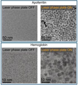
Cryo‑EM has transformed structural biology over the past decade, earning a Nobel Prize in 2017 for enabling high‑resolution structures without crystallization. But despite its impact, the technique still struggles with proteins below ~70 kilodaltons—a size range that includes about 90% of the human proteome. “Because of signal-to-noise limitations, the majority of human and animal proteins are too small to be analyzed by these methods [cryo-EM and cryoelectron tomography]. The increase in signal-to-noise ratio provided by this laser phase plate is expected to overcome these important limitations.”
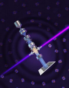
The new LPP begins to address that problem. The LPP uses an intense, continuous‑wave laser to shift the phase of the electron beam itself. This produces true phase contrast without dimming or destabilizing the beam. Mueller described the laser focus as “75 kilowatts focused to a few microns… That’s more powerful than what you use for welding. It has more power than a military laser. It builds up the brightest continuous laser focus ever.”
Installed in a custom Thermo Fisher Titan Krios, the LPP immediately improved the clarity and resolvability of small proteins, including hemoglobin, which sits at the lower limit of what today’s cryo‑EM instruments can handle. As the authors wrote in the abstract: “Here, we show that the laser phase plate (LPP)… enhances the resolution in single-particle reconstruction of small proteins by improving specimen-motion correction, recovery of information from the early frames, as well as particle visualization, 3D classification, and alignment.”
These improvements were achieved using standard defocus ranges and reconstruction workflows. “For the most challenging cases—small particles, bad specimens—the laser produces a very considerable advantage,” Mueller said.
The impact extends beyond single‑particle analysis. Cryo‑electron tomography (cryo‑ET), which assembles multiple angular views of a molecule or protein into a three-dimensional image, stands to benefit even more. “With cryo-ET, we’re looking at small, very complicated cellular material that’s incredibly crowded inside the cell,” said Bridget Carragher, PhD, founding technical director of imaging at Biohub. “It’s like a forest of trees, and you’re trying to find one leaf on one tree in there. Cryo-ET needs a dramatic step forward in contrast, so we can start to see what’s going on inside the cell. That’s what the laser phase plate promises to give us.”
Biohub is developing a dual‑laser version of the system, designed to reduce component wear and minimize aberrations. Meanwhile, Mueller’s team is pushing toward imaging proteins as small as 17 kilodaltons, a threshold that would open access to vast regions of the human proteome previously invisible to cryo‑EM.
“This technology is a step function change for biology,” said Stephani Otte, PhD, Biohub’s vice president of imaging science. “What was once invisible will become visible—and that changes everything about how we understand disease.”
“The bottom line is, if you have a large protein and a really good sample—a fresh one or one frozen without bubbles, for example—you may not need the phase plate to get a single, high-quality image. But for a small protein and a bad sample, laser-on is best,” Mueller said. “This could fill an enormous gap in our knowledge of protein structures that can’t be crystallized or are too small for today’s cryo-EM. And it will be revolutionary for cryo-ET.”
The post Laser‑Driven Phase Contrast Enhances Cryo‑EM Resolution of Small Proteins appeared first on GEN – Genetic Engineering and Biotechnology News.
Microglia at a key inflection point in Alzheimer’s disease
Nature Medicine, Published online: 11 June 2026; doi:10.1038/s41591-026-04409-3
We analyzed brains from octogenarians and cognitively resilient centenarians to understand why some individuals with substantial Alzheimer’s disease pathology develop dementia whereas others remain cognitively intact. Spatial transcriptomics revealed gene expression changes in discrete tissue domains surrounding amyloid plaques and tau pathology that distinguish early, clinically silent, disease from later stages associated with cognitive decline.
Targeting Mu opioid receptor neurons of the habenula to limit naloxone aversion
Opioid use disorder is a chronic, relapsing condition that continues to rise worldwide. Naloxone (Narcan®), an opioid antagonist, reverses overdose but triggers strong negative affect. The neuronal circuits underlying these aversive effects remain unclear. We previously identified mu opioid receptor–expressing habenular neurons (Hb-MOR) as key encoders of negative emotional states and hypothesized that they also mediate naloxone aversion.
Pioneering Innovation in Transplant Diagnostics
Sara Barrington is CEO of Verici Dx, a precision diagnostics company redefining how transplant clinicians understand and predict graft health. By combining multi-omic analysis with proprietary artificial intelligence (AI) models, Verici Dx delivers predictive, actionable intelligence that helps clinicians optimize therapy, inform biopsy decisions, and stratify risk for complex transplant patients.
In this edition of Asked & Answered, Barrington speaks with Inside Precision Medicine Editor in Chief Damian Doherty about pioneering innovation in transplant diagnostics to better reflect biological and patient complexity, and ultimately, improve health outcomes.
Q: Tell me about Verici Dx, its founding, mission, and products.
Sara Barrington: Verici Dx was founded with a very clear purpose: to fundamentally improve how transplant clinicians are informed and manage graft health. Transplant medicine requires high-stakes, time-sensitive decision-making to promote longevity of the graft, minimize the risk of complications, and improve the patient’s quality of life.

Kidney transplantation has long struggled with one fundamental challenge: accurately identifying rejection early and distinguishing it from other causes of injury and graft dysfunction. Clinicians often rely on conventional blood markers such as serum creatinine or, more recently, donor-derived cell-free DNA. While these tools are useful for signaling that “something is wrong,” they are injury markers and lack a level of precision important in rejection biology.
Creatinine levels, for example, increase only after functional damage has already occurred and cannot differentiate rejection from non-immune causes of injury such as ischemic reperfusion injury, drug toxicity, or viral infections like BK virus–associated nephropathy. Biomarkers can generate false positives, triggering biopsies or treatment escalation that may not be necessary and can expose patients to avoidable risk.
This lack of specificity has made rejection difficult to manage proactively. By the time rejection is clearly identified, tissue injury may already be established. At the same time, clinicians are understandably cautious about intensifying immunosuppression without clear evidence, given the associated risks of infection, malignancy, and toxicity.
Our mission is to close this information gap by delivering precision diagnostics that reflect the true complexity of transplant biology and translate that complexity into insights clinicians can proactively and confidently apply to patient care.
We operate at the intersection of multi-omic science, advanced analytics, and clinical reality. We know that transplant rejection and graft injury are not single-signal events, but are multifactorial, dynamic biological processes. We design our diagnostics to capture that complex biology in a way that mirrors how clinicians think about their patients, rather than forcing clinical decision-making to fit the limitations of older technologies.
Our lead product, Tutivia , is a blood-based test that assesses the immune and broader, relevant biological status of kidney transplant patients to identify the risk of all forms of acute rejection, including T cell (TCMR) and antibody-mediated rejection (ABMR). Tutivia combines transcriptomics with proprietary AI models to address this precise unmet need: biologically accurate insight into what is actually happening within the graft.
The test delivers a risk score that enables more precise immunosuppression management by distinguishing rejection from other causes of graft injury, such as BK virus–associated nephropathy and ischemia-reperfusion injury, which can confound conventional blood tests like serum creatinine and donor-derived cell-free DNA.
By focusing on the immune and other pathways associated with rejection, Tutivia moves beyond damage markers and toward a direct assessment of rejection biology itself. That shift, from injury detection to biological interpretation, is what makes the test such a valuable tool for transplant clinicians.
Thermo Fisher Scientific recently launched its One Lambda Pre-Transplant Risk Assessment (PTRA) assay, using pre-transplant prognostic technology licensed from Verici Dx. The assay provides a risk score for early acute rejection based on a patient’s unique gene profile, helping clinicians better balance the risk of rejection against the potential side effects of over-immunosuppression. Using a 29-gene mRNA signature, the PTRA assay stratifies patients into high- and low-risk categories, with clinical validation showing high-risk patients are seven times more likely to experience early acute rejection.
Q: Tutivia is built on RNA sequencing. Why is precision transcriptomic analysis well-suited for identifying rejection biology?
Barrington: The need for reliable biomarkers is crucial for individualizing therapy that offers the potential to extend allograft survival.
Early research underpinning the science leading to the development of Tutivia was led by principal investigators at The Mount Sinai Hospital in New York, one of the world’s leading transplant research centers, combining decades of clinical expertise with rigorous science. The underlying science reflects the fact that rejection in kidney transplant patients is not driven by a single molecule or pathway, it results from coordinated immune and other biologic pathways across multiple cell types, signaling cascades, and regulatory mechanisms. Injury-driven biomarkers capture only a narrow slice of this biology and can be influenced by non-rejection factors, limiting their specificity and reliability.
Precision transcriptomic analysis, on the other hand, measures gene expression patterns across thousands of genes simultaneously. In developing specific tests, this analysis is the basis for gene selection using unsupervised deep learning mathematical modeling, allowing the most impactful and reproducible gene expression signals and interactions to be included in the final signature.
Tutivia uses a peripheral blood next-generation sequencing assay to evaluate a 17-gene mRNA signature in combination with a proprietary artificial intelligence algorithm to categorize kidney transplant patients as at low risk or high risk of acute rejection.
This provides a comprehensive view of immune activation, regulation, and injury responses within the transplanted organ, allowing the identification of signature changes that are truly characteristic of rejection biology.
In short, RNA sequencing lets us ask better biological questions: not just whether the kidney is injured, but whether immune-mediated rejection is driving that injury. That distinction is critical for precision medicine in transplantation.
Q: What does earlier and more precise identification of rejection biology mean for patient management and outcomes?
Barrington: Earlier identification of rejection biology can create an opportunity to intervene before irreversible damage occurs. When clinicians have confidence in what the biology is telling them, they can make timely, targeted decisions that may preserve graft function.
This precision also supports more individualized care. Rather than applying broad treatment strategies, clinicians can tailor immunosuppression based on biological risk, potentially reducing over-treatment and its associated complications.
For patients, this can translate into fewer unnecessary biopsies, fewer adverse events related to immunosuppression, and a greater likelihood of long-term graft survival. Ultimately, better information leads to better decisions, and better decisions lead to better outcomes.
Our vision is that precision diagnostics like Tutivia become a standard component of transplant management, providing earlier insight into rejection biology, supporting more informed clinical decisions, and helping clinicians intervene at the right time for the right patient.
Q: AI is central to your platform. How does Verici Dx apply AI differently from others in diagnostics?
Barrington: We apply AI with rigor and restraint. We use advanced machine learning and mathematical modeling to analyze highly complex biological data, but always within a disciplined scientific framework. Our models are developed using unsupervised approaches and validated extensively on inclusive patient populations. Once validated, they are locked and reproducible; they are not evolving over time. That distinction matters because clinicians are making life-altering decisions based on these results, and trust, consistency, and validation are essential.
Q: Validation and standards are recurring themes for Verici Dx. How was Tutivia validated?
Barrington: Tutivia was validated through a rigorous clinical study that included a broad and diverse transplant patient population, rather than narrowly curated cohorts. We deliberately chose a more difficult validation path because clinical reality is complex, and diagnostics must perform under those conditions. While this approach requires more time and effort, it ensures the test is reliable, reproducible, and clinically relevant across the full spectrum of patients that clinicians see every day.
Q: What’s in the future for the company?
Barrington: Verici Dx is focused on deepening the clinical adoption of Tutivia as well as expanding the studies demonstrating the utility of an earlier and more precise biomarker in improving patient outcomes. Long-term outcomes for transplant patients involve more biological pathways, and significant complexity is involved in managing care. We are focused on developing and validating tests using both transcriptomic and proteomic precision tools to assist clinicians and their patients in this journey.
Damian Doherty has been in media and publishing for over 30 years, beginning at News Corporation. Damian has managed, edited, and launched life science titles in drug discovery and precision medicine. He was features editor of Drug Discovery World and founded the Precision Medicine Leaders Summit and the Journal of Precision Medicine. He edited AIMed magazine before launching Photo51Media, a platform for illuminating untold, compelling stories in precision healthcare. Damian joined Mary Ann Liebert in 2021 to help steer the new rebrand and relaunch of Clinical OMICS to Inside Precision Medicine.
The post Pioneering Innovation in Transplant Diagnostics appeared first on Inside Precision Medicine.