

Genetically Raised Pulse Pressure Predisposes to Dementia-Linked Death

People who carry a higher number of genetic variants for increased pulse pressure may be at increased risk of having dementia as a contributing factor in their deaths, research suggests.

The findings, in Neurology, point to shared genetics underpinning cardiometabolic disease and dementia.

Although it is known that cardiometabolic conditions such as high blood pressure, diabetes, and stroke are associated with the neurodegenerative disease, it has been unclear how genetic predictors for the former influence the latter.

Pulse pressure is the difference between systolic and diastolic pressure and a higher number is indicative of poorer cardiac health, such as hardened arteries or poor heart function.

“While having the APOE ɛ4 allele, a gene variant, is the strongest common genetic risk factor for Alzheimer’s disease, some people may inherit a combination of small effect gene variants linked to cardiometabolic disease that may also increase the risk,” explained researcher Laura Raffield, PhD, from the University of North Carolina at Chapel Hill.

“Our study found an association with genetic variants linked to high pulse pressure and an increased risk of death from dementia.”

Late-life dementia is strongly inherited, with genetics responsible for around 60% to 80% of Alzheimer’s disease.

While the APOE ɛ4 allele is a well-known genetic risk factor for Alzheimer’s disease, other common genetic variants have been implicated and there may also be vascular contributions.

The study investigated how polygenic risk scores for cardiometabolic disease related to dementia-related outcomes among more than 8000 participants in the Reasons for Geographic and Racial Differences in Stroke (REGARDS) study.

The authors note that REGARDS has particularly high numbers of Black participants, who are underrepresented in late-life dementia research, and eight in 10 in the current study identified as non-Hispanic Black.

Study participants had a mean age of 63.7 years at the start of the study and underwent screening for cognitive function annually. They were followed for up to 14 years for risk of death from dementia and nine years for cognitive impairment, on average.

A total of 619 people developed cognitive impairment during this time compared with 7159 with no such impairment. The 459 individuals who had dementia listed as a cause of death were compared with 8362 who died from other causes or for whom the reason was missing.

A higher polygenic risk score for pulse pressure was associated with a 16% increased risk of dementia as a contributing cause of death, after accounting for potential confounding factors.

This statistically significant result was not seen in polygenic risk scores for systolic or diastolic blood pressure, potentially supporting earlier research indicating that pulse pressure is a better predictor of coronary heart disease.

The polygenic risk score for pulse pressure was not associated with incident cognitive impairment, suggesting limited shared genetics between this dementia-related outcomes.

Overall, there were significant or nominal associations in polygenic risk scores for triglycerides, pulse pressure, type 2 diabetes and both the endpoint of Alzheimer’s disease and related dementias and also dementia as a contributing cause of death. However, this was not the case with incident cognitive impairment.

In addition, the results confirmed that the APOE variant was a predictor of both dementia as a contributing cause of death and incident cognitive impairment, and that local African ancestry was associated with nominally attenuated effect size.

The authors concluded that the inconsistent results between incident cognitive impairment and dementia as a contributing cause of death “imply that different dementia-related end points may capture distinct biological pathways.”

They elaborated: “For example, genetic risk factors of [cardiometabolic diseases] may be more strongly related to later disease progression, such as amyloid accumulation throughout later life, rather than to early cognitive impairment.”

The post Genetically Raised Pulse Pressure Predisposes to Dementia-Linked Death appeared first on Inside Precision Medicine.

NIIMBL to Support Vector Production and AI-Ready Training Projects

Viral vector production and training schemes designed to foster development of an AI-ready workforce dominate the list of projects selected for support by the U.S. National Institute for Innovation in Manufacturing Biopharmaceuticals (NIIMBL).

The institute, a public-private partnership focused on advancing manufacturing and solving industry challenges, announced its latest funding awards, explaining that the 39 recipients would support U.S. production and talent development.

Sandeep Kedia, NIIMBL senior technology fellow and project call program lead, says the projects “represent the kind of innovation needed to strengthen the nation’s biopharmaceutical manufacturing capabilities.

“By bringing together advanced process analytical technologies, AI-driven optimization, and next-generation production platforms, our members are helping accelerate the adoption of transformative technologies across the industry,” he adds.

Several of the selected projects focus on the production of adeno-associated viral (AAV) vectors—hollow viruses used to deliver genetic information—which play a crucial role in cell and gene therapy manufacturing.

For example, researchers at Michigan Technological University will work with industry partners on an aqueous two-phase continuous vector purification system. The aim is to boost yield while reducing cost, labor, and analytical complexity.

Similarly, a team at North Carolina State aims to develop “improved purification materials that can better capture full AAVs, along with machine-learning software that identifies optimal process conditions.”

The third vector-focused project will see an MIT group work with EMD Millipore, Landmark Bio, and Virica Biotech to try to reduce the number of empty viral capsids inadvertently made during vector production.

The researchers will combine an approach called decoupled replication-initiated vector encapsulation, or DRIVE, with various process control strategies to create a platform that makes high-titer, high-quality rAAVs.

According to the MIT team, “By reducing [the proportion of] empty capsids, the approach can streamline downstream purification, reduce time and cost, and improve the overall quality of gene therapy products.”

AI-ready workforce

In addition to the technology projects, NIIMBL will support several training programs with an emphasis on ensuring the next generation of biopharmaceutical engineers are AI-ready, according to workforce director John Balchunas.

“Our workforce initiatives are designed to meet talent needs head‑on by creating more innovative pathways into biomanufacturing careers,” he says, adding, “These new projects will strengthen partnerships and ensure that learners can gain the skills needed to thrive in a rapidly evolving biopharma industry.”

One such project will see a team at Texas A&M University’s National Center for Therapeutics Manufacturing expand an existing effort called NeuroPipes, which seeks to interest neurodiverse people in careers in biopharma. The aim is to provide technical skills training that prepares neurodivergent adults for careers in drug manufacturing.

Another project will see Wistar Institute researchers set up BioPATH, a national consortium focused on advancing workforce training in biomanufacturing, AI, and automation.

The idea, according to the Wistar team and collaborators at the International Academy of Automation Engineering, is to “bridge the gap between foundational bioprocess and GMP knowledge and the emerging needs of automation, data-driven manufacturing, and digitally enabled quality systems.”

The post NIIMBL to Support Vector Production and AI-Ready Training Projects appeared first on GEN – Genetic Engineering and Biotechnology News.

A novel early screening approach for MCI due to AD based on a “maze” hand-interaction kinetic paradigm

BackgroundMild cognitive impairment due to Alzheimer’s disease (MCI due to AD) is a crucial stage for the early identification of Alzheimer’s disease (AD), and timely detection at this stage may provide opportunities for earlier intervention and potentially delay disease progression.MethodsThis study proposed a digital early-screening method based on a touchscreen maze hand-interaction kinetic paradigm, which integrates digital biomarkers from the visuospatial/executive and episodic memory domains to support the screening of MCI due to AD. A customized maze task was administered to 40 patients with clinically diagnosed MCI due to AD and 40 healthy controls (HC). Behavioral data were collected, and two categories of digital biomarkers were extracted: (1) visuospatial/executive digital biomarkers, such as task completion time (VSETT) and average movement speed (VSES); and (2) episodic memory digital biomarkers, such as episodic memory total time (EMTT) and number of correct choices (EMCC). Significant digital biomarkers were identified through between-group comparisons, and their combined classification performance was evaluated using binary logistic regression and receiver operating characteristic (ROC) analysis.ResultsThe integrated digital biomarker model showed promising apparent discriminative performance in the full cohort, with an AUC of 0.899 (95% CI: 0.831–0.967). To reduce potential optimism associated with biomarker selection, model development, and model evaluation within the same dataset, internal validation was performed using full-pipeline repeated stratified five-fold cross-validation with all 16 candidate digital biomarkers entered into the validation procedure and biomarker selection repeated within each training fold. The internally validated model retained good discriminative performance, with a mean cross-validated AUC of 0.842, an empirical 95% interval of 0.779–0.878, an accuracy of 0.783, a sensitivity of 0.772, and a specificity of 0.795.ConclusionThese findings suggest that the proposed touchscreen maze-based digital assessment method may provide a promising and objective approach to supporting the early screening of MCI due to AD.

Prediction model for postoperative delirium risk in elderly hypertensive patients: machine learning-based development and validation

BackgroundPostoperative delirium (POD) is a severe complication in elderly hypertensive patients, associated with poor long-term outcomes. Existing models often rely on intraoperative data, limiting preoperative risk stratification. This study aimed to develop a non-invasive machine learning model to predict POD and investigate its preoperative markers’ impact on three-year mortality.MethodsPreoperative variables were selected using LASSO regression from a cohort of 1,782 patients. Ten machine learning models were trained and validated (7:3 ratio). Model performance was evaluated via AUC-ROC and decision curve analysis (DCA). The optimal model was interpreted using SHAP values. Long-term prognosis within the POD cohort was assessed using Kaplan-Meier curves and multivariable Cox proportional hazards regression.ResultsThe POD incidence was 10.9%. The Gradient Boosting Machine (GBM) demonstrated optimal performance (AUC = 0.868, 95% CI: 0.819–0.917). SHAP analysis identified MMSE score as the most influential predictor, followed by HADS score, age, CFS, frailty, and PSQI score. Multivariable Cox analysis revealed that lower MMSE, alongside elevated HADS, CFS, frailty, and PSQI scores—but not chronological age—were independent predictors of increased three-year mortality in POD patients (all P < 0.05).ConclusionWe developed a robust machine learning tool for individualized POD prediction. Cognitive impairment, psychological distress, frailty, and poor sleep quality serve as critical dual-prognostic markers for both acute POD occurrence and long-term survival. These findings underscore the necessity of routine multidimensional preoperative assessment to facilitate personalized interventions for vulnerable hypertensive populations.

Clinical practice guidelines and quality standards for early intervention in psychosis: an AGREE II appraisal and systematic review of service components

BackgroundEarly intervention in psychosis (EIP) is a key component of youth mental health care, yet recommended models of care for individuals at clinical high risk for psychosis (CHR-P) and first-episode psychosis (FEP) remain heterogeneous across jurisdictions. No previous study has combined a formal AGREE II appraisal with a structured synthesis of core service components for EIP.MethodsWe conducted a systematic review and AGREE II-based appraisal of international clinical practice guidelines (CPGs) and quality standards (QSs). MEDLINE (via PubMed), the Cochrane Library (CENTRAL), Web of Science, guideline repositories, and grey literature sources were searched to March 2025 in accordance with PRISMA 2020 guidance. Eligible documents were CPGs or QSs published in English or Italian from 2005 onward containing recommendations on the organization, assessment, and treatment of CHR-P and/or FEP. Methodological quality was appraised with AGREE II (inter-rater agreement assessed using ICCs); recommendations were extracted, and core components required endorsement by at least one third of relevant documents with at least one strong or mandatory recommendation. Twenty-six documents (24 CPGs and 2 QSs) were included. Methodological quality was moderate overall, with substantial variability across AGREE II domains: scores were generally strongest for scope and purpose, stakeholder involvement, and clarity of presentation, and more variable for Rigour of development, applicability, and editorial independence.ResultsWe identified 32 core recommendations. For CHR-P, the most consistent components were specialized services or dedicated pathways, comprehensive multidisciplinary assessment, cognitive-behavioural therapy, family interventions, and a cautious approach to antipsychotic use. For FEP, the most consistently endorsed components were specialized multidisciplinary teams, assertive and continuous follow-up, family work, psychoeducation, structured pharmacological monitoring, and supported education and employment.ConclusionThese findings provide a guideline-derived framework for youth EIP service delivery and model development, while highlighting the need for more methodologically robust and implementation-oriented guidance across settings.Systematic review registrationhttps://osf.io/cek7u, identifier cek7u.

Case Report: Multimodal clinical and neurophysiological effects of cranial electrotherapy stimulation in a patient with chronic stress and sleep disturbance

IntroductionCranial Electrotherapy Stimulation (CES) is a non-invasive brain stimulation (NIBS) technique used for anxiety and sleep disorders; however, case-level evidence linking clinical improvement with objective neurophysiological change remains limited. This case presents a multimodal examination of the therapeutic and neurophysiological effects of CES in a patient with sleep disturbances and chronic stress.MethodsA 51-year-old woman with chronic stress, anxiety, and persistent sleep disturbance following thyroid and parathyroid surgery, refractory to pharmacological treatment, underwent a 16-week CES intervention. Outcomes were assessed at baseline and follow-up using EQ-5D-5L, DASS-21, GAD-7, PSQI, and resting-state Electroencephalography (EEG).ResultsAnxiety, stress, and depressive symptoms improved to within normal ranges, alongside significant improvements in mental health-related quality of life. EEG demonstrated increased posterior alpha activity and improved frontal symmetry. Sleep quality showed modest improvement.ConclusionThis case suggests that CES may be a safe adjunctive intervention associated with symptomatic improvement and changes in neurophysiological patterns in stress-related conditions, supporting further controlled investigation.

FOCUS Bipolar: Families Opening Conversations for Understanding Signs

Conditions: Bipolar Disorder (BD); Bipolar Disorder I or II; Bipolar Disorder Family Members; Screening Tool

Interventions: Other: Co-design Workshop (Observational Procedure); Other: Alpha Testing (Observational Procedure); Other: Beta Testing (Observational Procedure); Other: Youth Screening Pathway & Brief Follow-Up Interview (Observational Procedure)

Sponsors: University of Texas Southwestern Medical Center; The Texas Child Mental Health Care Consortium (TCMHCC)

Recruiting

NeuroFinance Human Stress Trial During Financial and Informational Volatility

Conditions: Psychological Stress; Anxiety; Emotional Stress; Autonomic Nervous System Dysfunction; Sleep Disturbance; Insomnia; Behavioral Health; Cognitive Fatigue; Occupational Stress; Financial Stress; Burnout Syndrome; Heart Rate Variability; Neurobehavioral Manifestations

Interventions: Device: Wearable Physiologic Monitoring Platform

Sponsors: Truway Health, Inc.

Not yet recruiting

AI-Powered Blood Test Detects Early Retinal Damage in Diabetes 

Scientists have developed an AI-assisted prediction tool that can identify patients with type 2 diabetes at high risk of developing diabetic retinal neurodegeneration (DRN) before symptoms appear. Their findings were published today in the journal PLOS Medicine.

“Our study suggests that early retinal nerve damage in diabetes leaves measurable signals in the blood,” write the authors of the study, led by Wei Wang, MD, PhD, associate professor at the Guangdong Provincial Clinical Research Center for Ocular Diseases. “These findings suggest that a simple blood test analyzed with artificial intelligence may help identify people with diabetes who are at highest risk of early retinal nerve damage, well before visible damage appears on the retina.”

Type 2 diabetes affects more than half a billion people worldwide, carrying with it an increased risk of long-term complications including progressive neurodegeneration. Retinal nerves are among the earliest tissues to be damaged, which can eventually lead to severe visual impairment and vision loss. However, current diagnostic methods can only detect DRN once the retina has already suffered irreversible damage. 

Wang and colleagues developed a machine learning algorithm called Pro-DRN using data from 1,218 participants in the Guangzhou Diabetic Eye Study, all of whom were diagnosed with type 2 diabetes but had not yet developed DRN at the time of enrollment. The AI model integrated proteomics data from blood samples with yearly retinal images collected over a six-year follow-up period. 

This led to the identification of 71 proteins associated with the development of DRN. Among them, the proteins most consistently driving accurate predictions were ACTA2, COL6A3, and HSPG2, which are key structural components involved in maintaining the integrity of the nerve and muscle tissue in the eyes. These results were then validated in an independent cohort of 502 patients from UK Biobank, where the core effects and protein signals were reproduced. 

Pro-DRN has been deployed as an interactive, web-based risk assessment tool that doctors can use to support early DRN screening and monitor patient evolution over time. Individuals identified as being at high risk of DRN could benefit from more frequent checkups and early interventions aimed at preventing or slowing down progressive neurodegeneration. 

Because DRN is one of the first symptoms of nerve degeneration induced by diabetes, early detection could also signal the onset of nerve injury elsewhere in the body. Such damage can contribute to cognitive impairment, dementia, and peripheral neuropathy, which can cause loss of sensation and motor control in the hands, feet, and other extremities. A single eye test could therefore provide valuable insights into the overall health of the nervous system. 

In addition, the proteins identified to be involved in DRN progression could be investigated as potential targets for the development of novel therapies. Furthermore, the AI-based tool could also prove valuable for the selection and stratification of participants in clinical trials evaluating neuroprotective strategies designed to prevent or delay nerve damage. 

“Pro-DRN may help move diabetic eye care from detecting established damage toward earlier, molecularly informed risk stratification, so that closer monitoring and future neuroprotective interventions can be directed to the people most likely to benefit,” Wang and colleagues write. 

The post AI-Powered Blood Test Detects Early Retinal Damage in Diabetes  appeared first on Inside Precision Medicine.

Disease Detection Gets Boost from Keck’s New Brain Reference Map

Investigators at the USC Mark and Mary Stevens Neuroimaging and Informatics Institute (Stevens INI) at the Keck School of Medicine say they have created one of the largest reference models ever developed for the human brain, using diffusion MRI scans from more than 54,000 people to chart how the brain’s communication pathways develop, mature, and decline across the lifespan.

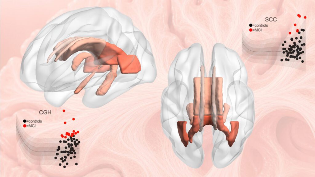
Published in Nature Communications, the study “Lifespan normative modeling of brain microstructure” provides the equivalent of growth charts for the brain’s white matter, the vast network of neural wiring that allows brain regions to communicate, according to the Keck team, which adds that the novel tool offers researchers a new way to detect subtle patterns linked to aging, Alzheimer’s disease, schizophrenia risk, and other neurological and psychiatric conditions.

“Just as pediatric growth charts help clinicians determine whether a child’s height or weight is developing as expected, these brain charts provide a reference for how the brain’s neural pathways typically change over the lifespan,” said Julio E. Villalón-Reina, MD, PhD, a postdoctoral researcher at the Stevens INI and the study’s first author. “That gives us a powerful new way to identify when an individual’s brain wiring falls outside the expected range.”

To study white matter, the team used diffusion MRI, an imaging method that tracks how water moves through brain tissue. Because water movement is shaped by microscopic features such as nerve fibers and myelin, diffusion MRI can reveal subtle changes in tissue organization not visible on standard brain scans.

After compiling diffusion MRI data from 54,583 individuals across 19 international datasets, the researchers built statistical growth and decline charts for the brain’s neural pathways.

The researchers focused on four widely used measures of white matter microstructure across 21 major brain regions. By modeling how these measures vary by age and sex, they generated lifespan curves and percentile ranges that show what is typical at different stages of life.

Statistical charts compiled from a large population allow brain abnormalities to be detected in new individuals. [Stevens INI]

The results revealed that white matter follows distinct developmental and aging trajectories, with some measures reaching peak maturity in early adulthood and others later in midlife.

“Brain development and brain aging are not uniform processes,” continued Villalón-Reina. “The brain’s neural pathways mature on distinct timelines, and some are more vulnerable to decline than others. Our model reveals this structure by merging data on a truly global scale.”

The scientists also discovered evidence for a longstanding theory of brain aging, sometimes described as last in, first out. According to this theory, brain pathways that develop last in childhood and adolescence tend to be more susceptible to decline in older age. The researchers observed that white matter regions that mature later did indeed decline faster in old age, offering new insight linking brain development and aging.

To demonstrate the model’s practical value, the researchers applied it to clinical datasets from people with mild cognitive impairment, dementia, and 22q11.2 deletion syndrome, a genetic condition that increases risk of schizophrenia.

In each case, the model identified alterations in the brain’s circuitry that deviated from age-expected norms. Importantly, these deviations were not identical across individuals with the same diagnosis, highlighting the value of a person-specific approach.

Paul M. Thompson, PhD, associate director of the Stevens INI and senior author of the study

“This monumental study took seven years to complete,” explained Paul M. Thompson, PhD, associate director of the Stevens INI and senior author of the study. “The vast scale of the data and the fine scale of the brain features assessed means we can now evaluate your neural pathways relative to other people of the same age, sex, and demographics. We can see how your brain differs from what we would expect for a person of your age and sex, giving us a tool to use in clinical trials of treatments for dozens of brain diseases.”

When applied to people with dementia and mild cognitive impairment, the model detected atypical white matter patterns in brain regions involved in memory and interregional communication. In people with 22q11.2 deletion syndrome, it identified deviations in multiple key neural pathways, helping researchers discover which brain systems develop differently.

The reference charts may also help researchers evaluate treatments by tracking whether a person’s white matter measures move closer to the expected range, or whether a treatment slows the shift away from healthy patterns over time. The charts will now be used to compare more than 30 brain diseases and conditions, offering a common framework for studying how different disorders emerge, progress, and respond to intervention.

The models are also a publicly available resource that can be extended as additional brain imaging data become available. The methods are now being used to study neurological, psychiatric, and neurodevelopmental disorders by providing a common reference standard for white matter microstructure across the lifespan.

“This study demonstrates the power of large-scale, international data sharing to create tools the entire research community can use,” pointed out Arthur W. Toga, PhD, director of the Stevens INI and provost professor at USC. “By establishing a lifespan framework for the brain’s communication pathways, this work opens new opportunities to detect subtle disease-related changes, compare conditions more rigorously, and move toward a more individualized understanding of brain health.”


The post Disease Detection Gets Boost from Keck’s New Brain Reference Map appeared first on GEN – Genetic Engineering and Biotechnology News.