

ObjectiveThis study aims to systematically analyze neuroimaging research on cognitive control in Autism spectrum disorder (ASD) from 2000 to 2025 using bibliometric methods, in order to reveal the evolutionary trajectory, core knowledge base, research hotspots, and future frontiers of the field.MethodsA search was conducted on the Web of Science Core Collection and Scopus databases, resulting in the inclusion of 1,581 relevant articles. VOSviewer and the Bibliometrix package in R were utilized to conduct a comprehensive visualization and quantitative analysis of annual publication volume, country/institution/author collaboration networks, keyword co-occurrence, document co-citation, and thematic evolution.Results(1) The volume of research literature showed exponential growth, with an annual growth rate of 21.61%, entering a period of rapid development particularly after 2012, which is closely related to the popularization of functional magnetic resonance imaging (fMRI) technology. (2) “Functional connectivity,” “executive function,” and “default mode network” were the most central keywords. “Functional connectivity” rapidly became a hub connecting various themes after 2010, marking a paradigm shift from “functional localization” to “brain network dysregulation.” (3) The “Triple network model” proposed by Menon was the most cited document, laying the core theoretical foundation for understanding ASD as a disorder of large-scale brain network dysfunction. (4) “Transdiagnostic” research has emerged as a new hotspot, while “multimodal imaging,” “machine learning,” and “dynamic connectivity” represent highly promising future directions.ConclusionOver the past two decades, neuroimaging research on cognitive control in ASD has undergone a profound paradigm shift: from focusing on abnormal activation in isolated brain regions to exploring the static and dynamic dysregulation of large-scale brain networks. The research perspective has also expanded from a single-disorder model to a transdiagnostic framework that includes comparisons with other neurodevelopmental disorders (e.g., ADHD). Future research should focus on the fusion of multimodal data, the application of computational psychiatry methods, and the translation of basic research findings into personalized clinical interventions.
Microglial mitochondria transfer to astrocytes via GPNMB-enriched extracellular vesicles alleviates cognitive deficits in tauopathy mice
Nature Neuroscience, Published online: 26 May 2026; doi:10.1038/s41593-026-02317-w
In PS19 mice, microglia-derived glycoprotein nonmetastatic melanoma protein B promotes mitochondrial extracellular vesicle secretion, enabling mitochondrial transfer to astrocytes. This improves astrocytic function and reduces Alzheimer’s disease (AD) symptoms, while GPNMB deficiency impairs it, highlighting new AD treatment avenues.
Virtual Forest-Based Mindfulness for Perinatal Mental Health
Conditions: Perinatal Anxiety; Perinatal Depression; Maternal Mental Health
Interventions: Behavioral: Virtual Reality Forest Mindfulness Intervention; Behavioral: Conventional Mindfulness Intervention
Sponsors: National Taiwan University Hospital
Not yet recruiting
Interventions: Behavioral: Virtual Reality Forest Mindfulness Intervention; Behavioral: Conventional Mindfulness Intervention
Sponsors: National Taiwan University Hospital
Not yet recruiting
Psychological Stress and Support Needs of Volunteer First Responders After Smartphone-based Cardiac Arrest Alerts
Conditions: Mental Health; Stress Disorder, Post Traumatic
Interventions: Behavioral: Structured Debriefing
Sponsors: University Hospital, Aachen; Alexianer Center for Mental Health Care, Aachen; Aachen Institute for Rescue Management and Public Safety (ARS), RWTH Aachen University
Not yet recruiting
Interventions: Behavioral: Structured Debriefing
Sponsors: University Hospital, Aachen; Alexianer Center for Mental Health Care, Aachen; Aachen Institute for Rescue Management and Public Safety (ARS), RWTH Aachen University
Not yet recruiting
Human Gut Organoids with Functional Nerves Developed that Can Be Mass Produced
Researchers at Cincinnati Children’s Hospital Medical Center and Nantes Université in France have designed 3D-printed scaffolding trays that will reportedly allow scientists to produce larger versions of functional human gut organoids twice as fast as previous methods—and these organoids grow their own nerve cells.
This improved technology could help accelerate production of human mini-organ tissues that are large enough to be useful in patching damage or restoring diminished functions of a person’s small intestine, stomach, or colon. Such tissues also would be valuable for future disease studies and to more accurately evaluate organ damage risks linked to oral medications, according to the investigators.
Details of the study “Large-scale and innervated functional human gut tissues for transplantation via transient spheroid confinement” appear in Nature Biomedical Engineering.
Using a confined culture system (CCS), the team grew small intestine, colon, and stomach organoids from tiny spherical forms into centimeter-scale tubular forms nearly 10 times larger than previous methods. Also, unlike methods that require a complex effort to introduce nerve cells, these organoids develop a nervous system on their own.
“By reaching transplantation maturity twice as fast and developing their own functional nerves, these organoids demonstrate how engineering principles can drive biological innovation,” said staff investigator Holly Poling, PhD. “Our confined culture system is more than a production method; it’s a scalable, flexible platform for building complex human tissues.”
New production system prompts faster growth
Experts at Cincinnati Children’s Center for Stem Cell & Organoid Medicine (CuSTOM) have been making miniature versions of digestive system organs for more than 15 years, working on improving the sophistication of the lab-grown tissues. More recently, the team has been developing methods to make enough customized tissue to transplant into patients to help patch organ damage or restore diminished specialized functions.
The new technique uses 3D printing technology to make tray-like scaffolding molds from surgical resin, then filling the molds with degassed polydimethylsiloxane—a flexible rubber-like type of silicone.
The new trays contain grooves designed to confine a collection of sphere-shaped organoids into a row, which encourages the spheroids to fuse together and mature. The fusions occur within a special mix of nutrients and other ingredients that support initial growth from induced pluripotent stem cells (iPSCs) into more complex organoids.
By day six, the discrete spheroids develop into unified constructs along the grooves of the trays. These are moved into another hydrogel medium for continued growth for another eight days.
By day 14, the organoid constructs have produced all the cell types and structures that previously required 28 days to achieve. These tissues are then transplanted into rodents that are genetically modified to minimize rejection risk.
All of the transplanted tissues engrafted in rodents, the co-authors state. After growing in the rodents, the team produced as much as eight cm of functioning small intestine tissue, compared to approximately one cm of tissue using previous protocols. Not only were the structures much larger than previous methods, but now their neuromuscular function was also similar to native human tissue, representing a major advance.
“We are now able not only to generate complex gastrointestinal organoids at scale, but also to guide their differentiation into functional tissues with integrated enteric neuronal networks,” noted senior author Maxime Mahe, PhD. “By leveraging a defined growth environment, the intrinsic self-organization capacity of the cells drives the formation of tissue structures that closely resemble the human gastrointestinal tract.”
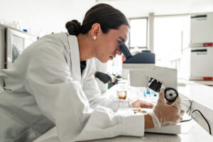
Jim Wells, PhD, a study co-author and chief scientific director at CuSTOM says the new technology overcomes key barriers to scale and function in organoid research and biomanufacturing.
“This platform’s simplicity, reproducibility, and versatility make it accessible for widespread adoption,” said Wells. “In addition, the emergence of a self-organized nervous system within these organoids is particularly important for further studies of neurodevelopmental disorders.”
Another step closer to human clinical trials
Michael Helmrath, MD, a surgeon-scientist at Cincinnati Children’s who co-directs CuSTOM, has been working for more than a decade to develop intestine organoids sophisticated enough for transplantation in human patients.
In 2017, Helmrath and colleagues demonstrated how to combine neural crest cells with intestinal tissue cells in a layered process to make the first human organoids with nerve function. His team also showed how intestine organoids could be grown larger by implanting them in a mouse to provide a blood supply. Ever since, intestine organoids have been getting more sophisticated, including versions with immune cells in addition to the specialized organ cells and nerves.
Now the new process—involving rats instead of mice—produces larger amounts of tissue.
“It is still not possible to grow complete, full-sized human organs in some sort of tank, but research like this has produced significant amounts of tissue that can be matched directly to individual patients,” explains Helmrath. “We believe such tissues, once transplanted, would further grow and multiply as part of the patient’s own organ to restore functions.”
More research and development is needed before “CCS organoids” will be ready for human clinical trials, according to Helmrath. But if successes continue, organoid medicine may allow more infants and children with dysfunctional organs to be treated without ever needing a full organ transplant.
The post Human Gut Organoids with Functional Nerves Developed that Can Be Mass Produced appeared first on GEN – Genetic Engineering and Biotechnology News.
Association between neurodegenerative plasma biomarkers and geriatric depression in older adults with and without clinical dementia in Kinshasa, Democratic Republic of the Congo
IntroductionPrevious Western studies have shown an association between neurodegenerative plasma biomarkers and geriatric depression. However, few studies have examined this relationship in sub-Saharan African countries. This article examines this association in older adults with or without clinical dementia in Kinshasa, Democratic Republic of Congo.MethodsThis cross-sectional study included eighty-one people aged 65 years and older assessed for dementia using the Community Dementia Screening Instrument (CSID) and the Alzheimer’s Disease Questionnaire. Depression and cognitive impairment were assessed using the Geriatric Depression Scale and the African Neuropsychological Battery (ANB), respectively. Plasma biomarkers measured included β-amyloid (Aβ) 40/42, phosphorylated tau 181 (p-tau), neurofilament light chain (NfL), glial fibrillar acid protein (GFAP), interleukins (IL-1β, IL-10), and tumor necrosis factor-α (TNF-α). Logistic regression models were used to estimate the likelihood that these biomarkers are associated with depression, adjusting for sex, age, and education level.ResultsThe mean age of the participants was 72.9 years. Of those suspected of suffering from Alzheimer’s disease (AD), 80% had geriatric depression. High blood pressure was common in participants with suspected AD (60%). People with dementia had significantly lower cognitive scores on all ANB tests than those without dementia. GFAP and NfL were significantly associated with higher odds of geriatric depression, with odds ratios of 1.98 (1.17–3.67; p = 0.02) and 1.76 (1.06–3.20; p = 0.04), respectively. Other biomarkers were not associated with depression.ConclusionThis study suggests a potential role of nonspecific biomarkers of AD, neurodegeneration, and astrocytic glial activation in geriatric depression in older adults in sub-Saharan Africa.
Validation of the CIDI 5.0 Against the SCID-5 for Lifetime Mental Disorders
Conditions: Major Depressive Disorder (MDD); Generalized Anxiety Disorder (GAD); Posttraumatic Stress Disorder (PTSD)
Interventions: Diagnostic Test: CIDI-5
Sponsors: The University of Hong Kong
Recruiting
Interventions: Diagnostic Test: CIDI-5
Sponsors: The University of Hong Kong
Recruiting
Multimodal Pharmacological Study of Clinical Cohorts for Major Depressive Disorder
Conditions: Major Depressive Disorder (MDD
Interventions: Drug: Therapeutic intervention
Sponsors: Shanghai Mental Health Center; Lingang National Laboratory
Not yet recruiting
Interventions: Drug: Therapeutic intervention
Sponsors: Shanghai Mental Health Center; Lingang National Laboratory
Not yet recruiting
Efficacy and Safety Study of Digital Cognitive Training and PCSK9 Inhibitor-Enhanced Lipid-lowering Strategy in Patients With Intracranial Atherosclerotic Stenosis: A 2×2 Randomized Controlled Trial
Conditions: Intracranial Arteriosclerosis; Cognitive Impairment
Interventions: Drug: Rosuvastatin; Drug: Ezetimibe; Drug: Recaticimab; Behavioral: Digital Cognitive Training; Behavioral: Active Control
Sponsors: Peking Union Medical College Hospital
Not yet recruiting
Interventions: Drug: Rosuvastatin; Drug: Ezetimibe; Drug: Recaticimab; Behavioral: Digital Cognitive Training; Behavioral: Active Control
Sponsors: Peking Union Medical College Hospital
Not yet recruiting
Effects of Emotional Intelligence Training on Marital Satisfaction and Mental Health Among Married People in Dhaka City
Conditions: Emotional Intelligence; Mental Health
Interventions: Behavioral: Emotional Intelligence Training
Sponsors: University of Dhaka
Not yet recruiting
Interventions: Behavioral: Emotional Intelligence Training
Sponsors: University of Dhaka
Not yet recruiting