

Background: University hospital employees face role-specific stressors that can impair mental well-being and work-related vitality. While worksite health promotion programs show potential for improving mental well-being by targeting lifestyle behaviors, most target single professions or hospital subunits, and evidence for mental well-being and work-related vitality remains mixed. Mobile apps offer unique advantages for delivering such worksite health promotion programs hospital-wide. However, accessible interventions tailored to a diverse workforce are lacking. Objective: This study aimed to investigate the feasibility of an app-based worksite health promotion program (the Recharge360 program [The Recharge Company]) targeting multiple lifestyle behaviors, including a team-based competition element, for improving mental well-being and work-related vitality of hospital employees over a 5-month follow-up period by evaluating two objectives: (1) the implementation process of the program, and (2) the preliminary effects of the program on mental well-being and work-related vitality. Methods: We included 532 employees (mean age 43, SD 12 y; n=482, 91% women; n=480, 90% highly educated) from a university hospital in Amsterdam, the Netherlands. The study had a single-arm, longitudinal pretest-posttest design lasting 5 months, during which employees participated in the 5-day Recharge360 program (Recharge week) 3 times—in weeks 1, 9, and 17. At baseline (T0) and after each Recharge week (T1-T3), we assessed mental well-being, work ability, need for recovery, and task performance. The process was evaluated by assessing recruitment, attrition, and survey completion rates, and the degree of participation. Preliminary effects were evaluated by linear mixed model regression analyses to assess changes in mental well-being and work-related vitality between baseline and follow-up. Results: Recruitment appeared feasible, but attrition rates were high (up to 70% in the final Recharge week), and the degree of participation decreased over time. We showed statistically significant, albeit small, increases in well-being at T3 (unstandardized β coefficient=2.08, 95% CI 0.33-3.84), with progressively larger improvements in the analyses among those who started at least 1, 2, and all 3 Recharge weeks (unstandardized β coefficient=3.27, 95% CI 1.09-5.45). Results for work-related vitality were mixed. The need for recovery remained unchanged, task performance increased slightly at T3 (unstandardized β coefficient=0.16, 95% CI 0.07-0.24). Work ability showed a small, but statistically significant, decline across follow-up (unstandardized β coefficient=−0.46, 95% CI −0.64 to −0.29). Conclusions: This app-based worksite health promotion program might be feasible to implement in a university hospital setting and shows potential to slightly improve mental well-being, but primarily for a selective group of highly educated, health-conscious women. While these findings support further investigation in a randomized controlled trial in similar university hospital settings, they also highlight the need for more participatory study designs to improve the tailoring of program components and engagement of underrepresented groups, as well as for a supportive culture and population-based approaches at the organizational level.
<img src="https://jmir-production.s3.us-east-2.amazonaws.com/thumbs/05362a750c53672d55df1ef541cf64a9" />
Mini Brains Help Identify Treatment for Rare Form Parkinson’s Disease
Brain organoids created in the lab using patient cells have allowed researchers based at the Wilhelmina Children’s Hospital in Utrecht to find out more about the progression of an ultra-rare type of childhood Parkinson’s disease.
Notably, as reported at the European Society of Human Genetics meeting in Gothenburg this week, the study also identified that an affordable and readily available form of vitamin B3 could help improve symptoms in these children.
Irena Muffels, MD, PhD, a clinical genetics resident at the Wilhemina Children’s Hospital, was working at the Icahn School of Medicine at Mount Sinai, New York, when the study began. Two parents contacted the lab where she was working to ask if they could look into the condition their children had been diagnosed with, a rare form of early Parkinson’s caused by mutations in the DHDDS gene.
“They contacted Professor Eva Morava, for whom I was then working. We started creating mini-brains—tiny blobs of brain tissue grown in the lab from patients’ own cells—thus avoiding the need to take samples directly from the children’s brains,” she explained.
Muffels and colleagues observed these mini brains in the lab and found there were significant signs of deterioration after four months mirroring that seen in the brains of children with this rare condition.
In healthy cells, DHDDS helps make dolichol, a small fat-like molecule that acts as a platform for attaching sugars to proteins. When DHDDS doesn’t work properly, dolichol levels fall. As a result, cells struggle to attach sugar chains, called glycans, to proteins in the usual way.
Those glycans are important because they help proteins fold correctly, reach the right place in the cell, and function as they should. In the patient‑derived mini brains, the team could see that these sugar chains were formed incorrectly.
Low dolichol also disrupts overall lipid regulation and causes cholesterol to build up in astrocytes, the brain’s supportive cells. “This accumulation builds over time, and this is why we think the disease progresses,” said Muffels, “since the accumulation of cholesterol leads to mitochondrial dysfunction, leading in turn to reduced energy production.”
Perlara, a rare‑disease biotech public benefit corporation that runs phenotypic screens in simple ‘patient avatar’ organisms, helped Muffels and colleagues identify nicotinamide mononucleotide, a naturally occurring type of vitamin B3, in a yeast‑based assay as a potential modifier of DHDDS‑driven cellular stress.
The vitamin appeared to have positive effects on the mini brains, and when the families of patients heard about it, they began using the supplement as it is readily available without a prescription and known to be safe. Within a few weeks, the patient’s walking got better and they had more energy and less tremors.
“The word about nicotinamide mononucleotide reached more DHDDS patients, and we currently have 12 patients taking it. We recently received funding from Congenital Disorders of Glycosylation UK… to start an international trial for nicotinamide mononucleotide supplementation in DHDDS-related disease,” said Muffels.
“Patients will take nicotinamide mononucleotide for a year and will be evaluated every three months. Although I have now left the U.S., I hope that the Wilhemina Hospital in Utrecht will be one of the official sites and that I may continue to work with these patients.”
The post Mini Brains Help Identify Treatment for Rare Form Parkinson’s Disease appeared first on Inside Precision Medicine.
ZoBio Introduces DNA-Encoded Library Service for Exploratory Drug Discovery Programs
Officials at Leiden, Netherlands-based CRO ZoBio say the company has launched a DNA-Encoded Library (DEL) discovery service, which is designed to help biotech and pharmaceutical companies generate validated, progressible hits against novel and challenging drug targets.
The new offering combines structure-grade protein production, quantitative biophysics, DEL screening, off-DNA hit validation and X-ray crystallography into a single workflow, according to a company spokesperson, who explains that it enables clients to move beyond hit identification toward high-confidence starting points for drug discovery programs.
ZoBio maintains that unlike transactional DEL screening approaches that focus solely on hit generation, its platform is designed to deliver biologically relevant, structurally characterized hit matter with clear potential for progression. The service is particularly suited to exploratory and difficult-to-drug targets, including protein–protein interactions (PPIs) and targets with poorly defined binding pockets, where conventional screening approaches often fail, notes Gregg Siegal, CEO of ZoBio.
“Drug discovery teams today are increasingly focused on highly validated but technically challenging targets, where traditional screening approaches can struggle to deliver meaningful starting points,” he continues. “Our approach combines DEL technology with the structural biology, biophysics, and assay expertise needed to generate hits that clients can confidently progress.”
Siegal also points out that the DEL service is library-agnostic, enabling clients to access commercially available DEL collections or apply ZoBio’s workflow to proprietary client-owned libraries. The integrated platform reportedly includes:
- Structure-grade protein production and characterization
- Quantitative biophysical assay development using techniques such as SPR
- Biophysically informed DEL selection design
- Interactive DEL data analysis and hit prioritization
- Off-DNA hit resynthesis and orthogonal validation
- Structural characterization through X-ray crystallography
- Mechanistic insight to support downstream optimization
The workflow is designed to support collaborative decision-making throughout the discovery process, helping clients rapidly establish whether difficult or exploratory targets are viable for further development, according to Siegal, who says that members of the ZoBio team will be available for meetings during the BIO International Convention in San Diego to discuss the new service.
The post ZoBio Introduces DNA-Encoded Library Service for Exploratory Drug Discovery Programs appeared first on GEN – Genetic Engineering and Biotechnology News.
This man with ALS is “the first power user” of a brain implant that lets him speak
Casey Harrell has had a set of electrodes embedded in his brain for almost three years. Harrell, who has amyotrophic lateral sclerosis (ALS) and is paralyzed, first used his brain-computer interface (BCI) to “speak” sentences with the help of a research team in 2023.
Since then, Harrell has clocked thousands of hours of use. He can use the device largely independently, once he’s been “plugged in” with the help of a carer. His team has added new features to it, and Harrell also uses it to surf the web and perform his job.
“Living with a disease like ALS, you are supposed to have diminished dreams. I do not,” Harrell tells MIT Technology Review. “Any one of these things would be an absolute godsend of improvement. To have all of them, and many, many more, is truly revolutionary.”
Within the first 22.6 months after the device was implanted, Harrell had used it for more than 3,800 hours at home without any researchers present, the team reported today in the journal Nature Medicine. “He’s the first power user of a speech BCI,” says team member Sergey Stavisky, a neuroengineer at the University of California, Davis.
Decoding speech
Three years ago, Harrell entrusted David Brandman, an associate professor of neurological surgery at the University of California, Davis, and his colleagues with his brain. Harrell, who was 45 at the time, had already been diagnosed with ALS, a degenerative disease that robs people of the use of their muscles.
Harrell was dependent on others to control his wheelchair and to dress and feed him. He had difficulty speaking; people struggled to understand what he was saying. Then Brandman and his colleagues asked if he’d like to trial a brain implant that might help him communicate. “The industry was [on the] cusp of a transformation, and I wanted to be part of it,” says Harrell. He signed up.
In July 2023, during a five-hour operation, doctors implanted four arrays of 64 electrodes each into his brain. Each pair of arrays was wired to a “pedestal” connection point—creating two docking locations on the exterior of his skull to connect the electrodes to a computer.
The team had long been working on developing algorithms to decode brain activity into speech. Their system works by recording activity from the speech motor cortex—a region of the brain responsible for the movements that allow us to speak.
“There are 39 phonemes that make up all the sounds in the [American] English language,” says Nicholas Card, a neuroengineer at UC Davis and member of the team. Mapping neural activity related to producing each of those phonemes can allow the team to create a personalized speech decoder and software that can “speak” those words. “We first go from brain data to phonemes, and then from phonemes to words,” he says.
They started using the device around a month after the surgery. The team got Harrell’s speech decoder working on the first day, says Card. On that day in August, Harrell used the device to speak with a 50-word vocabulary, and 99.6% of the words were as he’d intended. That vocabulary was later expanded to 125,000 words with 97.5% accuracy.
At the time, it was unclear how long the device might last. Brain-computer interfaces are still new—not many people have had them implanted for long periods of time. Scar tissue can form around electrodes in a person’s brain, interfering with their ability to pick up neural activity, for example. But that doesn’t seem to be the case for Harrell.
Power user
In another advance, Harrell is now able to use the device more independently. In 2023, members of the research team would have to visit Harrell at his home and physically connect and disconnect him from the device on the days he wanted to use it. Not anymore. The team has since automated more of the system—today, Harrell’s care partner can don and doff it for him. “He’ll wake up, get plugged in, and just get going,” says Stavisky.
This is important, says Mariska Vansteesel, a BCI researcher at Utrecht Medical Center who was not involved in the trial. “For these technologies to be relevant for patients, we really need to test them in settings in which they will eventually be used … to demonstrate that it has value, that it’s usable, and that it functions well without the constant involvement of a research team,” she says.
The team has also worked to improve the system itself. It is now 99% accurate, says Stavisky. Harrell can also control a cursor—a game changer that enables him to use his personal computer to send text messages and emails, surf the web, and keep up with his job as an environmental activist.
Over the years, the team has updated the system to accommodate specific requests from Harrell. He is now able to switch on a “privacy mode”—when active, any decoded text will be automatically deleted. He can also opt to use a “profanity filter” while he’s talking to his young daughter.
“We have been able to add on to the software side of the device … improving the accuracy and adding more bells and whistles to enable me to be more independent when using the device,” says Harrell. “We are making the road as we walk it, or roll it, so to speak.”
Nothing short of revolutionary
Vansteesel cautions that while the device is working well for Harrell, there’s no guarantee it will work as well, or as long, for other people with ALS. Over the last decade, she has worked with a woman with ALS who used a fully implanted device to communicate using “brain clicks”—cursor clicks made using brain activity. The woman used her BCI for seven years, but it stopped working toward the end of that period, apparently due to brain degeneration.
At any rate, not everyone with ALS will be willing to undergo invasive brain surgery, says Jane Huggins, who is developing noninvasive BCIs at the University of Michigan and was not involved in the trial. “Long-term, independent use with efficient and accurate communication is kind of the holy grail of BCI,” she says. “But we have been finding a consistent aversion to hospital stays among people with progressive conditions like ALS.”
Harrell, however, calls the device “nothing short of revolutionary.” “This has allowed me to keep working and earn money and insurance for my family. This is reconnecting me with friends and family who are too shy or too afraid to come over and not be able to understand me,” Harrell says. “With my seven-year-old daughter, I am able to create a bond that I wasn’t before able to forge. Now I can read to them and help them sharpen their own reading skills. By doing so, I am able to share the responsibility of parenting with my wife, who does so much caregiving for me and also our daughter.”
Stavisky and his colleagues hope to improve the device further still. “We’re never satisfied,” he says. One aim is to eventually restore Harrell’s “full voice.” They are working on a “brain-to-voice” system that could directly decode brain activity to a speaking voice, complete with natural-sounding cadence, inflection and intonation—a voice that could sound happy, angry, or sarcastic, for example.
“I was quietly confident that I could get some personal benefit from the system,” says Harrell. “Never in a million years would I think that I would achieve this much.”
Long-Read DNA Test Could Replace 15 Existing Tests for Rare Diseases
Researchers in the Netherlands have developed a DNA test for rare diseases that can provide much more comprehensive results than standard diagnostics in a shorter amount of time. A study published today in the New England Journal of Medicine reports that this new approach could replace 15 other genetic tests with a single analysis while increasing the number of patients who successfully receive a diagnosis.
Taken together, all rare diseases affect approximately 400 million people worldwide. Of the more than 7,000 rare diseases that have been identified so far, about 80% are caused by genetic mutations. Obtaining a diagnosis can be critical for people suspected of having a rare disease, offering them perspective for the future, better guidance and treatment, and less uncertainty. However, these patients often have to undergo multiple rounds of testing and wait years before receiving a definitive answer.
The new test is based on long-read genome sequencing, a technology that can read significantly longer stretches of DNA before assembling them into a complete genome. While conventional genomic tests typically read fragments around 300 nucleotides long, long-read sequencing can analyze stretches of up to 20,000 nucleotides at a time. The longer reads make it easier to accurately assemble the full genome, providing a more complete picture of the patient’s genetic makeup.
“Thanks to long reads, we obtain an even more complete view of DNA and can detect complex and hard-to-find abnormalities. We then link these to specific conditions,” says Alexander Hoischen, PhD, professor of genomic technologies at Radboud University Medical Center. “In this way, our knowledge grows and we can make more diagnoses.”
In addition, the test can detect epigenetic modifications in the genome that affect gene function without altering the underlying DNA sequence. Although these modifications can be responsible for some rare disorders, conventional testing methods are currently unable to detect them.
“With current diagnostics, this requires additional specialized tests, but with long reads we capture these modifications as a bonus—two in one,” explains Christian Gilissen, PhD, professor of genome bioinformatics at Radboud University Medical Center.
Earlier this year, the technology was used as part of the National Undiagnosed Hackathon, where over 140 experts across the Netherlands came together to search for a diagnosis for 33 families. Long-read sequencing was used to map their DNA in detail, leading to five new confirmed diagnoses within two days as well as strong suspicion of a diagnosis for another eight families.
As the number of rare disease diagnoses continues to rise, this new test could make the diagnostic process much faster and more efficient. Long-read sequencing could also help researchers identify and investigate the genetic drivers of rare conditions, many of which remain largely understudied.
Based on these findings, Lisenka Vissers, PhD, professor of translational genomics at Radboud University Medical Center, calls for the technology to be adopted worldwide as the first choice diagnostic approach when testing patients suspected to have a rare genetic disorder.
The post Long-Read DNA Test Could Replace 15 Existing Tests for Rare Diseases appeared first on Inside Precision Medicine.
Hantavirus One-Shot mRNA Vaccine Fully Protects in Syrian Hamster Model
Last month, the Andes virus outbreak on a Dutch cruise ship departing from Argentina brought a transmission context for hantavirus, that was previously unprecedented, to the forefront. The Andes virus is the only member of the hantavirus family that is capable of efficient person-to-person spread through close contact with respiratory secretions. Other hantaviruses are typically spread through contact with infected rodents, making the Andes virus a much more significant public health threat.
While at sea, the outbreak spread among passengers and crew, infecting 13 people and killing three. The cruise passengers have since returned to their home countries, 23 in total. Because a person can carry the virus for weeks before showing any symptoms, health agencies are facing a complex challenge of identifying everyone who was exposed. There are currently no vaccines or preventive treatments approved for the virus; this travel-related outbreak brought the need for vaccine development to the forefront.
Researchers at The University of Texas Medical Branch (UTMB) had previously developed and tested two mRNA vaccines against intramuscular Andes virus challenge in golden Syrian hamsters (“1-methylpseudouridine-modified or non-modified mRNA modalities encoding the envelope glycoproteins, Gn and Gc, in a single open reading frame.”)
When tested in the Syrian hamster model, both mRNA vaccines were efficacious in hamsters using a two-dose regimen. Recognizing that a fast-moving international outbreak doesn’t allow time for patients to wait weeks between shots, the team retested the vaccines to determine whether a single dose would be effective.
Now, a new report shares the finding that the vaccine provided full protection against the Andes hantavirus after a single dose.
This work is published in The Lancet in the paper, “Single-dose mRNA vaccines against Andes hantavirus.”
Alexander Bukreyev, PhD, head of the Laboratory of Viral Pathogenesis and Vaccine Development at UTMB, said that the group is working to fast-track these single-dose vaccines into human clinical trials.
The results exceeded expectations. When testing the vaccines in an animal model that mimics human disease, the scientists found that a single shot provided 100% protection against a lethal dose of the virus. Even when the researchers significantly lowered the dosage to a fraction of the original amount, the results remained definitive.
“Every vaccinated animal remained completely healthy and showed no symptoms or weight loss,” said Michelle Meyer, PhD, senior scientist in the Bukreyev Laboratory. “When we looked at the tissues from the vaccinated animals a month after infection, the virus was entirely gone. The vaccines triggered a powerful immune response, creating protective antibodies in as little as 14 days.”
Because the Andes virus can take a relatively long time to make a human severely ill, these fast-acting vaccines could serve a dual purpose, possibly functioning as an emergency tool for people who have already been exposed.
“If given quickly to high-risk contacts during an outbreak, such as the Andes virus situation on the cruise ship, the vaccines could theoretically jump-start their immune systems fast enough to intercept the virus—stopping it from replicating and preventing them from getting sick or spreading it further,” Bukreyev said.
The post Hantavirus One-Shot mRNA Vaccine Fully Protects in Syrian Hamster Model appeared first on GEN – Genetic Engineering and Biotechnology News.
Perceived organizational climate and turnover intention among young nurses from a humanistic care perspective: the mediating role of work engagement
BackgroundThe high turnover of young nurses poses a significant challenge to the stability of healthcare systems worldwide. While the relationships between perceived organizational climate, work engagement, and turnover intention are established, there is a lack of research integrating a humanistic care management perspective to elucidate the specific psychological mechanisms among young nurses at their uniquely vulnerable career stage. Drawing upon this contextual lens, this study aims to evaluate the status of perceived organizational climate, work engagement, and turnover intention among young nurses, and to further explore the potential mediating role of work engagement in the relationship between perceived organizational climate and turnover intention.MethodsA cross-sectional study was conducted from July to September 2025, surveying 366 young nurses from a tertiary Grade-A hospital in Shaanxi Province, China, using convenience sampling. Data were collected using a general information questionnaire, the Utrecht Work Engagement Scale, the Organizational Climate Questionnaire, and the Turnover Intention Questionnaire. A Structural Equation Model was employed to analyze the mediating effect.ResultsThe findings revealed significant interrelationships among perceived organizational climate, work engagement, and turnover intention. Work engagement was found to partially mediate the relationship between perceived organizational climate and turnover intention during young nurses, explaining 34.78% of the variance.ConclusionThese findings suggest that organizational climate functions as a critical job resource that may buffer turnover intention by fostering higher levels of work engagement. To maintain workforce stability, nursing managers should integrate humanistic care into organizational policies to cultivate a supportive environment. However, due to the cross-sectional design and convenience sampling from a single institution, causal inferences should be made with caution, and the generalizability of the findings may be limited.
Symeres Expands Spray Drying Capabilities at Its U.S. New Jersey Site
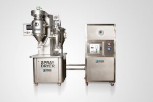
Netherlands CRDMO Symeres expanded its spray drying capabilities at its Cranbury, NJ CMC development site to support formulation development for poorly soluble and development-challenged small-molecule drug candidates.
The expanded capability is designed to support bioavailability enhancement strategies, including amorphous solid dispersions (ASDs), particle engineering and solubility optimization for compounds progressing from preclinical development through Phase II clinical activities.
The investment strengthens the company’s integrated CMC offering by combining spray drying, formulation sciences, analytical characterization, solid-state sciences, and process development within a single development environment, according to Henning Steinhagen, CEO of Symeres. This enables sponsors to progress from early formulation screening through to clinical-ready material within one coordinated scientific framework, reducing tech-transfer risk, accelerating decision-making, and improving development continuity, he adds.
“Our investment in spray drying reflects our commitment to supporting customers across the entire drug development journey,” continues Steinhagen. “While Symeres is widely recognized for its discovery expertise, this expanded capability further strengthens our ability to support complex molecules through development and into the clinic.”
“An increasing proportion of modern small molecule drug candidates require advanced formulation approaches to achieve acceptable bioavailability and clinical performance,” explains Paul O’Shea, managing director at Exemplify BioPharma, a Symeres company. “By expanding our spray drying capabilities within our integrated Cranbury CMC site, we can help clients address developability challenges earlier, reduce operational complexity, and support faster progression into clinical development.”
A Symeres spokesperson, who says the Cranbury site now supports laboratory-scale and pilot-scale spray drying workflows for a range of formulation development activities, including rapid material screening, process optimization and scalable process development, notes that the platform is particularly suited to Biopharmaceutical Classification System (BCS) Class II and IV compounds, highly lipophilic molecules and targeted therapies requiring enhanced oral exposure.
The post Symeres Expands Spray Drying Capabilities at Its U.S. New Jersey Site appeared first on GEN – Genetic Engineering and Biotechnology News.
Microglial State Shift May Determine Whether Alzheimer’s Disease Pathology Leads to Dementia
Researchers from the VIB-KU Leuven Center for Neuroscience, the UK Dementia Research Institute, and Muna Therapeutics have discovered a biological transition that occurs in Alzheimer’s disease (AD) that may influence whether the accumulation of amyloid-β plaques and tau pathology progresses to dementia. Using human brain tissue from octogenarians with and without dementia as well as cognitively healthy centenarians, the team found that a shift in the behavior of microglia occurs at a critical point between amyloid-driven inflammation and tau-associated neurodegeneration. The research, published in Nature Medicine, point to these microglial transitions as a potential new target for treating the disease.
“This has been an exciting journey with many partners,” said co-senior author Bart De Strooper, MD, PhD, a professor at VIB-KU Leuven Center for Neuroscience, Belgium. “The study, entirely based on human donor material, provides insight into one type of resilience mechanism in the progression of AD to dementia.”
Alzheimer’s disease affects more than 55 million people worldwide, but prior research has not been able to fully explain why some people with AD remain cognitively healthy despite having significant amounts of amyloid plaques and tau tangles in their brains. Current models of AD progression have assumed a linear path from amyloid accumulation to tau pathology, neurodegeneration, and cognitive decline. However, observations of cognitively intact older adults with substantial pathology have indicated there may be other biological factors that influence dementia development.
“AD is not an inevitable outcome of pathology but a dynamic process shaped by how brain cells respond to amyloid-β (Aβ) and tau,” the researchers wrote, adding that “Clinical symptoms may thus arise only when these compensatory mechanisms fail, crossing inflection points that shift the brain from adaptation to degeneration.”
Previous studies using single-cell and spatial transcriptomics had shown that microglia, astrocytes, oligodendrocytes, and neurons undergo stage-specific changes in response to amyloid accumulation. Those findings, combined with observations that some centenarians maintain cognition despite extensive pathology, suggested that immune-cell responses might influence disease outcomes and were the underpinnings of the current research.
To further understand these mechanisms, the investigators examined 24 well-characterized octogenarian brains and 20 brains from cognitively intact centenarians enrolled in the Dutch 100-plus Study. Using spatial transcriptomics, single-nucleus RNA sequencing, and in situ hybridization to tissue from the superior frontal gyrus, the team was able to analyze gene activity at single-cell resolution while preserving the spatial relationships between cells and pathological features in the brain.
Their analysis identified six distinct tissue domains representing a continuum of Alzheimer’s disease progression. Within this “spatial pathological” continuum of AD the researchers found a “key inflection point marked by a shift from Aβ-associated inflammatory changes to tau-associated cellular programs.”
This transition coincided with a significant change in microglial behavior. Early in the disease process, microglia adopted inflammatory states associated with amyloid plaques. Later, they shifted into antigen-presenting states linked to emerging tau pathology. The researchers described these as early and late plaque-induced gene, or PIG, programs.
Data from the study indicated that resilience to disease progression involved different microglial responses in the different groups studied. Octogenarians who accumulated amyloid plaques but remained free of dementia mounted the early inflammatory microglial response but did not progress to the later antigen-presenting state. This compared with cognitively intact centenarians in which the later microglial program was activated, but this activation occurred without the corresponding buildup of tau pathology typically associated with neurodegeneration.
“Resilient individuals showed distinct pathological patterns: octogenarians without dementia lacked late PIGs, whereas centenarians showed late PIG activation that was uncoupled from tau accumulation,” the researchers wrote.
According to the investigators, these findings indicate that resilience to development of dementia in AD is not simply a matter of avoiding pathology, but may depend on how the brain regulates the cellular consequences of AD pathology.
The research also revealed clues about how microglia might be targeted therapeutically. The researchers suggest that preserving beneficial early microglial functions involved in amyloid clearance and synaptic maintenance while preventing chronic antigen-presenting activation associated with tau pathology could help slow disease progression. Potential targets include pathways involving TREM2, CSF1R, and molecules associated with microglial state transitions.
“These findings open new opportunities to target microglial states—especially pathways such as TREM2—and extend resilience rather than simply focusing on plaque removal,” said co-senior author Niels Plath, PhD, chief scientific officer of Muna Therapeutics. “We are excited to continue this journey and understand the causal role of microglial transitions leading to the identification of novel therapeutic approaches to delay or prevent disease progression.”
Continued research will look to determine the causal mechanisms that drive these microglial state transitions and identifying the genetic, immune, or aging-related factors that allow some individuals to remain resilient despite significant Alzheimer’s pathology.
The post Microglial State Shift May Determine Whether Alzheimer’s Disease Pathology Leads to Dementia appeared first on Inside Precision Medicine.
Infant Brain Development Sheds Light on Parkinson’s Link with Autism
Changes in the brains of babies shortly after birth could help explain why people go on to develop Parkinson’s disease (PD) and why this is particularly common in people with autism spectrum disorder (ASD).
The findings, in Science Advances, suggest a reason why humans are vulnerable to neurodegenerative diseases in a way that is not seen among other primates.
Researchers were surprised to find increased expression in genes associated with PD among humans that was not found in chimps or rhesus monkeys.
Human changes in regulatory DNA operated across two cell lineages, one linked with autism and the other with PD.
“Patients with ASD are also more likely to develop PD, a link that is currently not understood,” commented the researchers, led by Menno Creyghton, PhD, from Erasmus University Medical Center in the Netherlands.
“Our analysis linking the two diseases through a common network could provide insight into common biology between the two diseases.”
Human brains have several differences to those of primates, including a larger volume, more projections between outermost areas, and greater complexity of neurons in the upper layer.
The researchers note that, soon after birth, babies experience protracted brain development that coincides with periods of extensive synaptic modeling.
To investigate differences with our close evolutionary relatives, they conducted a single-cell transcriptomic and epigenomic analysis of human, rhesus macaque, and chimpanzee brain tissue during early postnatal development.
Through this, the team identified human infant–specific programs and cell states that were not observed in adults and correlated these early life processes with susceptibility to PD in aged neurons.
Specifically, Creyghton and co-workers identified human gene expression changes in immature oligodendrocytes enriched in both ASD risk genes and gene expression losses in patients with ASD.
There were also gene expression gains in human babies enriched in PD risk genes for and among genes dysregulated in PD patients.
Both transcriptional programs were part of a core network that contained human-specific sequence changes in regulatory DNA. This lacked cell lineage specificity, operating in both the oligodendrocyte and neuronal lineages, with different downstream targets for each.
In the oligodendrocyte lineage, the downstream target genes were overrepresented in autism risk genes whereas in the neural lineage, PD risk genes were enriched.
“It is therefore possible that these pathologies could be maintained in the human lineage by complex antagonistic interactions across developmental stages,” the authors speculated.
They noted the “unexpected” correlation between human evolution and Parkinson’s disease.
“It has been suggested that humans are exceptionally vulnerable to neurodegenerative diseases including PD as similar disease pathology has not clearly been observed in other primate species,” the team pointed out.
“We show that the human infant–specific expression changes that are overrepresented in PD-deregulated genes are enriched in synaptic developmental genes.
“Imbalances in synaptic function can strongly affect neural vulnerabilities, especially in larger projection neurons. As such, pathological deregulation of such a program may play a role in PD susceptibility.”
The post Infant Brain Development Sheds Light on Parkinson’s Link with Autism appeared first on Inside Precision Medicine.