

BackgroundEarly intervention in psychosis (EIP) is a key component of youth mental health care, yet recommended models of care for individuals at clinical high risk for psychosis (CHR-P) and first-episode psychosis (FEP) remain heterogeneous across jurisdictions. No previous study has combined a formal AGREE II appraisal with a structured synthesis of core service components for EIP.MethodsWe conducted a systematic review and AGREE II-based appraisal of international clinical practice guidelines (CPGs) and quality standards (QSs). MEDLINE (via PubMed), the Cochrane Library (CENTRAL), Web of Science, guideline repositories, and grey literature sources were searched to March 2025 in accordance with PRISMA 2020 guidance. Eligible documents were CPGs or QSs published in English or Italian from 2005 onward containing recommendations on the organization, assessment, and treatment of CHR-P and/or FEP. Methodological quality was appraised with AGREE II (inter-rater agreement assessed using ICCs); recommendations were extracted, and core components required endorsement by at least one third of relevant documents with at least one strong or mandatory recommendation. Twenty-six documents (24 CPGs and 2 QSs) were included. Methodological quality was moderate overall, with substantial variability across AGREE II domains: scores were generally strongest for scope and purpose, stakeholder involvement, and clarity of presentation, and more variable for Rigour of development, applicability, and editorial independence.ResultsWe identified 32 core recommendations. For CHR-P, the most consistent components were specialized services or dedicated pathways, comprehensive multidisciplinary assessment, cognitive-behavioural therapy, family interventions, and a cautious approach to antipsychotic use. For FEP, the most consistently endorsed components were specialized multidisciplinary teams, assertive and continuous follow-up, family work, psychoeducation, structured pharmacological monitoring, and supported education and employment.ConclusionThese findings provide a guideline-derived framework for youth EIP service delivery and model development, while highlighting the need for more methodologically robust and implementation-oriented guidance across settings.Systematic review registrationhttps://osf.io/cek7u, identifier cek7u.
Developing a Virtual Reality-assisted Intervention for Emotion Regulation Difficulties in Psychosis
Conditions: Psychosis; SCHIZOPHRENIA 1 (Disorder); Schizophrenia and Related Disorders
Interventions: Other: MANOEUVRE therapy
Sponsors: King’s College London; South London and Maudsley NHS Foundation Trust
Not yet recruiting
Interventions: Other: MANOEUVRE therapy
Sponsors: King’s College London; South London and Maudsley NHS Foundation Trust
Not yet recruiting
Clinical correlates and cognitive associations of the neutrophil-to-lymphocyte ratio in first-episode psychosis and at-risk mental states
ObjectiveClinical high-risk for psychosis (CHR-P) and first-episode psychosis (FEP) individuals present immune alterations that precede treatment initiation. The neutrophil-to-lymphocyte ratio (NLR) has emerged as a simple, accessible marker of systemic inflammation. This study investigates differences across CHR-P, FEP, and healthy control (HC) populations and explores peripheral associations between NLR and demographic, clinical, and cognitive variables.MethodsData were collected from 63 FEP, 56 CHR-P, and 27 HC individuals from two early intervention services in Spain. Socio-demographic, clinical, and neurocognitive data were collected from all participants, along with peripheral blood samples for NLR calculation. Correlations between NLR and clinical and neurocognitive variables were analyzed using multivariate models to control for potential confounders.ResultsNo significant differences in NLR were observed between the FEP, CHR-P, and HC groups (F = 1.04; p=0.36). In the FEP population, NLR was positively correlated with higher levels of positive symptoms (β=0.035; p=0.01) and longer duration of untreated psychosis (β=0.003; p=0.04) after adjusting for sex and age. In CHR-P individuals, NLR was negatively correlated with antidepressant use (β=-0.664; p=0.02). No significant associations were found between NLR and neurocognitive performance or antipsychotic treatment in any clinical group.ConclusionsOur findings do not support the utility of NLR as a diagnostic biomarker in early psychosis. However, the observed association between elevated NLR and positive psychotic symptoms in FEP suggests that NLR could serve as a state biomarker, reflecting inflammatory status related to symptom severity. Further research is needed to explore NLR dynamics in larger samples and its potential role in monitoring clinical progression in psychosis.
Disease Detection Gets Boost from Keck’s New Brain Reference Map
Investigators at the USC Mark and Mary Stevens Neuroimaging and Informatics Institute (Stevens INI) at the Keck School of Medicine say they have created one of the largest reference models ever developed for the human brain, using diffusion MRI scans from more than 54,000 people to chart how the brain’s communication pathways develop, mature, and decline across the lifespan.
Published in Nature Communications, the study “Lifespan normative modeling of brain microstructure” provides the equivalent of growth charts for the brain’s white matter, the vast network of neural wiring that allows brain regions to communicate, according to the Keck team, which adds that the novel tool offers researchers a new way to detect subtle patterns linked to aging, Alzheimer’s disease, schizophrenia risk, and other neurological and psychiatric conditions.
“Just as pediatric growth charts help clinicians determine whether a child’s height or weight is developing as expected, these brain charts provide a reference for how the brain’s neural pathways typically change over the lifespan,” said Julio E. Villalón-Reina, MD, PhD, a postdoctoral researcher at the Stevens INI and the study’s first author. “That gives us a powerful new way to identify when an individual’s brain wiring falls outside the expected range.”
To study white matter, the team used diffusion MRI, an imaging method that tracks how water moves through brain tissue. Because water movement is shaped by microscopic features such as nerve fibers and myelin, diffusion MRI can reveal subtle changes in tissue organization not visible on standard brain scans.
After compiling diffusion MRI data from 54,583 individuals across 19 international datasets, the researchers built statistical growth and decline charts for the brain’s neural pathways.
The researchers focused on four widely used measures of white matter microstructure across 21 major brain regions. By modeling how these measures vary by age and sex, they generated lifespan curves and percentile ranges that show what is typical at different stages of life.
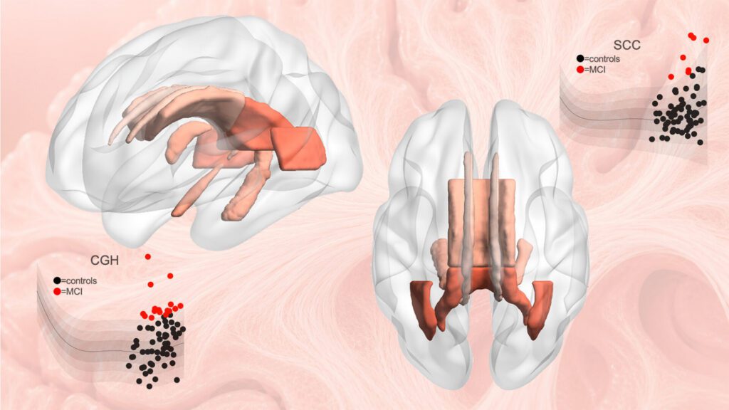
The results revealed that white matter follows distinct developmental and aging trajectories, with some measures reaching peak maturity in early adulthood and others later in midlife.
“Brain development and brain aging are not uniform processes,” continued Villalón-Reina. “The brain’s neural pathways mature on distinct timelines, and some are more vulnerable to decline than others. Our model reveals this structure by merging data on a truly global scale.”
The scientists also discovered evidence for a longstanding theory of brain aging, sometimes described as last in, first out. According to this theory, brain pathways that develop last in childhood and adolescence tend to be more susceptible to decline in older age. The researchers observed that white matter regions that mature later did indeed decline faster in old age, offering new insight linking brain development and aging.
To demonstrate the model’s practical value, the researchers applied it to clinical datasets from people with mild cognitive impairment, dementia, and 22q11.2 deletion syndrome, a genetic condition that increases risk of schizophrenia.
In each case, the model identified alterations in the brain’s circuitry that deviated from age-expected norms. Importantly, these deviations were not identical across individuals with the same diagnosis, highlighting the value of a person-specific approach.

“This monumental study took seven years to complete,” explained Paul M. Thompson, PhD, associate director of the Stevens INI and senior author of the study. “The vast scale of the data and the fine scale of the brain features assessed means we can now evaluate your neural pathways relative to other people of the same age, sex, and demographics. We can see how your brain differs from what we would expect for a person of your age and sex, giving us a tool to use in clinical trials of treatments for dozens of brain diseases.”
When applied to people with dementia and mild cognitive impairment, the model detected atypical white matter patterns in brain regions involved in memory and interregional communication. In people with 22q11.2 deletion syndrome, it identified deviations in multiple key neural pathways, helping researchers discover which brain systems develop differently.
The reference charts may also help researchers evaluate treatments by tracking whether a person’s white matter measures move closer to the expected range, or whether a treatment slows the shift away from healthy patterns over time. The charts will now be used to compare more than 30 brain diseases and conditions, offering a common framework for studying how different disorders emerge, progress, and respond to intervention.
The models are also a publicly available resource that can be extended as additional brain imaging data become available. The methods are now being used to study neurological, psychiatric, and neurodevelopmental disorders by providing a common reference standard for white matter microstructure across the lifespan.
“This study demonstrates the power of large-scale, international data sharing to create tools the entire research community can use,” pointed out Arthur W. Toga, PhD, director of the Stevens INI and provost professor at USC. “By establishing a lifespan framework for the brain’s communication pathways, this work opens new opportunities to detect subtle disease-related changes, compare conditions more rigorously, and move toward a more individualized understanding of brain health.”
The post Disease Detection Gets Boost from Keck’s New Brain Reference Map appeared first on GEN – Genetic Engineering and Biotechnology News.
Reduced left dorsolateral prefrontal activation and right inferior frontal de-oxygenation differ between psychotic and non−psychotic adolescent depression during verbal fluency
BackgroundAdolescents with major depressive disorder (MDD) who exhibit psychotic symptoms (MDD-Psy) often show greater clinical severity and higher suicide risk than those without psychosis (MDD-NonPsy). It remains unclear whether prefrontal hemodynamic responses during cognitive challenge can differ between these subgroups.MethodsWe recruited 273 adolescents aged 14–16 years (110 MDD-NonPsy, 163 MDD-Psy). All completed a 60-s verbal fluency task (VFT) while 53-channel functional near-infrared spectroscopy recorded oxy- and deoxyhemoglobin (HbO, HbR) from six frontal ROIs. Whole-brain FDR correction identified baseline-deviated channels, and ROC analyses tested the discriminative value of significant channels/ROIs. Clinical severity was indexed by the Kutcher Adolescent Depression Scale (KDAS) and the Brief Psychiatric Rating Scale (BPRS).ResultsCompared with MDD-NonPsy, adolescents with MDD-Psy exhibited higher KDAS (median 16 vs 13; z = -7.07, p < 0.001) and BPRS total scores (median 35 vs 28; z = -11.02, p < 0.001). After FDR correction, MDD-NonPsy showed positive deviations at channels 8 and 23 (t = 2.10-2.70, pFDR=0.038), whereas MDD-Psy exhibited negative deviation at channel 2 (MNI –60, 19, 18; left middle frontal gyrus; t =-2.89, pFDR = 0.004). Across ROIs, only right inferior frontal gyrus HbR differed between groups (median –0.03 vs 0.00 mM·mm; z = -2.18, pFDR = 0.030). No ROI-level HbO differences survived correction. At the single-channel level, HbO at channel 2 and HbR in IFG_R correlated modestly with lower KDAS and restlessness scores (ρ = –0.14 to –0.16, p <0.050). ROC analyses indicated modest but significant classification accuracy for psychosis (AUC = 0.577-0.581, p = 0.030).ConclusionsLeft dorsolateral and right inferior prefrontal hemodynamic signatures differ between adolescents with MDD who present with versus without psychotic features. However, given the absence of healthy control or other psychiatric comparison groups, these findings cannot establish diagnostic specificity. Pending replication in independent samples, these fNIRS−derived hemodynamic signatures may inform future hypothesis−driven research.
Exploring the Feasibility of Transcranial Ultrasound Stimulation in the Treatment of Schizophrenia
Conditions: Schizo Affective Disorder
Interventions: Device: Transcranial Ultrasound Stimulation; Device: Transcranial Ultrasound Stimulation
Sponsors: Far Eastern Memorial Hospital
Recruiting
Interventions: Device: Transcranial Ultrasound Stimulation; Device: Transcranial Ultrasound Stimulation
Sponsors: Far Eastern Memorial Hospital
Recruiting
<![CDATA[New analysis finds postpartum depression and postpartum psychosis have distinct underlying biological mechanisms.]]>
Neuropixels Opto Integrates Electrophysiology and Optogenetics to Probe Neuronal Function
High-resolution extracellular electrophysiology is typically used to record from neurons in order to understand brain function. Combining electrophysiology with optogenetics allows researchers to test the causal role of specific neurons by activating or inactivating those populations while recording the effects of neural activity.
Now, a new technology, co-developed by UCL scientists, simultaneously records and manipulates neuronal activity deep within the brain. The device, known as Neuropixels Opto and researched in mice, integrates electrophysiology and optogenetics in a single probe, enabling unprecedented insight into how individual neurons in the brain function and interact. By packing around 1,000 closely spaced recording sites onto an ultra-thin probe, it is possible to capture high-resolution signals from individual brain cells while monitoring large neural networks at the same time. The device could transform our understanding of neural circuits and neurological conditions, such as Alzheimer’s disease and schizophrenia.
“This makes it possible, for the first time, to directly test how specific neurons influence the activity of surrounding circuits—revealing causal relationships between neuronal activity and brain function,” notes Matteo Carandini, PhD, a professor at the UCL Institute of Ophthalmology. “The ability to both record and control neuronal activity in the same experiment represents a significant advance for neuroscience.”
This work is published in Nature Methods in the paper, “Neuropixels Opto: combining high-resolution electrophysiology and optogenetics.” The device, which packs 960 electrical recording sites and two sets of 14 light emitters onto a 70-μm-wide, 1-cm-long shank, allows spatially addressable optogenetic stimulation with blue and red light. The device allows researchers to monitor the electrical activity of hundreds of neurons while also selectively activating or silencing specific cells using light.
“The brain processes information through complex patterns of electrical activity, with billions of neurons communicating via rapid electrical signals,” explains Carandini. “Understanding how these signals give rise to behavior, thought and disease requires tools that can both observe and influence neuronal activity.”
“Until now, scientists have typically relied on separate approaches: electrophysiological probes to record neural activity, and optogenetics to control it,” Carandini adds. “Combining the two has proved challenging, particularly in deeper brain regions, where delivering light without disrupting sensitive recordings is technically difficult. Neuropixels Opto overcomes these limitations by integrating both capabilities into a single device, enabling simultaneous measurement and manipulation of neural circuits.”
Karolina Socha, PhD, research fellow at UCL Institute of Ophthalmology, has used the probes to investigate the function of the cerebral cortex. “We were surprised to discover that the activity of neurons in the cortex can be remarkably localized. Up to now, we thought that neurons are so interconnected that there would be no way to activate some of them without activating many others,” she said. “The new Neuropixels Opto probes revealed that these neurons can operate not only in concert but also rather independently.”
The technology may also have important implications for understanding neurological and psychiatric conditions. Many disorders, including schizophrenia, Alzheimer’s Disease and Parkinson’s Disease, are associated with disruptions in how neurons communicate. By providing a clearer picture of how neural circuits function in both healthy and diseased states, Neuropixels Opto could support the development of more targeted treatments.
The post Neuropixels Opto Integrates Electrophysiology and Optogenetics to Probe Neuronal Function appeared first on GEN – Genetic Engineering and Biotechnology News.
A 20-Year Follow-up of First Episode Psychosis: Longitudinal Effects of Early Treatment Strategies and Relapse
Conditions: Schizophrenia
Sponsors: The University of Hong Kong; Research Grants Council, Hong Kong
Not yet recruiting
Sponsors: The University of Hong Kong; Research Grants Council, Hong Kong
Not yet recruiting
<![CDATA[Phase 3 at-home data show sublingual dexmedetomidine cuts severe agitation in bipolar disorder and schizophrenia, supporting FDA label expansion.]]>