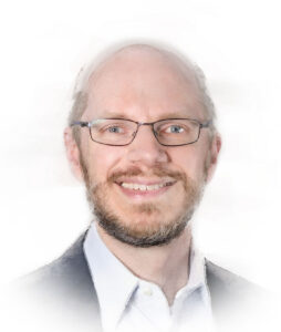
The Digital Path to AI in Cancer Care

David West
David West
CEO, Proscia

As cancer care becomes data-driven, artificial intelligence (AI) will play an increasingly central role across the treatment continuum, from biomarker identification and drug development to clinical trial recruitment and diagnostics. In this corner of healthcare, the ability of AI to interpret and annotate tumor sample slides that have been digitized is taking center stage. While the promise is great, and AI interpretation is already influencing some clinical care, it has not yet reached critical mass.

“There’s something like a billion slides created every year for diagnostic purposes, and today most of those, about 85%, are still read by a pathologist with a microscope on physical glass slides,” said David West, CEO and co-founder of digital pathology company Proscia. In practice, that means pathologists manually examine slides, identify cancer, grade tumors, and dictate reports in a traditional approach to diagnosing cancer that has seen little change in decades.

Mohamed Omar
Mohamed Omar, MD
Associate Professor
Cedars-Sinai Medical Center

But that foundation is now shifting. Advances in slide scanning, cloud storage, and AI are turning digital pathology images into data that can be analyzed at scale. At Memorial Sloan Kettering Cancer Center, large archives of digitized slides helped launch Paige AI, one of the earliest companies to train deep learning systems on pathology images linked to clinical and genomic outcomes. This yielded the first U.S. Food and Drug Administration (FDA)-approved diagnostic using AI and digital pathology: Paige Prostate Detect. The company, which was acquired last year by AI-enabled precision medicine company Tempus, now combines Paige’s digital pathology-based AI with Tempus’s broad genomic sequencing data platform.

Researchers in the field say the implications of AI in digital pathology extend beyond image analysis. Mohamed Omar, MD, an associate professor of computational biology at Cedars-Sinai Medical Center, Los Angeles, noted that large language models can help clinicians navigate a research landscape that produces “hundreds of papers every single day” to inform ongoing cancer research. Multimodal AI tools promise to unlock even more insights from digital pathology data by combining it with genomic, radiomic, and clinical data to build powerful new models of both common and rare cancers for diagnosis, drug development, and clinical trial enrollment.

Razik Yousfi
Razik Yousfi
SVP and GM, Tempus

While adoption is in its early stages, the advent of faster and less expensive scanners is bringing digital pathology within reach of both regional and rural hospitals. Razik Yousfi, senior vice president and general manager of AI products at Tempus, and a co-founder of Paige, predicts that within the next 10 years, the majority of pathology workflows will be digital. The ultimate goal of the application of AI here is not to replace human pathologists, but to empower them with a capable assistant while spreading adoption beyond major medical centers.

Building the foundations

As the field of applying AI to digital pathology progresses, it needs to build the groundwork for a wider range of potential applications that could address rare cancers and other areas without an abundance of data. One such project is called Atlas, a collaboration between researchers in Korea, Germany, and the United States to build a foundation model trained using 1.2 million histopathology whole-slide images from 490,000 cases sourced from the Mayo Clinic and Charité – Universitätsmedizin Berlin.

Foundation models like Atlas allow large-scale pre-training of data to develop numerical representations called embeddings that capture both the structural and contextual features of slides in the dataset. Atlas incorporates a diversity of diseases, staining types, and scanners, and uses multiple image magnifications during training. This broad approach confers power and utility. It allows the digitized representations of the histology to be adapted, queried, or fine-tuned to very specific downstream tasks using much less data than would be needed to build a one-off model.

As such, a foundation model provides a reusable digitized computational backbone that can be tapped across a wide range of uses, like tumor classification, detection of morphologic structures, biomarker quantification, and outcome prediction. In short, foundational models make the process of querying digital pathology images more efficient compared with past approaches.

Andrew P. Norgan
Andrew P. Norgan, MD, PhD
CMO, Mayo Clinic

“In the case of pathology, the successful AI models developed using ‘conventional’ neural network approaches before the advent of FMs (foundation models) typically required huge amounts of training data to achieve high performance and generalizability—the ability to work across datasets distinct from the training data,” said lead Atlas researcher Andrew P. Norgan, MD, PhD, CMO of Mayo Clinic Digital Pathology and assistant professor of laboratory medicine and pathology. “We think of FMs as [an] enabler that allows model development in pathology … to move from artisanal or craft processes to more scalable and reproducible processes that should allow for the rapid development of high-quality models to address problems in pathology.”

At Paige AI, the company’s early work resulted in the first FDA-approved AI diagnostic, Paige Prostate Detect. Its algorithm was built using a technique called multiple instance learning instead of traditional supervised neural network techniques that require detailed human annotation of slides, a time-consuming and expensive method that could expose the learning to human error. The difference between the two methods is that traditional neural networks expose AI to a slide with cancer and tell it that there is cancer present. In multiple instance learning, the model is shown unannotated slides and is tasked with finding the cancer.

Even this approach, however, required a very large dataset. It became apparent to company leaders that the heavy lifting required to get Paige Prostate Detect to work wasn’t scalable.

“We had kind of cracked this recipe,” said Yousfi. “We know how to use a lot of GPU (graphics processing unit) compute, and if we get a ton of data and a lot of compute, we can build anything. But GPU infrastructure is very expensive, and it takes a lot of time to train a very large system.”

Perhaps the most important factor moving Paige away from this model is that it will not work when there is only a small amount of data available. This blocks the ability to train AI to recognize rare cancers for which sample counts are low. The company needed a different approach.

“We had this idea [for] a new system that was basically trained on all of the images we had access to, independent of the organ and indication and tissue and task,” Yousfi said. “Back then, we didn’t know what that thing was called. But ultimately, that became what everyone is calling today a foundation model.”

Originally trained on 200,000 slides, Paige’s new model now includes 3.5 million images and roughly two billion parameters, making it the backbone for other downstream applications the company builds today. This ability to use foundation models as the AI and data encyclopedia for smaller applications will ultimately propel the field of digital pathology forward by widening the playing field.

Going multimodal

To address more complex predictive problems, additional data types can be integrated. Clinical, radiologic, or genomic data can be combined with morphologic embeddings or used during training to help the model learn which tissue features carry a signal of disease or identify a biomarker. These approaches aim to support precision oncology by making morphologic data computable and aligning slide-derived features with other cancer-focused datasets. “These approaches can surface subtle or ‘latent’ patterns in pathology slides and align them with other data sources,” Norgan said. Pathologist and oncology care teams can then evaluate and interpret the features identified by the models within the clinical and biological context.

“In this way, pathologists and oncology teams use these outputs as decision-support tools, while clinical judgment remains central to diagnostic interpretation and therapeutic decision making,” Norgan added.

Atlas has now been succeeded by Atlas2, which was trained on 5.5 million pathology images and is now a two billion-parameter model, making it one of the largest pathology foundation models to date. The team has explored distilling methods to create smaller, more efficient, and targeted versions of the model that retain performance, with an eye toward finding a balance between scale and deployability.

Proscia is embarking on a different multimodal approach that combines vision models with language models, with the intent of creating methods to query the morphology of digitized slides. Their efforts in vision-language models (VLMs) combine textual data with visual data and allow the model to describe the morphology of a slide, answer questions about what it contains, find images in a database based on a text query, and even follow multimodal instructions such as “circle the tumor area on this image.”

In short, a VLM can be engaged in the same way you can engage a human. “I could go ask a pathologist to point out all the areas of tumor-infiltrating lymphocytes,” West said. “Now, because language-vision models are encoding language and images in the same space, they can do that, too. You can ask the model to describe what is happening in an image, and it will tell you exactly what it sees.”

At Cedars-Sinai, Omar’s work with large language models takes a less direct route of leveraging queries to gather information from research studies or even images. “Basically, you could go to the tool, ask questions, and the tool will provide you with pieces of code,” he explained. “These pieces of code are what you use on the slide to get more information.”

Atlas provides a similar function at the Mayo Clinic, Norgan noted. Because the model-generated embeddings in the digitized slide also encode semantic information, the Atlas team is now building a slide search function, which would allow researchers or clinicians to identify and access slides, or regions of slides, with related features.

Democratizing care

Although it will take time to disseminate the tools needed for AI-enabled digitized models of cancer care to smaller health systems, the future is now at Moffitt Cancer Center, where the research hospital is engaged in a top-to-bottom digitization of its system.

Marilyn Bui
Marilyn Bui, MD, PhD
Senior Member
Moffitt Cancer Center

According to Marilyn Bui, MD, PhD, senior member of the departments of pathology and machine learning, the comprehensive cancer center plans for full digital adoption across clinical and research labs by 2027. Last August, it entered a multi-year collaboration with integrated AI and digital pathology company PathAI to deploy its cloud-based digital pathology image management system for both research and clinical applications.

Within the pathology department, the transition will mean that all glass slides will be scanned and reviewed digitally, providing the basis for applying AI computational tools to assist pathologists. Bui said that the cancer center is accelerating its move toward clinical AI adoption: “Just today I received an email asking which AI algorithms we plan to incorporate for clinical utility—prostate cancer, breast cancer, general tumor detection,” she said. “For us, it’s no longer just research.”

Moffitt is taking a hybrid approach to algorithm development and deployment within the system. Some AI tools will come from commercial vendors and will be validated internally, while others will be developed by investigators through the center’s translational pathology work. Taking this approach will allow it to apply AI to both common cancers and the rare tumor types Moffitt frequently encounters.

While the digital initiative will be transformational, Bui emphasized that the goal is not to replace pathologists but to enhance their capabilities. She prefers to refer to AI as augmented intelligence to reflect this. “Artificial intelligence suggests a robot replacing us,” she said. “But what we mean is augmented intelligence—tools that assist and enhance our ability to make clinical decisions.”

Further, Moffitt intends to integrate digitized slide data with genomic, proteomic, and clinical outcome data to build a multimodal data environment that could advance precision oncology. “Digital pathology and AI will allow us to extract far more information from tissue samples,” Bui said, “making our diagnoses more actionable for the clinical team and ultimately improving patient care.”

The promise of AI in oncology isn’t just better algorithms, it’s broader access. The maturation of computational pathology and its dissemination from large cancer centers like Moffitt to regional and rural health systems has the potential to provide levels of care typically only available at large research hospitals in community settings as well.

“It’s about democratizing access to care,” said Omar. “For a person in Maine or Wisconsin or another place to have access to the same high-quality care that you would get from a larger academic medical center in LA or New York, slides have to be digitized.”

Over the next 10 years, there could be a compelling business case for hospitals to embrace digital pathology. As the cost of scanners comes down and a broad range of diagnostic tools becomes available, digitizing routine H&E slides could become common.

While genetic cancer testing can cost hundreds of dollars, Omar pointed out that pathology slides “cost $5 [and] they are available universally, in all patients with cancer.” As AI models increasingly identify genomic-level insights directly from those inexpensive images, it represents a “huge win for accessibility, making AI work for patients who cannot afford genetic tests,” Omar said. If there is broad adoption of digital pathology “it is very easy to roll out any kind of AI models and computational tools across the board, across situations and locations that don’t have access to care.”

“At the end of the day, all slides will be digitized,” he concluded. “It’s just a matter of time.”


Chris Anderson, a Maine native, has been a B2B editor for more than 25 years. He was the founding editor of Security Systems News and Drug Discovery News, and led the print launch and expanded coverage as editor in chief of Clinical OMICs, now named Inside Precision Medicine.

The post The Digital Path to AI in Cancer Care appeared first on Inside Precision Medicine.

From Reactive to Proactive: Reimagining Hypertension Management in the Precision Medicine Era

According to the World Health Organization, an estimated 1.4 billion adults aged 30–79 worldwide had hypertension in 2024, representing around one-third of the global population of that age. Of these, 44% were unaware that they were living with a leading risk factor for premature death and poor health worldwide due to its association with myocardial infarction, stroke, and kidney disease.

Despite the size of the hypertension problem, its diagnosis and treatment pathway has remained largely the same for decades.

A 60-year-old pathway

“The current pathway in hypertension diagnosis and treatment has really not changed in over 60 years,” said Sandosh Padmanabhan, MD, PhD, chair of pharmacogenomics and professor of cardiovascular genomics and therapeutics at the University of Glasgow in Scotland.

He explained that it is based on opportunistic detection of hypertension, which has traditionally been defined as a blood pressure (BP) of 140/90 mmHg in the clinic, although thresholds vary by measurement method and guideline. For example, out-of-office measures typically use lower cut-points (e.g., home/daytime ambulatory averages) of 135/85 mmHg.

Sandosh Padmanabhan
Sandosh Padmanabhan, MD, PhD
Professor
University of Glasgow

Diagnosis typically occurs when a patient visits their primary care physician (PCP) or has a pharmacy BP check. Confirmation follows, ideally with out-of-office BP monitoring to avoid misclassification caused by one-off measurements.

Patients are then stratified by predicted 10-year cardiovascular risk, using risk calculators such as Q-risk or the PREVENT score, and treatment is based on a stepwise algorithm. First, patients are generally given lifestyle advice like reducing salt, alcohol, and caffeine intake, improving sleep, managing stress, and increasing exercise. This may give them a chance to reduce their BP without pharmacologic intervention.

If unsuccessful, depending on local guidelines, patients may be offered an angiotensin-converting enzyme (ACE) inhibitor or angiotensin receptor blocker if under 55 years of age. Those over 55 years or of Black African or Caribbean origin are started on a calcium channel blocker. The next steps combine ACE inhibitors and calcium channel blockers, then add a thiazide-like diuretic, followed by spironolactone or other drugs.

However, this approach uses “a population-level logic,” said Padmanabhan. Although age and ethnicity are considered, “these are broad demographic proxies that don’t include any understanding of the individuals’ underlying pathophysiology or the genetic makeup.”

He stresses that, on a public health basis, the system works. There are multiple effective, low-cost antihypertensive drug classes and many generic options available that effectively lower BP. Despite this, control rates are poor. “Fewer than one in four hypertensive adults globally have their BP adequately controlled,” he said.

The measurement problem

Part of the issue lies in how BP is measured. “To give you an idea about the scale of inertia, we diagnose BP using a device that was introduced in the late 19th century,” Padmanabhan noted, referring to the sphygmomanometer invented by Scipione Riva-Rocci in 1896. Not only that, the technique can also be flawed. Variables such as incorrect cuff size, improper positioning, and patient movement can distort readings. Even talking during measurement can increase BP values by 5–9 mmHg or even higher.

Crucially, a single measurement provides little insight into cumulative lifetime exposure to high BP and can be skewed by issues like white coat hypertension or masked hypertension. “We look at the BP number, but the patients don’t experience that number. What they experience is a lifelong vascular risk,” Padmanabhan explained. “Treatment is not about a short-term reduction in a number. It’s about long-term sustained risk reduction.”

Yet the current system remains reactive and is not working well enough. “We have to move away from reactive diagnosis to proactive identification,” Padmanabhan said. “The earlier we measure accurately and respond systematically, the fewer surprises we’ll see later.”

Continuous monitoring

The pitfalls of opportunistic, or even planned, BP measurement are driving the emergence of new technologies capable of continuous monitoring.

Josep Solà
Josep Solà, PhD
CTO and Co-founder
Aktiia

Josep Solà, PhD, began working on optical sensing technology in 2004 at the Centre for Electronics and Microtechnology in Switzerland. By analyzing subtle changes in reflected light caused by arterial dilation, it became clear that BP could be measured using these light signals. In 2018, this research was spun out into Aktiia, where Solà is CTO and co-founder. The company has developed and commercialized the Hilo™ band: a CE-certified wearable medical device designed for continuous, cuffless, BP monitoring that has been clinically validated against traditional ambulatory BP monitoring.

The band tracks BP and heart rate automatically, about 25 times per day, without requiring any action from users. Paired with an app, the device shows users daily, nightly, and long-term BP trends. It is currently available as a certified medical device across Europe, Australia, and Canada, and, following FDA approval in July 2025, the company is preparing for a U.S. launch.

Solà said he and co-founder Mattia Bertschi, PhD, were convinced they could change how hypertension is being managed today. He believes there is no good reason why most people with hypertension cannot control the condition. The medication is cheap and effective; the problem is that there has been no technology that patients can use to properly manage their condition.

“No one wants to use a cuff every day for the next 30 years,” said Solà. “They’re just so inconvenient, and you cannot expect people to proactively measure something they don’t feel.”

The Hilo band gives wearers a feedback loop that has historically been missing from BP measurement. Users can immediately see that reducing their salt or alcohol intake, for example, lowers their BP. “We are empowering people,” said Solà. “We are empowering them to look at the intervention, or combination of interventions, with or without medication, to see what is effective for them, and this reinforces their willingness to continue with the changes they are making.”

Hilo product
Credit: Hilo

Data published by Aktiia has shown that this approach works. A study of 8,950 U.K.-based Hilo users indicated that individuals who monitored their BP continuously showed better control over time. Specifically, users over 50 years of age appeared able to prevent the age-related rise in systolic BP typically seen in the general population, which the researchers say “may reflect greater awareness, stronger treatment adherence, and lifestyle changes prompted by continuous feedback.”

Wearables at scale: Opportunity and caution

Beyond dedicated monitoring devices like the Hilo band, smartwatches and other devices are increasingly capable of detecting physiological signals associated with cardiovascular risk. The Apple Watch can detect potential signs of chronic hypertension by analyzing heart rate sensor data over 30-day periods, the Huawei Watch D provides on-demand and 24-hour ambulatory BP monitoring using an air-filled strap, while the team behind the Oura ring is developing a “Blood Pressure Profile” feature to detect early signs of hypertension.

Although this represents a significant step toward embedding cardiovascular monitoring into everyday life, the increasing use of these devices raises important questions about accuracy, interpretation, and clinical integration, particularly as they often rely on indirect signals rather than direct BP measurement.

Adam Bress
Adam Bress, PharmD
Researcher
University of Utah

As Adam Bress, PharmD, from the Spencer Fox Eccles School of Medicine at the University of Utah, and colleagues have recently shown, translating wearable-derived signals into meaningful clinical information is not straightforward.

They evaluated the hypertension alert feature of the Apple Watch, which has a published sensitivity of 41% and specificity of 92%, meaning that approximately 59% of individuals with undiagnosed hypertension would not receive an alert, while about eight percent of those without hypertension would receive a false alert.

“The problem there, is that this data only tells you how the alert works in a very controlled, limited population,” said Bress. “In order to understand how it’s going to work in the real world, we need to know how the true prevalence of undiagnosed hypertension varies in the population and in subgroups and to what degree.”

Using data from nearly 4,000 adults in the U.S., Bress and colleagues showed that the pretest probability of having hypertension has a significant impact on the reliability of the alert. For example, among adults under 30 years of age, the pretest probability of having hypertension is 14%. A positive alert on the Apple Watch would increase this probability to 47%, whereas no alert reduces the probability to 10%.

However, for adults aged 60 years and older, an alert increases the probability of an individual having hypertension from a pretest level of 45% to 81%, whereas the absence of an alert only lowers it to 34%. This translates to large numbers of false negatives when applied across millions of users.

In Apple’s validation study, the company stresses that the watch is not intended to replace traditional diagnosis methods or to be used as a method of BP surveillance, and that the absence of a notification does not indicate the absence of hypertension.

“The concern is, if you’re not getting an alert, will people interpret that as them not having hypertension,” said Bress. “That’s the worry. … The groups in which the negative alert is the least trustworthy contain the people with the highest risk. We’re most worried about people being falsely reassured.”

At the same time, he is clear that wearables should not be dismissed. “This technology is an important step forward; we need more wearable tech that can screen,” he said.

Unfortunately, access to these devices is not universal. Advanced monitoring technologies are often first adopted by the “worried well”—people who are more affluent and health-conscious—rather than those at highest risk.

“The only thing that can change this is a clear political decision to make awareness of hypertension large scale,” said Solà. Devices like the Hilo band could be used much like the continuous glucose monitors for diabetes. The difference is that if someone with diabetes doesn’t keep their blood glucose levels under control through regular monitoring, they can become ill very quickly. With hypertension, the effects of poor control don’t become apparent for decades.

“We need the policymakers to understand that investing in this technology today will have a return on investment in 10 years from now, not in one year from now,” Solà remarked.

Targeted drug selection

Even when hypertension is detected early and monitored closely, treatment remains largely empirical and can lead to therapeutic inertia, one of the biggest current challenges in hypertension care. “BP is not like diabetes, it doesn’t cause symptoms, and because of that, we don’t escalate treatment often enough,” said Padmanabhan.

At the same time, treatment selection remains largely trial-and-error. Clinicians cycle through medications sequentially, adjusting regimens based on response rather than underlying biology. The issue is that failed attempts risk side effects and can erode trust. That lack of trust can then impact adherence and, therefore, cardiovascular risk.

Instead, Padmanabhan believes that we need to move toward mechanistically informed drug selection.

This approach is common in oncology, where targeted therapies have been matched to specific mutations, but the picture is more complex for BP. Genome-wide association studies (GWAS) have identified more than 30 genes associated with monogenic forms of hypertension or hypotension and more than 2,100 single nucleotide polymorphisms linked to BP regulation, underscoring its highly polygenic nature.

This, combined with the strong influence of environmental factors, means that there is no single pathway or biomarker that can be easily targeted to reduce BP.

Padmanabhan’s work on the uromodulin gene (UMOD), however, shows that GWAS data can translate into therapy. His team identified a signal on chromosome 16 linked to uromodulin, a protein that is only expressed in one part of the kidney and plays a role in salt regulation. In a clinical trial  comparing people with low BP to those with high BP, they found that people with the UMOD allele that increases protein expression experienced a sustained reduction in BP when treated with the loop diuretic torasemide, whereas the effect was only temporary and followed by rebound in those carrying the UMOD allele that lowers protein expression.

Approximately two-thirds of the population carry the UMOD allele that increases protein expression, meaning that loop diuretics like furosemide or torasemide, which are more commonly used to treat heart failure, could potentially be used in hypertension personalized by the patient’s genotype.

So far, “this is the only clinical trial from a GWAS-identified genetic variant in hypertension,” Padmanabhan noted, highlighting both the promise and challenge of pharmacogenomics in hypertension.

Although clinical translation from GWAS of hypertension has been limited, research has shown that genetic variation in drug-metabolizing enzymes can significantly impact hypertension treatment efficacy and toxicity. For example, variants of CYP2D6 affect metoprolol metabolism whereas those in CYP2C9 influence responses to losartan. Research is needed to determine whether testing for these variants or others could reduce trial-and-error prescription, minimize side effects, and thus increase patient confidence and long-term engagement.

Teresa Castielo
Teresa Castielo, MD
Director
MIAL Healthcare

On a more fundamental level, biological sex differences remain a significant consideration in cardiovascular medicine. “Biological factors are an integral part of the clinical picture,” noted Teresa Castiello, MD, consultant cardiologist and director of MIAL Healthcare in London. She points out that clinical trials have historically seen a predominance of male participants; as a result, many standard medication dosages are based on data primarily derived from men.

This can lead to challenges with tolerability and a higher incidence of side effects in women as the therapeutic dose required for efficacy often tends to be lower in female patients.

Castiello suggests that this area of management warrants further refinement in clinical practice. She also emphasizes that key aspects of female cardiovascular risk, including reproductive history, menopause, and conditions like polycystic ovary syndrome, are nuances that may not always receive the necessary focus in routine care.

Toward a precise, preventative system

Ultimately, transforming hypertension care will require more than new technologies or therapies. It will require a fundamental change in how care is delivered.

Padmanabhan argues that hypertension should be managed through a “precision prevention service,” that integrates early detection, continuous monitoring, and personalized treatment, and involves more than just PCPs.

This approach recognizes that the disease is not just a clinical condition but a societal one, influenced by factors such as diet, socioeconomic status, work patterns, and access to care. Equity remains another critical issue. “We treat the ideal average patient under ideal circumstances but that’s not reality,” said Padmanabhan.

There also needs to be a cultural shift, said Castiello. “It’s not just the doctor’s responsibility; we also need to take responsibility for our own health.”

Solà shares a similar vision for the future: he would like to see BP measurement to become as routine as brushing your teeth, supported by technologies that empower individuals and reduce the burden on healthcare systems.

If realized, this shift could transform hypertension from a silent, progressive disease into a manageable, preventable condition, saving millions of lives in the process.


Laura Cowen is a freelance medical journalist who has been covering healthcare news for over 10 years. Her main specialties are oncology and diabetes, but she has written about subjects ranging from cardiology to ophthalmology and is particularly interested in infectious diseases and public health.

The post From Reactive to Proactive: Reimagining Hypertension Management in the Precision Medicine Era appeared first on Inside Precision Medicine.

Biobanks Set the Stage for Scaling Precision Medicine

Dating back more than a century, biobanks have outgrown their beginnings as small, local collections to become large, global facilities that store and handle millions of samples and serve thousands of researchers at any given time. Over the years, biobanks have transformed from passive repositories into active research infrastructures that are increasingly bridging the gap between medical research and clinical applications.

“Today’s biobanks have evolved far beyond sample storage,” said Yan Zhang, PhD, president of proteomic sciences at Thermo Fisher Scientific. “They are automated, digitally connected systems integrated with hospitals and health networks to ensure appropriate consent, longitudinal clinical context, and the ability to re-engage participants over time.”

Yan Zhang
Yan Zhang, PhD
President
Thermo Fisher Scientific

As safeguards of clinical samples, biobanks fulfill a central role in the advancement of precision medicine. Access to the right samples can make or break a research project, with most researchers reporting that they have had to limit their scope of work because of difficulties obtaining the samples they need.

“Robust, population-scale biobanking enables precision medicine to move from isolated findings toward broader clinical relevance,” said Zhang. “Modern biobanks combine genomics, proteomics, and other high-dimensional omics platforms with robust data architecture, high-performance computing, and artificial intelligence (AI)-driven modeling. Dedicated data science teams integrate molecular data, longitudinal health records, and curated public datasets to generate biologically meaningful interpretations.”

Biobanks now provide the infrastructure needed to support population-scale, longitudinal studies that allow scientists to uncover molecular drivers of disease and understand their evolution over time to ultimately identify biomarkers, develop targeted treatments, and inform clinical decisions.

“We’re seeing researchers design studies with scale in mind,” Zhang noted. “They’re combining proteomics, genomics, and clinical data to generate insights that are both statistically powerful and relevant to real-world populations. There’s also a clear shift from searching for a single biomarker to building a more complete, systems-level understanding of disease.”

To navigate today’s rapidly shifting landscape and meet their core purpose of supporting cutting-edge clinical research, biobanks have to keep up with fast-moving targets. Going forward, moving from initial discovery to translation will remain the number one challenge in precision medicine. “Generating discovery insight is no longer the limiting factor,” said Zhang. “Validating, standardizing, and implementing those insights at scale is.”

A matter of scale

Martin K. Rutter
Martin K. Rutter, MD
Deputy Chief Scientist
UK Biobank

One of the most transformative shifts in biobanking over the past decade has been an exponential increase in the scale of data collection and sample storage. At the forefront of this expansion is the UK Biobank, which currently stores around 18 million samples from 500,000 participants, together with imaging and biomarker data, healthcare records, questionnaires, physical measurements, demographics, lifestyle, and environmental data collected over the course of 20 years. This depth of phenotyping is what makes the data so valuable to researchers worldwide, said Martin K. Rutter, MD, professor of cardiometabolic medicine at the University of Manchester and deputy chief scientist at the UK Biobank. “When you link all that together, you can get amazing insights into the biology of disease.”

To keep up with increasing storage needs and researcher requests, the UK Biobank is now getting ready to move more than 10 million samples currently stored in its main laboratory to a new building in central Manchester by the end of the year. The new storage facility is designed to quadruple sample retrieval speed while making the whole infrastructure more energy-efficient and environmentally friendly.

The scale at which facilities like the UK Biobank operate today would have been unthinkable when it was established two decades ago. Such massive growth has been driven by rapid technological advances across genomics, transcriptomics, and proteomics, with costs continuing to fall while coverage, speed, and accuracy keep surging.

Partnerships with the pharmaceutical industry have also been instrumental in nurturing this exponential growth. This can be seen in initiatives like the UK Biobank Pharma Proteomics Project (UKB-PPP), a collaboration between the UK Biobank and 14 biopharmaceutical companies with the goal of analyzing proteomics data from 600,000 samples.

In the long run, scale provides the backbone to enable increasingly ambitious, statistically powerful studies. However, as they grow, biobanks face the challenge of navigating a constantly shifting landscape while making sure the samples and data they collect, store, and maintain are valuable to the entire research community they serve.

“Our job is to make the data available to researchers,” said Rutter. “We are involved now more than ever in connecting with research teams and trying to understand what their needs are.”

Through surveys and consultations, the UK Biobank actively gathers information to design prospective data collection programs that anticipate researcher needs. Next year, the biobank is planning a repeat assessment of its whole cohort, focusing on measurements of aging. The goal is to support researchers looking into causal pathways and mechanisms driving age-related diseases, empowering the development of preventive interventions and new diagnostics and treatments for age-related conditions.

Keeping pace with the evolving demands of researchers, industry, and the broader public is essential for biobanks to secure the funding necessary not only to operate but also to expand such vast enterprises, which remains a major challenge across this resource-intensive field.

Diversity takes the spotlight

Historically, samples collected by biobanks are biased in favor of participants who are white, middle-class, and have a higher education. This creates major disparities in the applicability of clinical research. In fact, studies have shown that patients from non-European ancestry backgrounds have not benefited equally from precision drugs approved by the U.S. Food and Drug Administration (FDA) to treat a range of cancer indications.

Even within biobanks dedicated to sampling the population of a specific region, ethnic minorities, low-income, or elderly people are often underrepresented, skewing results against the real-world populations they strive to serve. As the research community increasingly recognizes the importance of more diverse and representative patient cohorts, demand is rising for resources that address these barriers.

Representation is at the heart of All of Us, a program launched by the National Institutes of Health in 2018 to address the gap present at the time in many biobanks and sample repositories. This precision medicine initiative was designed to enroll participants who reflect the full range of populations found within the U.S., including individuals of varied ancestry backgrounds as well as those living in rural commmunities, which are rarely represented in biorepositories due in part to longstanding barriers to research participation, such as the logistical challenges of collecting samples and data from participants in remote locations.

Joshua Denny
Joshua C. Denny, MD
CEO
All of Us

“A lack of diversity impoverishes discovery and applicability of findings for all,” said Joshua C. Denny, MD, CEO of the All of Us Research Program.

For instance, data collected by All of Us has been used to investigate APOL1 gene variants linked to kidney disease, which are more common among people of West African ancestry. This research led to the identification of a novel APOL1 variant that can reduce the risk of kidney disease in individuals carrying high-risk variants.

The program has so far enrolled about 870,000 participants across all U.S. states, with about 80% of them representing communities that have historically been underrepresented in biomedical research. This has been achieved by emphasizing accessibility and flexible participation models; participants can enroll digitally and choose whether to share access to their electronic health records, donate biospecimens, and complete demographics and lifestyle surveys. They may also opt to provide saliva samples, simplifying logistics in rural areas with limited access to blood collection facilities.

“What works in a rural location is different from what works in a big city like New York,” said Denny. Whether it comes to location, age, or language, he emphasized the importance of adapting how the program approaches and engages each population.

Democratizing access to patient data across the research ecosystem is another major biobanking challenge that All of Us is committed to addressing. The program has established a streamlined access model that enables researchers to access the data they need in less than two hours if they belong to one of the 1,300 already approved institutions across the world. Together with central data storage and cloud-based analysis tools, their setup is designed to make the data accessible to researchers lacking the resources and local infrastructure for high-performance computing.

Towards global integration

With precision medicine studies steadily escalating both in size and complexity, researchers increasingly seek to bring together data stored across diverse biobanks to power larger, more ambitious studies with broader scientific and societal impact. However, building the infrastructure needed to enable cross-biobank studies is still a challenge, starting with convening stakeholders to harmonize data collection standards and establish international guidelines.

Anticipating this need, in 2013 the European Union established the Biobanking and Biomolecular Resources Research Infrastructure – European Research Infrastructure Consortium (BBMRI-ERIC), which currently coordinates the activity of about 500 biobanks across 32 countries.

Jens K. Habermann
Jens K. Habermann, MD, PhD
Director General
BBMRI-ERIC

“Precision medicine can only move forward with a strong starting point for research,” said Jens K. Habermann, MD, PhD, professor for translational surgical oncology and biobanking at the University of Lübeck and director general of the BBMRI-ERIC. “It can be very difficult for scientists to get all the information they need in one place, and this is what biobanks can enable.”

Pulling together data from all its members, the BBMRI-ERIC has set up a central catalogue for biobanks, biomolecular resources, and other data and sample collections, which users can employ to identify relevant resources and build virtual cohorts tailored to their research needs. The consortium also works with international committees to set guidelines and support members working towards compliance with international standards.

Despite ongoing progress, there are still obstacles ahead when it comes to harmonizing biobanking practices worldwide, including data collection, annotation, storage, and sharing. Tackling differences in data protection, consent, ethical standards, and regulatory requirements across borders will be another necessary step towards broader standardization. Finally, biobanks will need to invest in cybersecurity to ensure patient data can be shared between institutions safely.

Funding will be key to successfully addressing all these challenges. On this front, biobanks face the difficult task of maintaining their existing infrastructure, staying up to date and relevant to the research community, and investing in cross-biobank initiatives. All this must be balanced with growing financial pressure on research centers, hospitals, and the governments supporting them.

As part of its 10-year roadmap, the BBMRI-ERIC is setting the goal of forming international networks that bring together more diverse biobank types, such as environmental, wildlife, veterinary, and plant biodiversity repositories. The overarching aim is to move towards a One Health approach to biobanking, where samples and data that expand beyond monitoring human populations are brought together to tackle overlapping challenges that simultaneously affect human, animal, and environmental health.

Data-driven horizons

As the field forges ahead, biobanks are undergoing broad transformations in the way they operate. On the technology side, these changes are being propelled by the rise of multi-omics techniques in precision medicine research, as well as by rising demand from the research community for non-invasive patient monitoring data and longitudinal sample collection. All of these will be critical for the development of the next generation of personalized therapies and diagnostics.

“Over the next decade, biobanks are expected to become increasingly integrated into clinical and translational workflows,” said Zhang. “Proteomics, in particular, will play a growing role in helping us understand the dynamic biology of disease, enabling earlier detection, better prediction of recurrence, and more precise therapeutic strategies.”

A key driver of this shift will be AI. No longer just a supporting tool, AI is now becoming an integral part of biobank operations, contributing to real-time sample monitoring, predictive maintenance, risk management, and decision making.

On the data analysis side, Zhang has seen how AI is redirecting the focus from data generation to data interpretation. She said, “Biobanking has already enabled the collection of high-quality biospecimens linked to large-scale molecular and clinical datasets. The challenge now is extracting meaningful biological insight from that complexity.”

Although still in its early days, AI is becoming central to how researchers make use of biobank data, noted Rutter. Drawing from the UK Biobank data, recent studies have developed AI models that can predict a patient’s risk of stroke based on retinal images, calculate the risk of future disease by looking at an individual’s disease history, or spot neurodegenerative diseases like Alzheimer’s and Parkinson’s early using brain scans and physical activity data.

Going forward, Rutter expects to see biobanks moving away from static cohorts and in favor of continuous data collection, enabling more powerful predictions. For example, the UK Biobank is developing a mobile app that can track a participant’s physical activity and monitor their location and sleep patterns, offering an in-depth look at how a variety of factors affect their health with much more accuracy than self-reported surveys.

Over time, all these advances will steer clinical practice from treatment to prevention, allowing healthcare professionals to act early in the patient journey, when interventions are most effective, and eventually, even before disease develops. Ultimately, addressing complex diseases will require coordinated contributions from all stakeholders, including AI innovators, drug developers, clinicians, technology providers, and policymakers.

“The next decade will be incredibly exciting,” said Denny. “It will be all about leveraging the huge scale of resources that are just emerging today.”


Clara Rodríguez Fernández is a science journalist specializing in biotechnology, medicine, deeptech, and startup innovation. She previously worked as a reporter at Sifted and editor at Labiotech, and she holds an MRes degree in bioengineering from Imperial College London.

The post Biobanks Set the Stage for Scaling Precision Medicine appeared first on Inside Precision Medicine.

STAT+: Novo Nordisk launches high-dose Wegovy

Want to stay on top of the science and politics driving biotech today? Sign up to get our biotech newsletter in your inbox.

Good morning. Here in Chicago, cherry blossoms are blooming, rat birth control is working, and it finally feels like spring may be upon us.

Budget proposals aim to boost U.S. drugmaking

As part of President Trump’s 2027 budget blueprint, the FDA has proposed policies aimed at encouraging domestic development and manufacturing of drugs, such as making it easier for drugmakers to move into clinical testing in the U.S. and giving an “exclusivity” period to U.S.-based generics manufacturers.

Continue to STAT+ to read the full story…

STAT+: Trump budget’s ‘America First’ drug policy proposals

You’re reading the web edition of D.C. Diagnosis, STAT’s twice-weekly newsletter about the politics and policy of health and medicine. Sign up here to receive it in your inbox on Tuesdays and Thursdays.

The 2026 STAT Madness competition was stacked with research on topics like smart dental floss that monitors stress, Baby KJ’s personalized gene therapy, and an artificial intelligence model designed to predict cell behavior. Check out the winner, unveiled this morning. And as always, send news tips to John.Wilkerson@statnews.com or John_Wilkerson.07 on Signal.

Budget reruns

The 2027 budget that the Trump administration released on Friday is in many ways a repeat of last year’s proposal: It includes deep cuts to the National Institutes of Health, the elimination of a health research agency, and the creation of a new agency devoted to chronic diseases called the Administration for a Healthy America.

Continue to STAT+ to read the full story…

U.S. Tumor Testing Suffers Under Access Disparities

Genomic testing for advanced cancer in the U.S. is hampered by unequal access across demographic groups and needs targeted policy solutions, researchers report.

Next-generation sequencing (NGS) was not carried out for the five most prevalent types of solid tumor among thousands of patients studied, the team revealed in JAMA Network Open.

And among those who did undergo this testing, wait time significantly varied according to race, insurance status, and practice setting.

“Our findings highlight the underrepresentation of certain patient demographics in tumor genomic profiling, revealing disparities in access to standard-of-care diagnostic modalities,” reported researcher Chadi Hage Chehade, MD, from the University of Utah, and coworkers.

“These results emphasize the need for healthcare policies to mitigate these gaps.”

Precision oncology has defined a new era in cancer treatment, enabling clinicians to tailor care based on the specific clinicogenomic features of a patient’s tumor, enabling more effective and less toxic treatment strategies.

NGS has emerged as a transformative technology, enabling comprehensive genomic profiling and uncovering alterations for targeted therapies.

To examine equity of care in the field, the researchers studied electronic health record data for patients with common advanced or metastatic cancers that spanned over 800 U.S. community and academic sites across the U.S. between 2018 and 2022.

The team examined time to first NGS testing and frequency of testing for 63,294 patients, including those with metastatic breast (19.1%), prostate (6.9%), pancreatic (9.7%), colorectal (21.6%), and non–small cell lung cancer (42.7%, NSCLC).

The median age in the group was 68 years and 53.7% was female. In terms of ethnicity, 2.7% were Asian, 10.0% were Black, 6.0% were Hispanic, 61.0% were White, and 20.3% were other races and ethnicities.

The frequency of testing increased over the four-year span across all cancer types, but by the final year of study up to 40% to 50% of patients were still not receiving NGS testing.

Results showed there were differential rates of testing and longer waiting times to NGS testing in some groups.

Patients with lower socioeconomic status (SES), non-Hispanic Black or Hispanic patients, those covered by Medicare, Medicaid, or other government programs, and those treated at an academic practice setting were significantly less likely to be tested in some of cancers than patients with high SES, who were non-Hispanic White, those covered by a commercial health plan, or those treated in community practice, respectively.

Among specific cancers, Hispanic patients were significantly less likely to be tested in metastatic breast or prostate cancer, and non-Hispanic Black patients were less likely to receive NGS in advanced NSCLC, metastatic colorectal or metastatic pancreatic cancer.

The findings highlight the need to improve access to standard-of-care diagnostic modalities and serve as a call to improve NGS testing rates nationwide, said Igor Makhlin, MD, in an accompanying Commentary article.

“While the accelerating pace of research and AI-driven technology is poised to herald the next generation of discoveries that translate into greater survival for patients with cancer, we cannot ignore the increased burden to stay up to date, largely born by community oncologists who manage a wide gamut of solid and liquid cancers,” he maintained.

“Creation and adoption of innovative strategies to support clinicians in implementing breakthrough advances into their practice regardless of zip code, practice site, or other factors will require a concerted effort by all relevant stakeholders, but closing this gap in GCC is absolutely necessary. Our patients are depending on us.”

The post U.S. Tumor Testing Suffers Under Access Disparities appeared first on Inside Precision Medicine.

Rewriting Life Before Birth: Entering the Fetal Genetic Intervention Era

A woman lies on an exam table, holding her partner’s hand tightly with anticipation, as a technician glides an ultrasound probe across her abdomen. On the screen, shifting staticky shadows resolve into a skull, a liver, and the flicker of a beating heart. For many families, this moment brings joy and relief. For others, it’s paralyzing, as doctors detect signs that something is wrong.

A single nucleotide change can cause neurodevelopmental delays and dimorphism, failing livers, and arrhythmia-ridden hearts. For decades, medicine could only identify these conditions, usually after birth. Prenatal screening has made it easier to detect progressive diseases like Duchenne muscular dystrophy, which degenerates and damages muscles before symptoms typically appear in childhood. But treating before birth could preserve tissue prior to the onset of irreversible deterioration.

Once unthinkable, genetic diseases can now be treated before birth. Fetal genetic intervention—including early screening, in utero gene therapy, stem cell transplantation, and even embryo editing—aims not just to diagnose disease but to correct it at its earliest stages. It is a rapidly advancing frontier, defined by technological promise and profound ethical questions.

It starts with detection

Jennifer Hoskovec, vice president of medical affairs at BillionToOne, has spent more than 20 years in prenatal genetics, an era dominated by risk assessment rather than intervention.

Historically, prenatal genetic screening has fallen into two main categories. Aneuploidy testing determines the risk of Down syndrome and other trisomies, sex chromosome abnormalities, and specific microdeletions. Screening is essential for these de novo mutations, which have no U.S. Food and Drug Administration (FDA)-approved genetic interventions. High-risk Down syndrome patients may receive a fetal echocardiogram, closer ultrasound monitoring, or tertiary care delivery with neonatal support. The standard practice is to screen, monitor, and manage.

Billion to One | Headshots
Jennifer Hoskovec
Vice President
BillionToOne

The second category involves inherited recessive conditions like cystic fibrosis (CF), spinal muscular atrophy (SMA), and phenylketonuria. If both parents are carriers for the same genetic mutation, then their child has a 25% chance of being affected. Testing typically requires samples from both parents. If both are carriers, chorionic villus sampling (CVS) and amniocentesis can detect fetal abnormalities in the first and second trimesters, respectively. However, getting each partner to follow up is a major hindrance. “When people go through a screening process and are found to be carriers, less than 50% of their partners complete the testing,” Hoskovec told Inside Precision Medicine. “Half of U.S. carriers of these genetic conditions, whether common or rare, don’t know what it means for their pregnancy. That limits their ability to get diagnostic testing because we do not have all the pieces of the puzzle.”

Hoskovec’s team developed a workaround: a single-gene noninvasive prenatal test that analyzes fetal cell-free DNA (cfDNA) circulating in maternal blood. Around nine weeks into pregnancy, fragments of fetal DNA shed from the placenta can be sequenced and quantified. If a mother is a carrier for a condition like CF or sickle cell disease, the test looks for a second variant that is not present in her DNA and forms evidence of paternal contribution.

“For example, if a mother has [the] sickle cell trait, we first sequence the full beta-globin gene in the cfDNA, which contains a mixture of maternal and fetal DNA,” Hoskovec said. “We look for a second variant not present in the mother that would indicate paternal contribution.”

Despite not replacing CVS or amniocentesis, Hoskovec said the result is highly sensitive, identifying 95% of affected pregnancies in the conditions it covers. Crucially, it does not require partner testing. “This is a stepping stone,” Hoskovec explained. “This earlier detection will likely accelerate the field by increasing the number of eligible patients for clinical studies and registries, improving equitable access across ethnic groups, and advancing precision medicine in prenatal care.”

Avoiding germline editing

David H. Stitelman
David H. Stitelman, MDr
Associate Professor
Yale-New Haven Children’s Hospital and Yale School of Medicine

As screening opens the door, fetal surgeons and gene therapy researchers are taking their first steps through it. A pediatric surgeon at the Yale School of Medicine, David H. Stitelman, MD, believes prenatal treatment has benefits. The fetus is small, so it can receive higher doses based on weight. As its immune system is still developing and more tolerant, stem cells are growing quickly and organs are still being formed, so problems can be fixed before they become permanent. Because the placenta exchanges oxygen, lung conditions like congenital diaphragmatic hernia can be treated during fetal life. But once a newborn takes a first breath, defective lungs can spell immediate crisis.

Fetal therapy is not new. Specialized centers have performed open fetal surgery for spina bifida and diaphragmatic hernia lung growth, and blood transfusions for fetal anemia dating back to the 1960s. What is new is the molecular toolkit. Stitelman’s lab is investigating gene editing methods that use the cell’s repair machinery to fix one- to three-base-pair DNA errors. Another team, led by pediatric and fetal surgeon Tippi MacKenzie, MD, at the University of California, San Francisco, is using viruses to replace genes for lysosomal storage diseases and fetal stem cells for alpha thalassemia.

Some diseases require only modest correction. In hemophilia, one percent normal clotting factor expression improves outcomes greatly. Increasing the expression of functional CFTR protein to 15% of wild-type levels may cure CF or at least make it manageable. Even a small number of liver cells corrected in hereditary tyrosinemia can boost growth and repopulate the organ. However, some situations, such as congenital cancer syndromes, may require nearly 100% correction. At present, Stitelman’s team achieves single-digit percentage editing in models of CF and beta thalassemia. “We’re in the optimization phase,” Stitelman told Inside Precision Medicine. “We are testing different nanoparticles and generations of editing strategies to incrementally reach therapeutic levels.”

Stitelman draws a clear ethical boundary: this is somatic editing, not germline editing. The aim is to treat the fetus as a patient, not to create heritable genetic changes. Instead of editing embryos in vitro, systemic therapeutic agents are delivered to avoid reproductive cell damage.

Unintended germline modification remains a concern. Editing a target gene could inadvertently disrupt developmental genes and affect future generations. But, Stitelman argues, medicine always carries risk. “In 1950, children with leukemia all died,” said Stitelman. “Today, some forms have a 98% long-term survival rate with chemotherapy. We know chemotherapy can cause germline mutations, yet we accept that risk because it saves lives. With gene editing, the issue is not zero risk but understanding and quantifying the risk. Ideally, there would be no measurable off-target effects. In the places we have examined, we have not seen off-target effects.”

One pregnancy, two patients

In a landmark trial in 2011 known as the Management of Myelomeningocele Study, investigators found that fetal surgery for severe spina bifida (myelomeningocele) achieved better results than postnatal repair. Surgically closing the spinal defect in utero improved motor function and reduced the need for shunting to relieve hydrocephalus. The benefit was so clear that the trial was stopped early and influenced how doctors treat structural birth defects.

Aijun Wang
Aijun Wang, PhD
Professor
University of California, Davis

At the University of California, Davis, biomedical engineer Aijun Wang, PhD, is working closely with fetal surgery pioneer Diana L. Farmer, MD, to evolve fetal intervention from heroic surgery to cellular and molecular therapy. Wang and Farmer launched the Cellular Therapy for In Utero Repair of Myelomeningocele (CuRe) trial, combining fetal surgery with stem cell transplantation. The goal is to not only close the spinal defect but also restore neural tissue and improve long-term function.

The lens that Wang has used to focus his research is fetal and maternal safety. “The fetus is the patient, but treatment inevitably carries some risk to the mother,” Wang told Inside Precision Medicine. “Open fetal surgery, in particular, poses significant maternal risk. Genetic treatments introduce additional uncertainties because the long-term effects of DNA modification are not fully understood. Safety must remain the highest priority.”

Genetic medicine delivery is a critical challenge for all life stages, but the stakes are particularly high for a developing fetus. In fetal development, targeting stem cell populations is especially important because these cells are highly active, proliferating, and migrating. If edited successfully at the right developmental window, their progeny will carry the correction. The problem would be if the edit was not just unsuccessful but detrimental.

Wang’s lab focuses on delivery systems, particularly lipid nanoparticles carrying mRNA-encoding gene-editing enzymes. For genetic manipulation and high-throughput screening, Wang’s lab utilizes mouse models. Fetal sheep are used for scaling and dosing, while human organoids are used for human-specific editing and functional outcomes.

“In our clinical work, we have engaged with the FDA and conducted extensive preclinical studies,” said Wang. “Using multiple complementary models is essential. Combining small animal models, large animal translational models, and human organoid systems provides a comprehensive framework for product development, from early screening to human-focused therapeutic design.”

Although the field is highly exciting and progressing rapidly, Wang warns against premature application, which could be dangerous. Safety, developmental biology, ethical considerations, and multidisciplinary collaboration are all essential. “Despite the excitement in the field, we must proceed cautiously,” said Wang. “There is strong potential for correcting specific mutations, especially point mutations, using precise gene editing approaches such as base editing. However, safety evaluation must precede rapid clinical application.”

Effective progress requires a village of physicians, surgeons, researchers, engineers, and ethicists working together. Scientific progress requires caution, responsibility, and thorough evaluation before clinical use.

The earlier, the better

If fetal intervention treats a diagnosed fetus, embryo editing operates even earlier—at the blastocyst stage in in vitro fertilization (IVF). Norbert Gleicher, MD, a fertility specialist known for treating some of the oldest and most difficult IVF patients in the United States, approaches genetic technologies with caution. Due to biological mosaicism, sampling limitations, and his belief that many abnormal embryos self-correct or develop normally, Gleicher opposes preimplantation genetic testing for aneuploidy.

Norbert Gleicher
Norbert Gleicher, MD
Founder & Medical Director
Center for Human Reproduction

But when it comes to single-gene diseases, he sees a different calculus. Couples with recessive mutations may have one-in-four embryos affected, and in dominant or X-linked diseases, half may carry the mutation. For patients who produce few embryos—especially older women—discarding affected embryos can mean losing precious chances at pregnancy. “If you can cure an embryo rather than discard it,” Gleicher told Inside Precision Medicine, “that makes a lot of sense.”

For single-gene diseases, Gleicher believes genetic editing with CRISPR or other platforms is the most straightforward intervention. He points to the 2025 work at the Children’s Hospital of Philadelphia on Baby KJ as a recent milestone. Even partial correction, which Gleicher believes is likely the case with Baby KJ—though no liver biopsies have been extracted—can transform prognosis. Gleicher said, “Correcting some cells was enough to clinically cure the baby, at least for the time being, from symptoms of a disease that historically kills affected children within a few years. However, we do not know whether the treated baby, who likely still has many affected cells, might become symptomatic again later in life.”

To Gleicher, success in a newborn is all the more reason to apply genetic intervention to fetal stages. “If this can be successful in a full human being, imagine how much easier it would be at the blastocyst stage, or even earlier at the cleavage stage, when the embryo consists of only six to eight cells,” said Gleicher. “If [CRISPR] is applied at that point, correcting those six to eight cells would mean that all their daughter cells would also be corrected. The result would be a normal baby at birth. That is the much stronger argument in this case.”

Just because something is possible, it doesn’t necessarily mean it should be done, and Gleicher establishes a clear ethical boundary. Editing to prevent a devastating single-gene disease is one thing. Editing for traits—eye color, intelligence, polygenic risk scores—is another. Polygenic predictions explain only a fraction of trait variance, and embryo implantation itself is uncertain. To him, offering polygenic selection in IVF is not only scientifically dubious but also ethically troubling. “It is surprising that professionals, particularly in genetics, would suggest such an approach,” said Gleicher. “It is worse than snake oil, because while snake oil may occasionally work by accident, this carries a real risk of causing serious harm.”

A pretty penny

What ultimately restricts fetal genetic intervention is timing. Early screening increases experimental trial eligibility, and early treatment may preserve organ development before irreversible damage. In conditions like CF and SMA, where postnatal gene therapies are expensive and delivered after injury, fetal intervention could change outcomes. Frontline screening can identify high-risk pregnancies at 11 weeks without family history or ethnicity, expanding trial access.

Yet, fetal genetic interventions require specialized teams, advanced delivery systems, counseling, and long-term follow-up. Without careful planning and reimbursement policies, only a few top-tier centers could progress, widening the gap. Ethical scrutiny remains inseparable from progress. Innovation must balance maternal risk, fetal benefit, and future consequences with safety, appropriate use, and clear limits. As prenatal care shifts from prediction to prevention, restraint and evidence will determine its future.


Jonathan D. Grinstein, PhD, North American editor for Inside Precision Medicine, investigates the most recent research and developments in a wide range of human healthcare topics and emerging trends, such as next-generation diagnostics, cell and gene therapy, and AI/ML for drug discovery. He is also the host of the Behind the Breakthroughs podcast, featuring people shaping the future of medicine. Jonathan earned his PhD in biomedical science from the University of California, San Diego, and a BA in neural science from New York University.

The post Rewriting Life Before Birth: Entering the Fetal Genetic Intervention Era appeared first on Inside Precision Medicine.

Enabling agent-first process redesign

Unlike static, rules-based systems, AI agents can learn, adapt, and optimize processes dynamically. As they interact with data, systems, people, and other agents in real time, AI agents can execute entire workflows autonomously.

But unlocking their potential requires redesigning processes around agents rather than bolting them onto fragmented legacy workflows using traditional optimization methods. Companies must become agent first.

In an agent-first enterprise, AI systems operate processes while humans set goals, define policy constraints, and handle exceptions.

“You need to shift the operating model to humans as governors and agents as operators,” says Scott Rodgers, global chief architect and U.S. CTO of the Deloitte Microsoft Technology Practice.

The agent-first imperative

With technology budgets for AI expected to increase more than 70% over the next two years, AI agents, powered by generative AI, are poised to fundamentally transform organizations and achieve results beyond traditional automation. These initiatives have the potential to produce significant performance gains, while shifting humans toward higher value work.

AI is advancing so quickly that static approaches to task automation will likely only produce incremental gains. Because legacy processes aren’t built for autonomous systems, AI agents require machine-readable process definitions, explicit policy constraints, and structured data flows, according to Rodgers.

Further complicating matters, many organizations don’t understand the full economic drivers of their business, such as cost to serve and per-transaction costs. As a result, they have trouble prioritizing agents that can create the most value and instead focus on flashy pilots. To achieve structural change, executives should think differently.

Companies must instead orchestrate outcomes faster than competitors. “The real risk isn’t that AI won’t work—it’s that competitors will redesign their operating models while you’re still piloting agents and copilots,” says Rodgers. “Nonlinear gains come when companies create agent-centric workflows with human governance and adaptive orchestration.”

Routine and repetitive tasks are increasingly handled automatically, freeing employees to focus on higher value, creative, and strategic work. This shift improves operational efficiency, fosters stronger collaboration, and generates faster decision-making—helping organizations modernize the workplace without sacrificing enterprise security.

Download the article.

This content was produced by Insights, the custom content arm of MIT Technology Review. It was not written by MIT Technology Review’s editorial staff. It was researched, designed, and written by human writers, editors, analysts, and illustrators. This includes the writing of surveys and collection of data for surveys. AI tools that may have been used were limited to secondary production processes that passed thorough human review.

The Download: AI’s impact on jobs, and data centres in space

This is today’s edition of The Download, our weekday newsletter that provides a daily dose of what’s going on in the world of technology.

The one piece of data that could actually shed light on your job and AI 

Within Silicon Valley’s orbit, an AI-fueled jobs apocalypse is spoken about as a given. Now even economists who have downplayed the threat are coming around to the idea.  

Alex Imas, based at the University of Chicago, is one of them. He believes that any plan to address AI’s impact will depend on collecting one vital piece of data: price elasticity. 

Imas argues that “we need a Manhattan Project” for this. Read the full story to find out why. 

—James O’Donnell 

This article is from The Algorithm, our weekly newsletter giving you the inside track on all things AI. Sign up to receive it in your inbox every Monday. 

Four things we’d need to put data centers in space 

In January, Elon Musk’s SpaceX applied to launch up to 1 million data centers into Earth’s orbit. The goal? To fully unleash the potential of AI—without triggering an environmental crisis on Earth. 

SpaceX is among a growing list of tech firms pursuing orbital computing infrastructure. But can their plans really work? Here are four must-haves for making space-based data centers a reality. 

—Tereza Pultarova 

This story is part of MIT Technology Review Explains, our series untangling the complex, messy world of technology to help you understand what’s coming next. You can read more from the series here. 

The must-reads 

I’ve combed the internet to find you today’s most fun/important/scary/fascinating stories about technology. 

1 Trump has again proposed major cuts to US science and tech spending 
He wants to slash nearly every science-focused agency. (Ars Technica) 
+ If Trump gets his way, the US could face a costly brain drain. (NYT $)  
+ Top research talent is already fleeing the country. (Guardian)  
+ Basic science deserves our boldest investment. (MIT Technology Review) 

2 Sam Altman lobbied against AI regulations he publicly welcomed  
A bombshell report reveals many OpenAI insiders don’t trust him. (The New Yorker $) 
+ Some have called him a sociopath. (Futurism) 
+ OpenAI’s CFO fears it won’t be IPO-ready this year. (The Information $)  
+ A war over AI regulation is brewing in the US. (MIT Technology Review) 

3 NASA’s Artemis II has broken humanity’s all-time distance record 
The astronauts have flown farther than any humans before them. (BBC) 
+ Their mission includes MIT-developed technology. (Axios) 

4 Chinese tech firms are selling intel “exposing” US forces 
It comes from combining AI with open-source data.. (WP $) 
+ AI is turning the Iran conflict into theater. (MIT Technology Review) 

5 War is pushing countries to ditch hyperscalers 
Driven by Iran naming tech giants as military targets. (Rest of World) 
+ No one wants a data center in their backyard. (MIT Technology Review) 

6 OpenAI, Anthropic, and Google have united against China’s AI copying 
They’re sharing information on “adversarial distillation” (Bloomberg $) 

7 Anduril and Impulse Space are working on Trump’s “Golden Dome” 
They’re developing space-based missile tracking for the project. (Gizmodo)  

8 OpenAI has urged California to probe Elon Musk’s “anti-competitive behavior.” 
It accuses Musk of trying to “take control of the future of AGI.” (Reuters $) 
+ And claims he coordinated attacks with Mark Zuckerberg. (CNBC) 
+ A former Tesla president has revealed how he survived working for Musk. (WP $) 

9 DeepSeek’s new AI model will run on Huawei chips 
It’s expected to launch in the next few weeks. (The Information $) 

10 Memes have nuked our culture 
Internet “brain rot” has escaped our phones to take over everything. (NYT $) 

Quote of the day 

“I must say, it was actually quite nice.” 

 —Astronaut Victor Glover tells President Donald Trump what it was like when Artemis II was out of communication with the rest of humanity, The New York Times reports. 

One More Thing 

eucalyptus forest

PABLO ALBARENGA

Inside the controversial tree farms powering Apple’s carbon-neutral goal  

In 2020, Apple set a goal to become net zero by the end of the decade. To hit that target, the company is offsetting its emissions by planting millions of eucalyptus trees in Brazil. 

Apple is betting that the strategy will lead to a greener future. But critics warn that the industrial tree farms will do more harm than good. 

Find out why the plans have sparked a backlash. 

—Gregory Barber 

We can still have nice things 

A place for comfort, fun and distraction to brighten up your day. (Got any ideas? Drop me a line.) 

+ Japan’s automated bike garage is a cyclist’s dream come true.  
+ This deep dive into bird behavior reveals the secrets of their dining habits. (Big thanks to reader Terry Gordon for the find!) 
+ The first photo from the Artemis astronauts vividly captures the glow of our atmosphere. 
+ There’s a new contender for the world’s most gorgeous website: RobertDeNiro.com. 

STAT+: FDA backs proposals to entice pharma companies to test, make drugs domestically

WASHINGTON — The Food and Drug Administration used the president’s budget to propose policies aimed at encouraging domestic development and manufacturing of drugs.  

FDA Commissioner Marty Makary has said the agency needs “giant, big ideas” to counter China’s dominance in early-stage clinical development of drugs. Among the FDA’s ideas are proposals to make it easier to run early-stage trials in the U.S. and to hand an advantage to U.S.-based generics manufacturers.

The Trump administration has been using a variety of policy levers to try and bring drug manufacturing to the U.S. For example, many of the brand drugmakers that struck deals to lower U.S. prices also promised to increase domestic manufacturing, under the threat of tariffs.

Continue to STAT+ to read the full story…