
Despite an estimated 190 million women and girls around the world living with endometriosis, a chronic and painful gynecological condition, no disease-modifying therapy has yet been approved to treat it. Léa Wenger, PhD, and her colleagues at Cyclana Bio are aiming to fix this.
Endometriosis occurs when endometrial tissue grows outside the uterus, causing inflammation, pain, and sometimes scarring and fertility problems. Although this condition was historically neglected in terms of research and development, Cyclana is now one of a small but growing group of companies trying to develop more effective endometriosis treatments. After completing a veterinary degree, Wenger shifted away from clinical practice when she discovered a passion for biomedical research during her PhD at the University of Cambridge. During this time, Wenger was diagnosed with endometriosis, which led her to co-found Cyclana Bio in 2024 with Kevin Chalut, PhD, who was her colleague at Altos Labs at the time.
The company joined the Babraham accelerator program last year and has already raised an oversubscribed £5 million ($6.8 million) pre-seed round. Wenger spoke to Inside Precision Medicine’s senior editor, Helen Albert, about her inspirations, career, and what she and her colleagues are hoping to achieve at Cyclana.
Q: What inspired you to become a scientist?

Léa Wenger: I was always very curious as a child. What drove me directly to science, rather than going for any other subjects, was my desire to be a vet. I wanted to be a vet and knew vets needed to know about science, so I decided to learn all the science I could. The irony of that was that I didn’t end up practicing a single day of veterinary medicine, but it got me into the doors of institutions where they teach you veterinary medicine in a way that was very scientific and research-driven. I really discovered a passion for science at that point, a passion for actually understanding things that we don’t know. I was exposed to this idea of driving knowledge where it isn’t present, and that was really what got me excited about research. That’s when I effectively shifted from the veterinary medicine career to the more traditional biomedical research route.
Q: What made you decide to go into biotech rather than staying in academia?
Wenger: I think the frustration I had in academia was that the system was set up to do a one-person, one-project type of research. That can be fun in some ways, but for me, it didn’t really address impact in the way that I really wanted it to. I wanted to feel like I was working towards creating discovery, translating it, and being able to improve patient lives. I just felt that biotech was a better conduit for that because it was based on faster-moving collaborative teamwork.
I was working in neurodegeneration at the time and on organoid models made of 3D stem cell-derived complex architectures. Organoid models are incredibly good at reproducing human development. But when you’re looking at neurodegenerative diseases that happen with age, it’s a lot harder. Aging in a dish is really hard to reproduce.
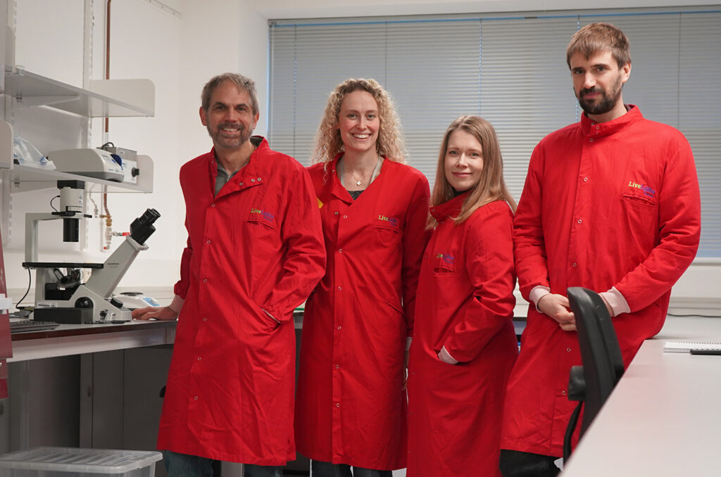
Siiri Salooma, PhD, founding scientist, and Tom Wyatt, PhD, founding scientist.
It was at exactly then that I wanted to go down this research route in more detail that Altos Labs opened in Cambridge. The company had a thesis of “Let’s try and do real discovery science, deep, groundbreaking science,” but in a biotech environment where you’re much more collaborative. That really attracted me at the time, and so I applied to work there after my PhD. Luckily enough, they took a chance on me and believed in me.
I loved it and I learned a huge amount. Not just in terms of how you build discovery programs from the ground up, but also how you work in a team, how you focus, and how you align incentives in biotech. I think it completely shifted my mindset away from simple academic curiosity to, “How do we drive that curiosity towards impact as quickly as possible?” The bar, in my opinion, is somewhat higher than in academia because you’re not just saying, “Is this good enough to publish?” You’re saying, “Is this a therapy? Is this actually good enough to put into a human and help them and not harm them?”
Q: What made you decide to found Cyclana Bio?
Wenger: I was in an epigenetics lab within Altos, and my co-founder was actually one of the group leaders there working on the extracellular matrix. The more I worked with him, the more I realized how massively important it is in guiding how cells behave. You can get completely different responses from a cell depending on what environment it’s in. I got really interested in that interface between the epigenetics, the gene level regulation, and the [extracellular] matrix.
I was doing a lot of discovery science there, but during that time, I also developed endometriosis. I was in my mid-to-late 20s when my symptoms started, and they got worse very quickly. Like every scientist who gets diagnosed with a condition, I nerded out on the disease. In my spare time, I downloaded all the data and looked into what research was there, and very quickly realized that there wasn’t much information available.
There’s not much that we know about the disease and how it happens. People are still debating the causes and drivers of endometriosis. That was interesting and another area of unknown, which has always been what I was attracted to. I’d always been passionate about women’s health, but never really had the opportunity to do something about it.
There’s an easy, non-invasive way of getting access to cells to study endometriosis because menstrual fluid is built and shed every month from your endometrium. It’s built and shed in healthy women, in women with endometriosis, and in women with other conditions. On top of that, biopsies are actually way more common in gynecology than in a lot of other conditions. So I realized this was a huge opportunity to do this tissue-level discovery that we were so passionate about, but for a cause that I really believed in, in a field that was unknown.
I spoke to the CEO at Altos at the time and explained what I wanted to do. He was supportive and thought it was an interesting idea, but ultimately, the indication didn’t align with the priorities of Altos—of looking into age-related diseases. So that’s when we left and started Cyclana Bio.
Q: How easy was it to start the company?
Wenger: We were very lucky in that we got into the Babraham accelerator program very quickly, last May. That was important because not only did it give us validation that someone had actually picked us and said this is a good idea, but it also gave us lab space and access.
We did take quite a bit of a risk. Both my co-founder and I left without having raised funding or grants to start the company. For a short while, we were living off our savings and also paying for some very preliminary science and our first scientist to try and get some data going. That was in May [2025], but quite quickly, we got a bit of traction. By July, we had our first investment term sheet because we started fundraising immediately. Then by September, we were oversubscribed. We finalized the closing in October–November for a £5 million pre-seed round.
I think along the way, we basically just had to assume it was going to happen. We were building the company as if we had the money already, although we were always very open with Tom [Wyatt], the scientist who joined us first, about how much funding we had when he joined.
We’re nine people now and have some amazing scientists who have joined the team, including a great CTO who was also at Altos beforehand. We are still growing as we speak. It’s funny how science brings so much more science.
Q: What are you trying to achieve at Cyclana?
Wenger: Our main aim is to get at least one therapy that’s truly disease-reversing to the clinic. Based on a lot of research, we know that the extracellular matrix can guide how cells respond. It can act as a sink for particular signal factors. Sometimes it can sequester or deliver things like growth factors or inflammatory signals, but it also massively changes how the cell is interacting with its neighbors.
It’s a key component of a positive runaway effect that happens in lots of chronic inflammatory diseases and in some cancers. A lot of the time, when trying to develop treatments, we focus on the cells and whether we can stop that inflammation. What we think is, if you don’t address problems with the matrix, you are not going to cure the disease. You’re effectively just going to mitigate the side effects, and this matrix is going to act like a memory of the disease. This means that if you stop the treatment, it comes back because the matrix issue hasn’t been solved.
We think that that’s a big element of what’s going wrong with endometriosis. Lesions are removed surgically and then they come back. We really think the diseased extracellular matrix is very much driving the pro-inflammatory phenotype, and that if we don’t address that, we don’t actually get to the point where we are curing the disease. We want to see if we can effectively reverse the phenotypes and if we can effectively get to a cure by stopping this recurrent feedback loop.
We haven’t settled on an exact target or modality yet. We’re exploring a few different targets, and I think based on exactly what mechanism we want to go after, we will determine what the best modality is. We want to be sure about the science, very sure about the target, and then make that target work.
Q: Where does precision medicine come into your strategy?
Wenger: Our overall strategy is based on how we see endometriosis as a whole, but I think endometriosis hopefully won’t be viewed like that much longer. We hope that there’s going to be much better stratification and classification of the disease, because it manifests very differently in different women.
Although we think the extracellular matrix might be a common mechanism, we’re building a research platform where we will hopefully find out for sure because we’re collecting data. We’ve got an ongoing observational clinical study where we’re collecting biopsy tissue, menstrual fluids, clinical data, and blood from women, either with or without endometriosis.
We’re collecting that data, looking at the tissue, the proteins, and the architecture, but also isolating cells to test in our models. Then, when we start perturbing with particular interventions that we think might reverse the disease’s impact on the matrix or have different effects on the cells, we might start seeing patterns as to which types of women with endometriosis respond well to different treatments.
It’s going pretty well so far, thanks to our clinical collaborators and participants who have donated samples. Menstrual fluid is a very good way of getting samples from seemingly healthy women, because they don’t need to go to the doctor, [they] just send us a sample. We are also collecting tissue biopsies during routine gynecological procedures to minimize invasiveness and inconvenience.
So far, there’s lots of variability, which was what we expected and which is why we want to collect [samples from] a high number of donors. Not because we think that variability is noise, but because variability is signal. It can tell us more about the nuances of the disease in these different manifestations.
Q: What has the experience of being a CEO and biotech founder been like so far?
Wenger: It’s definitely been a steep learning curve. I think that’s also why it’s been so fulfilling, because I do love being in an environment where I’m not complacent, where I’m always learning.
To some extent, because we had so much freedom at Altos to drive our own projects, I had exposure already to the pure project management side of science, so that didn’t seem quite as much of a step up.
Obviously, there’s a huge business, commercial, and legal dimension that I never had thought about before. But I have been trying to learn as much as I can, as quickly as I can, from others. One thing that the biotech field is quite good at is volunteering information. You go to any sort of networking event, or you meet someone from the industry, and they are often very willing to talk to you about what they’re interested in, but also about what you’re doing, and give any advice they might have.
I’ve met many people who have helped me along the way and who have shared their opinions with me. I walked in expecting academia to be way more collaborative than the biotech industry, but actually, I’ve been very pleasantly surprised with my experience.
Q: Can you share any key learning experiences from the last year?
Wenger: If you have scientific training or you can think in a scientific way, going into the field of business or building a company is somewhat similar. There’s lots of information and lots of alternative paths that you can take, just like in scientific discovery, and there is differently weighted evidence as to which paths are the best ones to take. Once you have a certain amount of information, you can then take the best educated guess. That’s how I’ve gone about building the company. For example, when I started, I was told by a friend, “If you’re starting a biotech, you’re going to need to raise venture capital.” They gave me a book called Venture Deals, which is a very good book that explains how funds work. I read that book and felt I was better equipped to talk to people at the fundraisers. I think the first thing I’d say is, when going into any sort of field, try to really understand how and why that field exists and what are the structures that define its environment. Then you can put context into how people work. As a first-time founder, you might think, “I’m going to find investors, and if they believe in me, they’ll invest.” But there’s so much more to running a venture capital fund. Those things are important to know when framing your discussion.
Something I would do differently is not do everything at once. I left my job, started the company, started the science, started building the network, and started fundraising at the same time. There was always this pressure when I was meeting people that I also had to get them to invest. I think looking back on it, if I could start over, I probably would have spent a few more months trying to build my network and understanding the field better before I started having those investment conversations. It still worked out for us, we still raised funds, but it was stressful. Networking events were very high stakes!
Q: How has the endometriosis space changed in recent years?
Wenger: Gedeon Richter purchasing the Celmatix portfolio and backing FimmCyte are very good signs that people are trying again. I think endometriosis has been plagued by failures in clinical trials, and I think now we’re finally seeing some non-hormonal options being tested, which makes me hopeful. Some will fail, some will succeed, and the successes will drive more interest and availability of funding and hopefully, more successes in the future. I’m really looking forward to seeing the results from some of those clinical trials because I think the more solutions we have for women, the better.
Q: Is funding in the overall field of women’s health changing for the better?
Wenger: Absolutely. I think the funding environment is more open to women’s health. I think that’s been helped by the World Economic Forum and McKinsey Health Institute driving the message of value there. There is excitement, I think, and more funding, especially privately.
I’m not sure about public funding. I do think that on the public funding side, we have a trend of saying, “We should fund women’s health, let’s look for quick wins.” I think that’s a bit of a problem with any field that’s been somewhat left behind, once we realize that we need to bring it back. The risk will be funding the wrong things or putting too much of the money into solutions that may not be revolutionary because they don’t have the foundational science to back them up.
It can also be easy to get stuck in the valley of death between seed and late-stage funding. But I do think that there are incredible scientists moving into the field, and there are some great companies starting up. So even if there is this bias towards pre-seed or seed funding, you only need a couple of those companies to have some really promising data, and they will be funded. The bar might be higher than in other fields, but if you produce groundbreaking discoveries, there will be money.
Q: What advice would you give other new founders starting to build their companies?
Wenger: Just follow your gut and your dream. That’s the most important thing. I started Cyclana because I thought this needed to happen and we needed to look into endometriosis. I thought it was a bit hypocritical of me to think we needed to do something and not do it, despite having the training and the skills to try and find a solution. If you really believe something needs to happen in the world, startups are the best way to feel like you are driving that change and contributing to seeing the change that you want to happen. Whether it succeeds or not, you won’t wake up thinking, “What am I doing this for?” You’ll just be thinking, “I really hope that we don’t fail!”
Helen Albert is senior editor at Inside Precision Medicine and a freelance science journalist. Prior to going freelance, she was editor-in-chief at Labiotech, an English-language, digital publication based in Berlin focusing on the European biotech industry. Before moving to Germany, she worked at a range of different science and health-focused publications in London. She was editor of The Biochemist magazine and blog, but also worked as a senior reporter at Springer Nature’s medwireNews for a number of years, as well as freelancing for various international publications. She has written for New Scientist, Chemistry World, Biodesigned, The BMJ, Forbes, Science Business, Cosmos magazine, and GEN. Helen has academic degrees in genetics and anthropology, and also spent some time early in her career working at the Sanger Institute in Cambridge before deciding to move into journalism.
The post Cyclana Bio Is Exploring the Extracellular Matrix to Treat Endometriosis appeared first on Inside Precision Medicine.


![Guangjun Nie, PhD, professor at the National Center for Nanoscience and Technology, Beijing, China, with his team of scientists in the lab. [Guangjun Nie]](https://www.genengnews.com/wp-content/uploads/2026/06/Guangjun-2-300x201.jpg)
![Wei Tao, PhD, professor at the Harvard Medical School (far right, first row) and his research team. [Wei Tao]](https://www.genengnews.com/wp-content/uploads/2026/06/Wei-Tao-300x219.jpg)