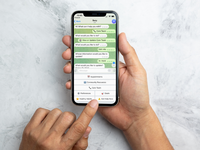
Impact of Restriction-Resumption Protocols on Mood and Anxiety in Healthy Adults: Randomized Controlled Trial
Efficacy of a World Health Organization–Guided Self-Help Intervention for Reducing Psychological Distress in Afghan Refugees: Randomized Controlled Trial
Collaborative Drug Discovery Inks Deal with Eli Lilly to Accelerate Biotech Innovation
Lilly created Lilly TuneLab to accelerate biotech innovation by enabling participating companies to access models trained on Lilly’s proprietary research data. Through this agreement, biotech companies that use CDD Vault will be able to utilize select Lilly predictive models within their natural scientific workflows, according to Barry A. Bunin, Collaborative Drug Discovery (CDD) president and CEO.
“By integrating TuneLab directly into CDD Vault, we are advancing CDD’s core vision to enable collaboration across drug discovery teams and organizations. We believe that solving the most complex challenges in drug discovery will depend on innovative collaboration models that provide broad access to research data and empower chemists and biologists to make informed, data-driven decisions,” said Bunin. “TuneLab’s ADMET models will fit in our secure CDD Vault software environment in natural workflows for experimental and computational scientists and with our growing CDD Vault ecosystem of biopharmaceutical companies.”
This agreement paves the way for the planned integration of Lilly TuneLab in both the core and AI modules within CDD Vault for biotech companies that opt into the program. A company spokesperson explained that the agreement builds on CDD’s founding vision from 2004 to demonstrate the economics of efficiency of web-based collaboration.
“TuneLab’s models are synergistic with our innovations such as Zero Click Models, Generative Bioisosteres, as well as Ultrafast Deep Learning similarity to SureChEMBL for novelty and Enamine libraries for convenient SAR-by-catalog,” noted CDD research informatics senior scientist Peter Gedeck, PhD.
The post Collaborative Drug Discovery Inks Deal with Eli Lilly to Accelerate Biotech Innovation appeared first on GEN – Genetic Engineering and Biotechnology News.
Early Medical Care Linked to Rapid Gut Microbiome Shifts in Amazonian Indigenous Communities
Researchers from Rutgers University have shown that even limited exposure to modern medical care is associated with rapid changes in gut microbial diversity among remote Indigenous communities in the Amazon. The research, published in Cell Reports, studied the effects on Indigenous people in Venezuela where subsistence lifestyles have remained largely unchanged until the introduction of a World Health Organization–supported medical program for the treatment of the parasitic infectious disease onchocerciasis, also known as river blindness. After receiving medical care, the researchers discovered that gut microbial communities in the people began to shift toward patterns more commonly seen in industrialized populations after only a few medical visits and included measurable declines in microbial diversity.
“The study gives us a better idea of how sensitive human gut microbes are,” said senior author Maria G. Dominguez-Bello, PhD, a professor of microbiome and health at Rutgers. “It opens the door for future research on how we can restore our microbiota after using medicines like antibiotics, which can deplete organisms in our gut.”
Prior research has established that urbanization-related changes in diet, lifestyle, and environment can alter the gut microbiome, but those factors are often intertwined. In this research, the unique setting and population allowed the Rutgers team to isolate the effects of repeated medical exposure in populations with minimal prior contact with modern healthcare.
“Many factors contribute to reduced microbial diversity associated with Westernization, complicating efforts to identify early drivers of microbiome change,” the researchers noted, adding that the Amazonian villages provided a setting where “low-exposure villages show higher baseline gut microbiota diversity than the medium-exposure village, and microbiota diversity declines over time in association with repeated exposure, particularly in children.”
The study followed 335 participants across multiple villages, collecting fecal samples and body-site swabs before and during repeated medical visits between October 2015 and February 2016. The villages were categorized by prior exposure to outsiders and medicine, allowing comparisons between low-exposure communities and a medium-exposure village that had a longer history of modern medical services. Researchers collected samples from the gut, mouth, nose, and skin in conjunction with the WHO’s quarterly visits that delivered antiparasitic treatments and basic care.
“This study leverages a rare longitudinal dataset from remote Amazonian Indigenous communities to examine how the human microbiome shifts during the earliest stages of contact with external institutions, prior to major dietary or lifestyle urbanization,” the researchers wrote.
In the gut microbiome, data from the collected samples showed a decline in taxa commonly associated with fiber metabolism and an increased abundance of bacterial groups more commonly seen in industrialized populations. Functional gene profiles also shifted, with increased representation of genes linked to simple carbohydrate metabolism and antimicrobial resistance, and reduced representation of genes involved in fiber fermentation and other metabolic processes.
The study also found that microbial changes were most pronounced in children, pointing toward a heightened sensitivity of developing microbiomes to repeated medical exposure. In addition to gut changes, oral, nasal, and skin microbiomes also shifted: oral microbial diversity declined; nasal diversity increased after initial visits; and skin communities showed reductions in diversity and shifts in composition.
Previous research cited by the authors has linked industrialized lifestyles with reduced microbial diversity, altered microbial networks, and increased prevalence of genes associated with antimicrobial resistance. However, the current study provides a unique glimpse at the effects of the earliest stages of treatment by modern medicine before shifts in diet or infrastructure occur.
This new data highlights the need to find the balance between essential medical interventions and preservation of microbial diversity. While treatments such as those for river blindness remain critical for reducing infectious disease burden, the researchers suggest that repeated exposure to medical care may coincide with microbial restructuring that reduces diversity and alters functional capacity.
“Understanding and navigating this balance is essential not only for microbiome science but also for ethical, culturally informed approaches that respect both biological and social dimensions of wellbeing,” the researchers noted.
Future research will examine ways to protect microbial diversity during necessary medical interventions and expanding longitudinal studies that assess resilience and recovery of the microbiome after repeated exposure to medicines.
The post Early Medical Care Linked to Rapid Gut Microbiome Shifts in Amazonian Indigenous Communities appeared first on Inside Precision Medicine.
HELIX AI Model Accurately Predicts RNA Splicing, Unlocks Precision Medicine
RNA splicing, in which different coding RNA, or exons, are joined together after noncoding regions, or introns, are removed, allows for a large array of RNA transcript isoforms with distinct sequences, and functions in tissue- and cell-type-specific patterns. Conversely, transcript isoform alterations can sensitively reflect dynamic changes in cellular states. Aberrant splicing is closely associated with major diseases, such as cancer.
In a new study published in Nature Computational Science titled, “HELIX: a scalable model for predicting context-dependent regulation of RNA splicing and isoform usage,” researchers from the Chinese Academy of Sciences have developed an AI-driven framework that enables highly accurate prediction of RNA splicing and isoform usage by integrating genomic sequence features with tissue-specific RNA binding protein (RBP) expression profiles. The work offers valuable insights for splicing regulatory patterns, pathogenic variant interpretation, and precision medicine research.
Isoform usage is jointly regulated by multiple layers of control, including regulatory elements, such as splicing enhancers and silencers on exons and introns, and tissue microenvironments. Scientists have been challenged to accurately characterize and predict RNA splicing and isoform usage across tissues, cell types, and disease states.
The study’s AI framework, Hierarchical Explainable LSTM for Isoform eXpression (HELIX), overcomes the limitations of conventional approaches via a two-layer deep-learning architecture.
First, the framework integrates DNA sequence information with the expression profiles of 1,499 RBPs. Long short-term memory (LSTM) networks are then employed to effectively capture the complex dependencies and competitive relationships among multiple splice sites.
This design enables precise, reliable prediction of RNA splicing and transcript isoform usage. The model was trained and optimized on large-scale short- and long-read RNA-seq datasets covering 30 distinct human tissues, allowing accurate quantification of complex transcript structures and isoform usage. Results show that HELIX substantially outperforms existing mainstream methods in both splicing strength prediction and overall isoform usage prediction.
In disease-related studies, HELIX deciphered aberrant RNA splicing and transcript isoform alterations. Notably, the researchers identified widespread splicing dysregulation and abnormal isoform usage in tumor cells using large colorectal cancer cohorts.
The results reveal strong correlations among such alterations and genomic mutations, RBP dysregulation, and patient clinical profiles. Results support that splicing abnormalities can serve as key molecular signatures for tumor progression and guiding patient stratification.
The team also developed scHELIX, a single-cell RNA sequencing extension of HELIX. scHELIX supports high-resolution profiling of transcript isoform usage across different cell types and tumor subpopulations, which offer a refined view of intratumoral heterogeneity.
The findings reveal distinct RNA splicing and isoform usage patterns among tumor subclones, providing new clues for tumor evolution research and potential therapeutic target discovery.
The post HELIX AI Model Accurately Predicts RNA Splicing, Unlocks Precision Medicine appeared first on GEN – Genetic Engineering and Biotechnology News.
Transmission Dominance Under Random-Contact Intensification in Epidemic Networks: Multilayer Contact Network Simulation Study
Background: In the context of COVID-19, infection spread through human contact networks remains a major public health challenge. Beyond cumulative infections and deaths, it is necessary to understand which contacts matter most, and which population segments contribute most to transmission under different social conditions. In multilayer urban networks with community structure, routine contacts coexist with incidental encounters, and it remains unclear whether incidental encounters can alter epidemic burden and the main contributors to transmission when per-layer contact caps and routine-contact minima are unchanged (for the nonrandom layers). Objective: Under explicit daily-contact constraints, we examined (1) how changing overall contact opportunities affects epidemic speed and burden when incidental encounters are held fixed, and (2) whether increasing incidental encounters alone, per-layer contact caps, and routine-contact minima fixed (for the nonrandom layers), shifts the main contributors to transmission from a high-contact group to a medium-contact group, and the underlying network mechanism. Methods: We constructed a multilayer potential contact network for a synthetic urban population of 10,038 individuals, representing household, school, workplace, distance-driven activities, and incidental encounters as separate layers. Daily contact networks were sampled from the potential network each day, and transmission was simulated for 120 days using a Susceptible-Exposed-Infectious-Removed model with vaccination. Individuals were classified into high-contact and medium-contact groups based on baseline contact intensity, and group contribution combined each group’s share of infectious individuals and its per-infectious effective transmission yield. Contact-constraint parameters were calibrated using an online survey in Tokyo and Kanagawa (n=1089), and scenario comparisons and parameter sweeps were used to locate the transition point. Results: With incidental encounters held fixed, higher overall contact opportunities produced earlier and higher epidemic peaks and larger cumulative infections and deaths, whereas reduced opportunities slowed and prolonged spread. Holding overall contact opportunities and routine contacts fixed, increasing incidental encounters shifted the main contributors to transmission: higher-contact individuals accounted for more effective transmissions at low incidental contact, whereas medium-contact individuals accounted for more beyond a clear transition point. Network visualization and schematics suggest a bridge-allocation mechanism, where stronger incidental contact adds cross-community bridges that more often terminate at medium-contact individuals and carry infection into less-affected communities. Across R=30 replicate runs under fixed settings, the dominance flip was consistently observed, and the estimated threshold W∗ showed a narrow but nonzero distribution (reported as median and IQR). Conclusions: In multilayer urban contact networks with community structure, our results indicate that intensifying incidental encounters can change the main contributors to transmission even when overall contact opportunities and routine contacts are unchanged. We present an analysis framework under explicit daily-contact constraints to identify this contributor shift and its transition point, supporting comparisons of intervention priorities across social contact conditions.
<img src="https://jmir-production.s3.us-east-2.amazonaws.com/thumbs/1fbd573eb4e3a2afda02c2afd8220499" />
AI Shows Value for Breast Cancer Prediction
AI is increasingly being used to help determine breast cancer risk, with two studies showing its value for primary prevention and in women of diverse ancestry.
The first study, in Science Translational Medicine, revealed that AI could outperform standard clinical risk tools at identifying women of European ancestry who were at greatest risk of developing breast cancer within the next decade.
Because current AI-based models mostly predict breast cancer risk in the short term, Mikael Eriksson, PhD, from the Karolinska Institute, and fellow researchers created a model for invasive and in situ breast cancer that covered a 10-year period.
“Considering that a tumor can take five to 20 years to develop into a screen- or clinically detected cancer, a 10-year or lifetime risk projection time is reasonable,” they noted.
The researchers validated their image-derived AI-based 10-year risk model using digital mammograms from 8676 women in two cohorts from the U.S. and Sweden, of whom 1633 had breast cancer.
They compared their model with three other risk assessment tools: the Tyrer-Cuzick-v8, which uses personal and family history to determine the lifetime risk of breast cancer; the Breast Cancer Surveillance Consortium v3 tool, which estimates the risk of developing invasive risk cancer over five years; and the Mirai AI algorithm for predicting breast cancer risk.
The model calculated that the 10-year risk of breast cancer was 3.83% for the North-American group and 3.14% for the Scandinavian group, and it showed promise for predicting the risk of invasive tumors.
The AI model performed significantly better at predicting invasive breast cancer than the three comparator models both overall, in women aged 50 to 69 years and those with estrogen-receptor-positive breast cancers.
“An image-derived AI-based risk model developed for 10-year risk assessment for identifying individuals in mammographic screening who may benefit from primary prevention strategies identifies up to 40% of breast cancers to be at high risk at study entry in U.S. and Swedish validation case cohorts per clinical guidelines,” summarized Eriksson and co-workers.
The second study, in Science Advances, revealed how AI could be of value for women from more diverse backgrounds, and that it had value regardless of race and ethnicity.
The research was driven by the observation that conventional prediction models incorporating genetic and clinical factors including breast density underperform in women who do not have European ancestry.
Shu Jiang, PhD, from Washington University School of Medicine in St Louis, and co-workers set out to evaluate the generalizability of an AI-derived mammogram risk score (MRS), a summary of texture features which captures intrinsic breast-tissue characteristics that are the basis for cancer to develop.
To do this, they used data from two large North American cohorts that represented more than 226,000 racially diverse women undergoing routine breast screening with mammography.
This was then used to examine the generalizability of MRS with breast cancer risk across non-Hispanic white (NHW), non-Hispanic Black (NHB), East Asian, South Asian, and Indigenous women.
Jiang and team found that the MRS was a strong predictor of breast cancer risk independent of race or ethnicity, demonstrating its potential for broader clinical utility.
Across the two independent screening cohorts, the MRS increased with age in line with rising breast cancer risk. It was a strong and consistent predictor of breast cancer risk across race and ethnic groups as shown by the association study and comparison of distributions and demonstrated excellent calibration in all groups.
Its remained robust across full-field digital mammograms or synthetic two-dimensional digital breast tomosynthesis, although the limited numbers of images precluded an analysis of how performance varied across manufacturers.
“Overall, these results support that MRS is a powerful breast cancer risk predictor that does not depend on race or ethnicity, supporting its potential for broader clinical adoption and use in varied populations of women undergoing routine screening mammography to identify those at increased risk,” Jiang and coworkers concluded.
The post AI Shows Value for Breast Cancer Prediction appeared first on Inside Precision Medicine.
Ultra-Short Radiotherapy Schedule for Prostate Cancer Supported by HERMES Trial
U.K. research has shown that condensing prostate cancer radiotherapy into two sessions, but with a higher dose per session, rather than the traditional five sessions does not lead to increased side effects when treatment is delivered using state-of-the-art magnetic resonance imaging (MRI)-guided technology.
Lead researcher Sian Cooper, clinical research fellow at The Royal Marsden NHS Foundation Trust and the Institute of Cancer Research in London, told Inside Precision Medicine that the results of the HERMES trial “point toward a future where prostate radiotherapy is tailored to the disease, to the individual’s anatomy and symptom profile, whilst being mindful of the impact of treatment time on patients’ lives.”
Presenting at the 2026 Congress of the European Society for Radiotherapy and Oncology (ESTRO), Cooper said that the number of people diagnosed with prostate cancer is projected to double by 2040, meaning the demand for effective and efficient treatment has never been greater.
At present, localized prostate cancer is typically treated with stereotactic body radiotherapy over five sessions, but there has been increasing interest delivering the treatment in fewer sessions, with a larger dose each session.
“For patients, a two-session treatment course would be far less disruptive than the weeks of daily hospital visits that radiotherapy has traditionally required. This convenience comes with clear benefits for work, leisure, family life and travel. For clinicians and health systems, fewer fractions mean faster workflow throughput, and getting patients the right treatment, quicker,” said Cooper.
“We wanted to find out whether giving the equivalent dose in just two treatment sessions could be feasible and safe for patients, and to understand how it might affect the potential side effects patients can experience, such as problems with urinary and bowel function.”
The move toward giving higher radiotherapy doses in fewer sessions has been made possible by improvements in radiotherapy delivery technology over recent years.
“It has allowed us to harness the power of modern computing and discoveries in clinical physics, to create radiotherapy doses which conform very tightly to the edge of the prostate,” Cooper explained. “This results in vastly less dose to the normal, healthy tissues around the cancer.”
The HERMES trial used a Unity MR-Linac (Elekta AB, Sweden) machine, which Cooper describes as “the ultimate evolution of this progress,” to deliver radiotherapy to participants.
The device combines real-time MR scanning with a linear accelerator and is known as MRI-guided adaptive radiotherapy. It allows adaptation of the radiotherapy beam design to changes in patient anatomy on the treatment day as well as moment by moment motion management, meaning that if there are any changes in the target or healthy bystander organs, the radiation beam can be switched off.
“This level of precision was needed to safely deliver the high dose of radiation necessary to maintain the biologically equivalent dose in just two fractions,” Cooper noted.
In all, 46 participants (median age 74 years) with intermediate or lower high-risk prostate cancer were randomly assigned to receive radiotherapy at a dose of 24 Gy in two fractions over 8 days, with a 27 Gy integrated boost to the MRI defined tumor (n=22), or 36.25 Gy in five fractions over two weeks to the planning target volume, with 40 Gy to the prostate and proximal one cm seminal vesicles (n=24). All participants had androgen deprivation treatment for at least six months.
Cooper reported that, at two years, four (18%) participants in the two-fraction arm and six (25%) in the five-fraction arm experienced moderate (grade 2) urinary adverse events (AEs) such as increased frequency or urgency. Grade 2 gastrointestinal AEs occurred in one participant in each arm (5% and 4%, respectively).
There were no grade 3 or worse genitourinary or gastrointestinal events in either group.
The team also showed that quality of life, measured by the International Prostate Symptom Score and the International Index of Erectile Function, showed minimal change up to two years but will continue to be monitored up to five years.
Cooper said that the investigators will present efficacy data, a secondary endpoint, when it matures but she pointed out that as “the cancer control rate for prostate cancer is so high, it often takes many years for failures to appear, whereas trial data for ultra hypofractionation show that the genitourinary adverse event rate is the primary concern after treatment.”
ESTRO president, Matthias Guckenberger, MD, from University Hospital Zurich, Switzerland, who was not involved in the research said: “While the technology used in this trial is currently available in only a limited number of specialist centers worldwide, they are growing rapidly. These results can help guide how they are used and help us understand whether two-session radiotherapy should become a new standard of care.”
The post Ultra-Short Radiotherapy Schedule for Prostate Cancer Supported by HERMES Trial appeared first on Inside Precision Medicine.
STAT+: Scientists track cellular disruptions that lead to type 1 diabetes
The story of type 1 diabetes begins in the pancreas, long seen as a battleground between insulin-producing beta cells and misdirected immune defenders. Scientists have been searching for ways to spot this internal warfare early enough to prevent a lifelong disease that depletes the body’s source of insulin.
Two new papers published Wednesday in Science Translational Medicine offer new clues to what happens in these beta cells before type 1 diabetes emerges. Experiments in human cells and in mouse models used biosensors and genetic analyses to illuminate this pathway and detect possible ways to halt beta-cell destruction.
In the first study, a team from the Indiana University School of Medicine explored how certain immune cells involved in inflammation, known as signaling cytokine interferon-alpha, normally trigger beta cells to produce other molecules that play a role in inflammation, cell proliferation, and cell death. These reactive oxygen species, ROS for short, sometimes cause collateral damage. But cells from patients with type 1 diabetes did not have ROS-producing beta cells, suggesting they lacked the cytokines that stimulate their ROS production. That dearth might be useful in flagging the decline of beta cells early on in type 1 diabetes, the study authors surmised.

