StockWatch: Parabilis Medicines Makes Wall Street History with $770.5M IPO

In what’s shaping up to be a historic year for biotech initial public offerings, Parabilis Medicines (Nasdaq: PBLS) made its mark on Wall Street this past week by pricing the largest-ever IPO by a drug developer. This upsized offering raised an eye-popping $770.5 million in gross proceeds.

No sooner did Parabilis begin trading public shares on Wednesday, a day after offering 33.5 million shares of its common stock at $20 per share, than its stock price leaped 58% from the IPO price, closing its first full trading day at $31.60. Those shares slid 4% on profit taking Thursday, closing at $30.31, then fell another 10% Friday to finish the week at $27.26.

A day earlier, Parabilis announced the closing of its IPO, including a full exercise by underwriters of their 30-day option to buy an additional 5.025 million shares at the IPO price, less underwriting discounts and commissions. That added another $100.5 million to the initial $670 million in gross proceeds, which translates to another $93.5 million in net proceeds. When added to the $618.2 million net that Parabilis garnered from the original IPO, the company’s haul from the offering rises to a no-less-eye-popping $711.7 million, according to Parabilis’ IPO final prospectus.

Parabilis priced its IPO above its initial price range of $17–19 a share. The company initially planned to offer 25 million shares, then raised that offer to 33.3 million before adding 200,000 more shares to finalize its offering.

Parabilis’ IPO surpasses the $625 million IPO carried out in April by Kailera Therapeutics (Nasdaq: KLRA), which topped the previous record-high among U.S. biotechs, the $604 million offering of Moderna (Nasdaq: MRNA) in December 2018, two years before the messenger RNA (mRNA) vaccine developer won FDA emergency authorization for its COVID-19 vaccine.

Based in Cambridge, MA, Parabilis is a developer of drugs and antibody-drug conjugates (ADCs) targeting historically undruggable protein targets and based on stabilized helical peptides or Helicons™.  Parabilis says it has generated proprietary datasets, comprising millions of data points for hundreds of thousands of Helicons across dozens of drug-like properties, following a decade of Helicon drug discovery.

“Unlike Kailera, which formed in 2024 and quickly stockpiled private capital before going public, Parabilis took a longer and far less linear road,” commented Ben Zercher, senior biotech & pharma analyst with PitchBook.

Parabilis was founded in 2015 as FogPharma to commercialize technology developed in and in-licensed from the lab of Harvard University researcher and serial entrepreneur Gregory Verdine, PhD, who served as the company’s co-founder and CEO from 2015–2023.

Six venture rounds

While the company closed six venture rounds, Zercher noted, its valuation had fluctuated in the face of the post-pandemic period that saw private biotechs struggle, as well as leadership turnover and the rebrand through which FogPharma became Parabilis in 2024.

The rebranded company built momentum last year, according to Zercher, on the strength of its lead candidate zolucatetide (formerly FOG-001), a stabilized peptide built using the company’s Helicon platform. Zolucatetide is the first and only direct inhibitor of the elusive β-catenin:TCF interaction, according to the company.

Parabilis stated in its final prospectus that it plans to spend approximately $150 million in IPO proceeds toward continuing ongoing clinical development of zolucatetide in desmoid tumors, including continuation of dose expansion and the launch of a Phase III registrational trial to topline data.

Approximately $120 million is set to be spent on continuing the ongoing clinical development of zolucatetide across several additional indications, including dose escalation and expansion in familial adenomatous polyposis (FAP); hepatocellular carcinoma, the most common type of primary liver cancer; and other rare tumors, with the aim of collecting data to support a registrational trial.

Parabilis plans to use the largest share of its IPO proceeds, approximately $190 million, toward advancing its pipeline of additional programs—including its ETS-related gene (ERG) protein degrader, an allosteric androgen receptor in its active state (ARON), and beta-catenin degraders—to Phase I clinical data.

“Our current pipeline is focused on various cancers and tumor types; however, we believe Helicons could also have broad applicability against targets in many diseases with substantial unmet need outside oncology, and we plan to evaluate other therapeutic areas in the future,” Parabilis stated in the final prospectus.

The remainder of the proceeds would be used, Parabilis said, toward continued evolution of the Helicon platform for discovering and developing drug candidates, as well as toward general corporate purposes that include additional development efforts, working capital, and operating expenses.

Zolucatetide received the FDA’s Fast Track designation last year, followed in March by the agency’s Orphan Drug designation. In January, Parabilis closed on a $305.2 million Series F crossover financing round by selling 49,518,175 shares at $6.1644 per share to various investors, garnering $304.5 million in net proceeds. Parabilis finished the first quarter with $329.039 million in cash and cash equivalents as of March 31.

Fifteenth biopharma IPO so far in 2026

Parabilis is the 15th and latest biotech or pharmaceutical company to carry out an IPO this year, raising a combined $12.11 billion in proceeds, according to PitchBook data. Biotech and pharma accounted for 15 IPOs in all of 2025, raising a combined $10.49 billion—an improvement in dollars over 2024’s $8.83 billion, which was raised in 33 IPOs.

Seven of this year’s IPO companies have seen their shares rise since their initial offerings, led by the 466% share price increase of Veradermics (NYSE: MANE), a developer of treatments for dermatology and aesthetic conditions that closed Friday at $96.24 a share.

“With the biotech window reopened, the volume of IPOs reflects a backlog of quality companies that kept building through the biotech funding downturn rather than a wave of hype,” Zercher added. “Where the pandemic-era class sold preclinical optionality, Parabilis and the 2026 cohort are being priced on de-risked clinical programs with clear regulatory paths.”

In addition to its IPO, Parabilis said, it has also closed on selling 4,166,666 shares at $18 per share—90% of the IPO price per share—through a concurrent private placement to Regeneron Pharmaceuticals that has raised approximately $75 million in proceeds.

Parabilis’ initial IPO filing, dated May 19, came just a day after Parabilis inked an up-to-$2.3 billion-plus strategic research collaboration with Regeneron to discover and develop an initial five candidates encompassing “antibody-Helicon conjugates,” a new form of ADCs aimed at challenging and historically undruggable targets by combining the cell permeability of small molecules with the binding capabilities of larger biologics, in order to reach targets long considered undruggable.

Financial runway into H2 2029

Regeneron agreed to pay Parabilis $50 million upfront toward launching the collaboration. That upfront payment, plus proceeds from the IPO and the company’s existing cash and cash equivalents, “will be sufficient to fund our operations into the second half of 2029,” Parabilis stated in its IPO final prospectus.

According to that final prospectus, Parabilis ended the first quarter with a $45.316 million net loss, up 18% from its $38.326 million net loss of Q1 2025, as well as a net loss of $145.889 million for last year, up nearly 24% from its $117.914 million net loss for 2024. The company has no reported revenue.

Parabilis’ accumulated deficit rose 8% during Q1, to $586.82 million from $541.504 million at the end of 2025.

To fund its operations, Parabilis reported, it has raised a total of $876.8 million as of March 31. That total consisted of $811.8 million from sales of its convertible preferred stock, $15 million in borrowings under a term loan, and a $50 million Simple Agreement for Future Equity (SAFE).

Leerink Partners, BofA Securities, Evercore ISI, and Guggenheim Securities are acting as active book-running managers for Parabilis’ IPO, while LifeSci Capital is acting as a passive book-running manager.

Leaders and laggards

  • Propanc Biopharma (Nasdaq: PPCB) shares soared 80% from $1.35 to $2.43 Thursday after the Australian developer of therapies for pancreatic, ovarian, and colorectal cancers said it approved a share repurchase program authorizing the company to repurchase up to $5 million of its common stock. “The management team believes we are entering a transformative stage for the company,” CEO James Nathanielsz stated, citing recent progress by Propanc’s lead asset PRP, a first-in-class therapy designed to treat and prevent metastatic cancer from solid tumors, toward entering the clinic with a pivotal Phase Ib, first-in-human study in 30–40 advanced cancer patients. He also cited the company’s efforts to publish key scientific data, file patentable discoveries, and form partnerships with contract research organizations (CROs), contract development and manufacturing organizations (CDMOs), and suppliers: “The foundation is clearly there, and as a result, we believe we are undervalued significantly.”
  • Tango Therapeutics (Nasdaq: TNGX) shares rocketed 53% from $20.22 to $30.93 June 8 after the developer of precision oncology treatments based on synthetic lethality announced positive initial data from its ongoing Phase I/II trial (NCT06922591) assessing its next-generation, MTA-cooperative PRMT5 inhibitor candidate vopimetostat (TNG456) in combination with Revolution Medicines’ (Nasdaq: RVMD) RAS(ON) inhibitors daraxonrasib (RMC-6236) and zoldonrasib (RMC-9805) in patients with MTAP-deleted and RAS-mutant metastatic pancreatic ductal adenocarcinoma (PDAC). Tango reported that 92% of patients with PDAC in the trial’s vopimetostat plus daraxonrasib arm achieved an objective response, while patients with second and third line PDAC treated with the combination showed a six-month progression-free survival (PFS) rate of 90% (median PFS not yet reached), suggesting durability of clinical benefit. Tango said it plans to finalize the design of a Phase III randomized-controlled trial of the combination approach in front-line pancreatic cancer and disclose vopimetostat lung cancer monotherapy data in the second half of 2026.

The post StockWatch: Parabilis Medicines Makes Wall Street History with $770.5M IPO appeared first on GEN – Genetic Engineering and Biotechnology News.

Collagen Resides Inside Cells in Liquid Condensate-Like Form

A study by scientists at the Centre for Genomic Regulation (CRG) in Barcelona has found that collagen, the protein that builds skin, bones, tendons, and organs, exists inside cells as a liquid-like droplet rather than as the long, rigid rod-like structure we might find in textbooks.

The team used techniques including high-resolution live-cell imaging to generate what they say is the first direct observation of how the most abundant protein in the human body, which accounts for around a third of total protein mass, exists naturally inside living cells.

Collagen is built inside a cellular compartment called the endoplasmic reticulum (ER). The study specifically looked at a precursor form inside cells called procollagen 1 (PC1), which matures into type 1 collagen. Type 1 collagen is the most common type of collagen, consisting of around 90% of the body’s total collagen.

“Inside a cell, collagens are not rigid molecules as one had assumed,” said ICREA research professor Vivek Malhotra, PhD, senior author of the study at the CRG. They are, in fact, very pliable, taking a liquid condensate form much like oil in a drop of water.”

The liquid-like state may serve a protective function. Collagen’s job, once outside the cell, is to assemble into the rigid fibers that hold tissues together. The same process inside the cell would be catastrophic. “This is another way by which cells ensure that collagens probably never become fibrous inside the cell,” said Malhotra. “Because if it were to become fibrous, it would kill the cell.”

The new findings have implications for how the body exports its primary structural building block from production sites inside cells. The researchers suggest cells avoid using conventional receptors or vesicles, which is the route established by work carried out in the 1980s and 1990s and recognized with a Nobel prize in 2013.

Instead, they propose a “liquid extrusion” hypothesis, whereby collagen moves from its site of synthesis to the next compartment of the secretory pathway through capillary action. The new theory has important implications for wound healing, fibrosis, and cancer.

Malhotra and colleagues describe their study and results in a paper in the Journal of Cell Biology titled “Procollagen 1 assembles into phase-separated condensates in the endoplasmic reticulum.”

“Procollagen I (PC1) is assembled into a trimer within the lumen of the endoplasmic reticulum (ER),” the authors explained. Under a microscope, purified collagen looks like long, rigid rods of up to 400 nm in length, and this conformation is presumed to represent their assembled state in vivo, the team continued. “However, there is currently no direct experimental evidence demonstrating that PC1 adopts or is maintained in such a rigid, extended conformation within the ER lumen in vivo,” they wrote. Also, the vesicles that transport proteins out from their site of synthesis to the cell’s exterior are only 60 to 90 nanometers in diameter.

Since collagen’s structure was first described more than half a century ago, the field of cell biology has asked how such large molecules can be transported out of cells. The canonical picture of the protein describes collagen only after it has left cells and assembled into the fibers that hold tissues together. The newly reported findings suggest that inside the cell, collagen is not yet assembled into that rod structure.

Using high-resolution live-cell imaging of human hepatic stellate cells—the liver cells that produce collagen and drive scarring in liver fibrosis—the team showed that collagen inside the cell gathers into small droplets that merge, split, and exchange material with their surroundings. These are all signatures of a condensate, compartments of proteins that become so concentrated they disassociate from their surroundings, like droplets of oil in water.

Most of cell biology has focused on condensates in the nucleus and on stress granules in the cytosol, said first author Soumya Bhattacharyya, PhD, a postdoctoral researcher in Malhotra’s lab. “We’re just beginning to understand condensates inside the endoplasmic reticulum.”

The findings emerged from microscopy images taken by Bhattacharyya in May 2024. Bhattacharyya was using the liver cell system as a tool to study what happens when collagen production is increased in fibrotic cells. “I had no idea what it would lead to. But when we took the samples, what struck me were these bright spherical structures you can’t miss,” recalled Bhattacharyya.

The initial reaction in the laboratory to a finding that challenged cell biology dogma was sceptical. “I thought it must be an artefact,” said Malhotra. In the months that followed, the team had to settle whether the protein clumping they observed inside the endoplasmic reticulum was junk. Cells have an elaborate system for detecting badly folded proteins and either refolding them or marking them for destruction, centered on a chaperone called BiP.

If the collagen droplets were heaps of misfolded protein, the researchers would detect high levels of BiP. The droplets contained, instead, a mixture of helper proteins, including chaperones that specifically recognize properly folded collagen.

Human liver cells showing collagen droplets inside the cell (green clusters), held in place by TANGO1 (magenta), with extracellular collagen fibres visible as the surrounding network. Cell nuclei are stained blue. [Soumya Bhattacharyya / Centre for Genomic Regulation]
Human liver cells showing collagen droplets inside the cell (green clusters), held in place by TANGO1 (magenta), with extracellular collagen fibers visible as the surrounding network. Cell nuclei are stained blue. [Soumya Bhattacharyya/Centre for Genomic Regulation]

The study also clarifies the function of TANGO1, a protein discovered by the Malhotra lab roughly two decades ago and known to be required for collagen export. When the researchers depleted TANGO1, the collagen droplets still formed but were no longer positioned at the ER exit sites (ERES) where cargo leaves the compartment. Collagen secretion dropped accordingly. “PC1 condensates were still formed after TANGO1 knockdown, indicating that TANGO1 is not required for condensate formation per se,” the investigators stated. “However, TANGO1 depletion caused a marked reduction in the association of PC1 condensates with ERES …”

The discovery suggests TANGO1 acts as a mooring point that holds the droplet at the export site rather than as a conventional cargo receptor. The authors propose that collagen then leaves the cell by a physical process called wetting, in which the liquid droplet attaches to and flows through the exit site.

Malhotra offers two possible physical mechanisms for this transfer. “Imagine you have a rubber ball with a nozzle, filled with liquid. You squeeze it, you force the liquid to come out of this little orifice. Is that the mechanism? Or is the liquid rising by capillary forces, just like nutrients flow up against gravity in plants by capillary action?”

The proposed liquid extrusion mechanism remains a model, but the next experiments to obtain direct visualization of the export mechanism are already underway. The team also plans to develop a mouse model, in collaboration with external partners, to confirm the findings in living tissue. If the model is confirmed, the work has implications for several pathological conditions in which excess collagen secretion plays a central role, including liver, lung, and skin fibrosis, as well as for targeting the dense matrix that tumors use to shield themselves from chemotherapy and the immune system.

“One of the major problems in cancer is that the cells secrete so many collagens and other proteins out into the extracellular matrix that they hide in a shell made of these components and become chemo- and immuno-refractory, meaning they are not seen by the chemical therapeutics or by the immune system,” Malhotra said. “People are trying to find ways to break this tissue cement, and our study could help inform those strategies.”

The proposed collagen secretion model suggests that either degrading TANGO1 to prevent cargo from being captured at the exit site or dissolving the condensate itself to prevent the cargo from being properly organized in the first place could be new strategies worth exploring.

The post Collagen Resides Inside Cells in Liquid Condensate-Like Form appeared first on GEN – Genetic Engineering and Biotechnology News.

Hantavirus One-Shot mRNA Vaccine Fully Protects in Syrian Hamster Model

Last month, the Andes virus outbreak on a Dutch cruise ship departing from Argentina brought a transmission context for hantavirus, that was previously unprecedented, to the forefront. The Andes virus is the only member of the hantavirus family that is capable of efficient person-to-person spread through close contact with respiratory secretions. Other hantaviruses are typically spread through contact with infected rodents, making the Andes virus a much more significant public health threat.

While at sea, the outbreak spread among passengers and crew, infecting 13 people and killing three. The cruise passengers have since returned to their home countries, 23 in total. Because a person can carry the virus for weeks before showing any symptoms, health agencies are facing a complex challenge of identifying everyone who was exposed. There are currently no vaccines or preventive treatments approved for the virus; this travel-related outbreak brought the need for vaccine development to the forefront.

Researchers at The University of Texas Medical Branch (UTMB) had previously developed and tested two mRNA vaccines against intramuscular Andes virus challenge in golden Syrian hamsters (“1-methylpseudouridine-modified or non-modified mRNA modalities encoding the envelope glycoproteins, Gn and Gc, in a single open reading frame.”)

When tested in the Syrian hamster model, both mRNA vaccines were efficacious in hamsters using a two-dose regimen. Recognizing that a fast-moving international outbreak doesn’t allow time for patients to wait weeks between shots, the team retested the vaccines to determine whether a single dose would be effective.

Now, a new report shares the finding that the vaccine provided full protection against the Andes hantavirus after a single dose.

This work is published in The Lancet in the paper, “Single-dose mRNA vaccines against Andes hantavirus.”

Alexander Bukreyev, PhD, head of the Laboratory of Viral Pathogenesis and Vaccine Development at UTMB, said that the group is working to fast-track these single-dose vaccines into human clinical trials.

The results exceeded expectations. When testing the vaccines in an animal model that mimics human disease, the scientists found that a single shot provided 100% protection against a lethal dose of the virus. Even when the researchers significantly lowered the dosage to a fraction of the original amount, the results remained definitive.

“Every vaccinated animal remained completely healthy and showed no symptoms or weight loss,” said Michelle Meyer, PhD, senior scientist in the Bukreyev Laboratory. “When we looked at the tissues from the vaccinated animals a month after infection, the virus was entirely gone. The vaccines triggered a powerful immune response, creating protective antibodies in as little as 14 days.”

Because the Andes virus can take a relatively long time to make a human severely ill, these fast-acting vaccines could serve a dual purpose, possibly functioning as an emergency tool for people who have already been exposed.

“If given quickly to high-risk contacts during an outbreak, such as the Andes virus situation on the cruise ship, the vaccines could theoretically jump-start their immune systems fast enough to intercept the virus—stopping it from replicating and preventing them from getting sick or spreading it further,” Bukreyev said.

The post Hantavirus One-Shot mRNA Vaccine Fully Protects in Syrian Hamster Model appeared first on GEN – Genetic Engineering and Biotechnology News.

SonoThera Raises $125M to Develop Ultrasound-Mediated Genetic Medicines

Biotechnology company SonoThera has raised $125 million in an oversubscribed Series B financing round. The financing was led by Vida Ventures, with participation from ARK Invest, CureDuchenne Ventures, Leaps by Bayer, Otsuka Pharmaceutical, SymBiosis, UCB Ventures SA, Vivo Capital, and existing investors ARCH Venture Partners, Alexandria Venture Investments, Duquesne Family Office, Illumina Ventures, Johnson & Johnson Innovation – JJDC, Medical Excellence Capital, RA Capital, and Vertex Ventures HC.

SonoThera will use the funds to advance its lead programs in Duchenne muscular dystrophy (DMD) and autosomal dominant polycystic kidney disease (ADPKD) in the clinic. The funds will also support efforts to expand its pipeline of targeted redosable genetic medicines across multiple organ systems and scale its proprietary platform technologies for safe, targeted therapy delivery.

The company’s platform combines a proprietary ultrasound-mediated delivery technology dubbed RIPPLE™, with a payload engineering platform dubbed PORE™. The platforms are designed to support the development of DNA and RNA therapeutics, gene editing, and gene silencing approaches. SonoThera is using its tech to develop genetic medicines that it claims will address key limitations of conventional gene therapies including delivery challenges, payload size constraints, immune responses, safety events, and difficulties with redosing. 

As Kenneth Greenberd, PhD, SonoThera’s co-founder and CEO, stated “we founded SonoThera to take a fundamentally different approach, with a platform designed to broaden the therapeutic possibilities of the field. We believe our technology has the potential to expand the range of diseases addressable by genetic medicines while enabling more precise, durable, safer, and repeatable therapies for patients.”

SonoThera has already demonstrated the targeted delivery and expression capabilities of its platform across multiple tissues, including skeletal muscle, heart, liver, kidney, adipose, and brain. It has also shown that it can deliver large payloads such as full-length dystrophin for DMD and RNA-based payloads for gene silencing applications in preclinical studies. 

The company expects to initiate its first clinical trial in DMD in 2027.

Commenting on the financing, Rajul Jain, MD, managing director at Vida Ventures, said “we believe SonoThera, with its RIPPLE delivery and PORE payload engineering technologies, has the potential to unlock opportunities in diseases with significant unmet need that have been previously inaccessible to other genetic medicine approaches.” 

In connection with the financing, Jain and Rakhshita Dhar, MS, vice president & head of Healthcare Venture Investments at Leaps by Bayer, have joined SonoThera’s Board of Directors.

The post SonoThera Raises $125M to Develop Ultrasound-Mediated Genetic Medicines appeared first on GEN – Genetic Engineering and Biotechnology News.

The Download: “reprogramming” aging, and the hidden sense of interoception

This is today’s edition of The Download, our weekday newsletter that provides a daily dose of what’s going on in the world of technology.

Why “reprogramming” is the buzziest approach to reversing aging right now

Earlier this week, Life Biosciences, a biotech company focused on reversing age-related diseases, announced that it had dosed its first volunteer. A person with glaucoma has had an experimental treatment injected straight into their eyeball.

The idea is to treat the disease by regenerating healthy nerves in the eye—but the company already hopes to go further. If the treatment can reverse glaucoma, similar treatments could reverse other diseases of aging. Maybe, just maybe, they could reverse aging altogether.

The approach relies on “reprogramming” cells to a younger state. It’s one of many strategies being explored by biotech companies looking to slow and reverse aging. But of all of them, it seems to be the one that is truly taking off.

Read the full story on the pursuit of reprogramming for rejuvenation.

—Jessica Hamzelou

This story is from The Checkup, our weekly newsletter giving you the inside track on all things biotech. Sign up to receive it in your inbox every Thursday.

Inside Interoception: The hidden sense of how you feel inside

Scientists have a word for how we sense ourselves from the inside: interoception. Today, thanks to a 2021 Nobel Prize and new tools that can map internal signaling across the body, research into interoception is taking off.

As researchers decode how signals move between body and brain, a clearer picture is starting to take shape—with implications for how we understand and treat conditions from obesity to chronic pain to anxiety.

Find out how it’s leading to a “new continent of awareness.”

—Katherine W. Isaacs

This story is part of MIT Technology Review Explains, our series untangling the complex, messy world of technology to help you understand what’s coming next. You can read more from the series here. 

The must-reads

I’ve combed the internet to find you today’s most fun/important/scary/fascinating stories about technology.

1 SpaceX has officially delivered the largest IPO in history
It’s raised a record $75 billion at a $1.77 trillion valuation. (Axios)
+ Making Elon Musk the world’s first trillionaire (on paper). (Reuters $)
+ The IPO will now put his “extreme ownership” to the test. (Wired $)
+ While China attempts to build a Starlink rival. (Rest of World)
+ And other challenges to SpaceX emerge. (MIT Technology Review)

2 Jeff Bezos wants to build an “artificial general engineer”
Through his new industrial AI startup, Prometheus. (NYT $)
+ Which just raised $12 billion, valuing it at $41 billion. (TechCrunch)
+ Meanwhile, OpenAI is building a fully automated researcher. (MIT Technology Review)

3 Chinese regulators are dramatically intensifying tech enforcement
A spell of relative restraint has ended. (SCMP)
+ Regulators have admonished e-commerce giants Alibaba and JD.com. (FT $)
+ And blocked Meta’s acquisition of Chinese AI startup Manus. (BBC)

4 Google says Chinese cybercriminals used Gemini to scam Americans
It’s suing the network over the alleged AI-powered scams.(NYT $)
+ “Supercharged scams” are one of our 10 Things That Matter in AI Right Now. (MIT Technology Review)

5 Ukraine’s defense AI chief predicts a “new paradigm” of warfare
He expects AI systems to unify into a single battlefield network. (Reuters $)
+ AI chatbots could be used for targeting decisions. (MIT Technology Review)

6 Anthropic has rankled users with its safety-first Fable model
Stringent safety rules and refusals to help have sparked a backlash. (NBC)
+ Anthropic has backtracked on some policies. (Wired $)

7 Pokémon Go data trained AI that could assist military drones
It could help them locate themselves in war zones. (Guardian)
+ Pokémon Go data is also training delivery robots. (MIT Technology Review)

8 Orbital data centers are harder than Silicon Valley thinks
Shedding heat in space requires ingenious new designs. (IEEE Spectrum)
+ We need a few things to put data centers in space. (MIT Technology Review)

9 A toy universe shows time could be a quantum illusion
It could emerge from quantum interactions, rather than just existing by default. (New Scientist $)

10 Chatbots keep telling stories about a lighthouse keeper called Ella
And now we may finally know why. (404 Media)

Quote of the day

“People are paying a trillion dollars for Elon.” 

—Ross Gerber, the CEO of Gerber Kawasaki, which owns SpaceX stock, tells the New York Times why he believes the company’s IPO is overvalued.

One More Thing

a knight standing in a virtual space

GEORGE WYLESOL


How generative AI could reinvent what it means to play

I was immediately attracted to open-world games, in which you’re free to explore a vast simulated world and choose what challenges to accept. To make them feel alive, these games are inhabited by crowds of “nonplayer characters” (NPCs). But the illusion starts to weaken when you spend enough time with them.

It may not always be like that. Just as it’s upending other industries, generative AI is opening the door to entirely new kinds of in-game interactions that are open-ended, creative, and unexpected. The game may not always have to end.

Discover how generative AI could make games—and other worlds—deeply immersive.

—Niall Firth

We can still have nice things

A place for comfort, fun, and distraction to brighten up your day. (Got any ideas? Drop me a line.)

+ My feet have fallen for the Crocs x Super Mario collection.
+ Denmark’s 2026 Mullet Championship is the hottest hairdo contest of the year.
+ Hungry at half-time? Here are seven mouth-watering international recipes inspired by the World Cup.
+ Feast your eyes on a helicopter bound for Mars and a flowery Milky Way frame in Nature’s top images from last month.

Why “reprogramming” is the buzziest approach to reversing aging right now

Earlier this week, Life Biosciences, a biotech company focused on reversing age-related diseases, announced that it had dosed its first volunteer. A person with glaucoma has had an experimental treatment injected straight into their eyeball.

The idea is to try to treat the disease—which can cause vision loss—by regenerating healthy nerves in the eye. But David Sinclair, the chairman and cofounder of the company behind the trial, hopes to go further. If the treatment can reverse glaucoma, perhaps similar treatments can reverse other diseases of aging. Maybe, just maybe, they can reverse aging altogether.

The approach is designed to work by “reprogramming” cells to a younger state. It’s one of many strategies being explored by biotech companies looking to slow and reverse the process of aging. But of all of them, it seems to be the one that is truly taking off.

Aging is complicated. As we get older, we experience so many changes across pretty much all our biological systems. Scientists have tried to categorize these effects. In 2013, one team published a seminal paper describing nine “hallmarks of aging.” That list features many of the processes scientists have attempted to target. But some of those targets have fallen in and out of fashion over the years.

Take telomere attrition, for example. Telomeres are DNA sequences at the ends of our chromosomes, often likened to the plastic caps that stop the ends of our shoelaces from fraying. When cells divide, telomeres shorten until, eventually, the DNA is vulnerable to damage.

When I started reporting on aging, telomere shortening was all the rage. Shrinking telomeres had been linked to age-related diseases of the heart and brain. Shortened telomeres were considered a sign of premature aging. In 2017 Liz Parrish, CEO of the biotech company BioViva, injected herself with an experimental gene therapy that she hoped might lengthen her telomeres.

Then it suddenly seemed to go out of style. Research continued, but all the excitement within the aging and longevity community seemed to move on to another hallmark. (Parrish also continued with self-experimentation; she calls herself “the most genetically modified person on Earth.”)

That hallmark was cellular senescence. This happens when cells stop dividing but don’t die, instead entering a “zombie” state in which they churn out chemicals that can cause harmful inflammation.

Senescent cells gradually accumulate in pretty much every organ studied, where they are thought to contribute to age-related damage. Why not just periodically clear them out? When a team of scientists took that approach in mice in 2011, they found they could delay the onset of age-related conditions like cataracts and hunchback. The treated mice even looked younger.

But when scientists at Unity Biotechnology trialed a similar approach in people with osteoarthritis and an age-related eye condition in the late 2010s and early 2020s, the results were disappointing. The company laid off every employee in May last year and has since shuttered entirely.

Again, that doesn’t mean senolytic drugs that target “zombie cells” won’t work. But it feels as if many in the field have moved on. These days, the buzz is all about ✨reprogramming✨.

The idea here is to essentially return cells to a young state. It’s based on the Nobel Prize–winning discovery that four genetic factors can turn an adult cell into a stem cell, which can be encouraged to develop into pretty much any other cell type.

Some promising studies in mice suggest that this approach might help wind back the clock. It seems to improve tissue healing, restore vision, and even improve learning and memory.

Running parallel to all this research are repeated injections of hundreds of millions of dollars in funding. In 2021, my colleague Antonio Regalado reported on the founding of the biotech company Altos Labs to pursue reprogramming for rejuvenation.

Altos was funded by the billionaire Yuri Milner—reportedly along with Jeff Bezos, among others—to the tune of $3 billion, a previously unheard-of figure for a biotech startup. Other well-funded companies have since sprung up in this space.

There’s Retro Biosciences, for instance, which is pursuing reprogramming (among other approaches) in an effort to add 10 years of healthy life to human lifespans. Retro’s launch was supported by $180 million from OpenAI’s Sam Altman. Last month, the company announced a valuation of $1.8 billion.

NewLimit, another billionaire-backed biotech exploring reprogramming, says it has promising results from research in mice. It plans to trial a drug designed to rejuvenate the liver in people next year. Last week, the company announced it had raised $435 million toward reaching that goal, among others.

Life Biosciences, which was founded by the Harvard biologist David Sinclair, most recently secured $80 million to support its research. The eye trial is now officially underway, but Sinclair also has plans for whole-body rejuvenation. Earlier this week, he told my colleague Antonio that he plans to test a “highly, highly confidential” oral reprogramming drug as part of a $101 million competition organized by the XPrize Foundation. 

Reprogramming has certainly caught the attention of scientists, biotech companies, and investors. Studies in mice are hugely promising. Human trials are launching. And research in the field has billions of dollars’ worth of support.A lot of people in the field are really excited about reprogramming. But it comes with risks. And we still don’t know if it will work. The question now is: Do we finally have a rejuvenation drug within reach? And if not, what will the next research trend look like?

This article first appeared in The Checkup, MIT Technology Review’s weekly biotech newsletter. To receive it in your inbox every Thursday, and read articles like this first, sign up here.

Laser‑Driven Phase Contrast Enhances Cryo‑EM Resolution of Small Proteins

You know when you are at the eye doctor getting an updated prescription, and suddenly the world snaps into sharper focus? Physicists at the University of California (UC), Berkeley, have now done something similar for electron microscopy. By introducing phase contrast into a cryo‑electron microscope, they have delivered dramatically sharper images of some of biology’s smallest and most elusive proteins.

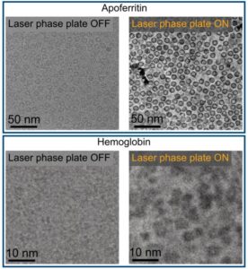
The advance comes from a new laser phase plate (LPP), described in the paper “Laser phase plate improves structure determination of small proteins by cryo‑EM,” which was published recently in Science. Led by physicist Holger Mueller, PhD, of UC Berkeley and Lawrence Berkeley National Laboratory, the team demonstrated that a laser‑driven phase plate can overcome one of cryo‑EM’s most persistent limitations: poor contrast for small proteins.

Cryo-EM images of two proteins, apoferritin and hemoglobin, taken without and with a laser phase plate. The images are analyzed in a computer to produce detailed 3D structures of the proteins. [Holger Müller, Jessie Zhang/UC Berkeley]

Cryo‑EM has transformed structural biology over the past decade, earning a Nobel Prize in 2017 for enabling high‑resolution structures without crystallization. But despite its impact, the technique still struggles with proteins below ~70 kilodaltons—a size range that includes about 90% of the human proteome. “Because of signal-to-noise limitations, the majority of human and animal proteins are too small to be analyzed by these methods [cryo-EM and cryoelectron tomography]. The increase in signal-to-noise ratio provided by this laser phase plate is expected to overcome these important limitations.”

The new LPP begins to address that problem. The LPP uses an intense, continuous‑wave laser to shift the phase of the electron beam itself. This produces true phase contrast without dimming or destabilizing the beam. Mueller described the laser focus as “75 kilowatts focused to a few microns… That’s more powerful than what you use for welding. It has more power than a military laser. It builds up the brightest continuous laser focus ever.”

Installed in a custom Thermo Fisher Titan Krios, the LPP immediately improved the clarity and resolvability of small proteins, including hemoglobin, which sits at the lower limit of what today’s cryo‑EM instruments can handle. As the authors wrote in the abstract: “Here, we show that the laser phase plate (LPP)… enhances the resolution in single-particle reconstruction of small proteins by improving specimen-motion correction, recovery of information from the early frames, as well as particle visualization, 3D classification, and alignment.”

phase plate cover Cryo-EM
A laser (purple) is powerfully amplified by highly polished mirrors and focused on the electron beam (blue) to shift its phase and increase the cryo-EM microscope’s contrast, allowing biologists to image smaller proteins and the crowded structures inside cells. [Sayo Studio]

These improvements were achieved using standard defocus ranges and reconstruction workflows. “For the most challenging cases—small particles, bad specimens—the laser produces a very considerable advantage,” Mueller said.


The impact extends beyond single‑particle analysis. Cryo‑electron tomography (cryo‑ET), which assembles multiple angular views of a molecule or protein into a three-dimensional image, stands to benefit even more. “With cryo-ET, we’re looking at small, very complicated cellular material that’s incredibly crowded inside the cell,” said Bridget Carragher, PhD, founding technical director of imaging at Biohub. “It’s like a forest of trees, and you’re trying to find one leaf on one tree in there. Cryo-ET needs a dramatic step forward in contrast, so we can start to see what’s going on inside the cell. That’s what the laser phase plate promises to give us.”

Biohub is developing a dual‑laser version of the system, designed to reduce component wear and minimize aberrations. Meanwhile, Mueller’s team is pushing toward imaging proteins as small as 17 kilodaltons, a threshold that would open access to vast regions of the human proteome previously invisible to cryo‑EM.

“This technology is a step function change for biology,” said Stephani Otte, PhD, Biohub’s vice president of imaging science. “What was once invisible will become visible—and that changes everything about how we understand disease.”

“The bottom line is, if you have a large protein and a really good sample—a fresh one or one frozen without bubbles, for example—you may not need the phase plate to get a single, high-quality image. But for a small protein and a bad sample, laser-on is best,” Mueller said. “This could fill an enormous gap in our knowledge of protein structures that can’t be crystallized or are too small for today’s cryo-EM. And it will be revolutionary for cryo-ET.”

The post Laser‑Driven Phase Contrast Enhances Cryo‑EM Resolution of Small Proteins appeared first on GEN – Genetic Engineering and Biotechnology News.

New mRNA Delivery Platform Restores Muscle Function in DMD Models

Although gene therapy has shown promise for the treatment of Duchenne muscular dystrophy (DMD), the limitations of viral vectors have proven challenging to clinical advancement. Now, a new treatment platform delivered skeletal-muscle-targeted full-length DMD mRNA systemically in a murine model of DMD, successfully restoring the production of dystrophin, and dramatically improve muscle strength, endurance, and function in vivo.

The approach uses allogenically engineered targeting extracellular vesicles (DMD t-EVs)— which offer distinct benefits over current viral-based gene therapies, including reduced side effects and the ability to transfer the entire DMD gene. The researchers engineered the EVs with special tags that directly target skeletal muscles after being injected into the bloodstream. The work also demonstrated the safety and biocompatibility of DMD t-EVs in non-human primates, supporting their translational potential.

“Our new platform overcomes the limitations of current viral-based gene therapies, allowing for the delivery of full-length mRNA, restoring wild-type translation of dystrophin and significantly improving muscle function,” said Betty Kim, MD, PhD, in the department of neurosurgery at UT MD Anderson. “We are highly encouraged by these results, which provide a blueprint for mRNA-loaded EVs as a next-generation therapeutic strategy.”

The study, published today in Nature Biomedical Engineering, is entitled, “Skeletal-muscle-targeted non-viral delivery of full-length DMD mRNA for Duchenne muscular dystrophy.”

DMD is a severe genetic disorder caused by mutations in the DMD gene that prevent dystrophin production, which helps stabilize and protect muscle cells during contractions in healthy individuals. Without dystrophin, the muscles become easily damaged, leading to eventual inflammation and cell death. DMD primarily affects males, with symptoms such as delayed walking and waddling usually appearing in early childhood. As the disease progresses, it leads to loss of walking ability, scoliosis, heart problems and eventual respiratory failure.

Because DMD is the longest known gene in the human genome, current viral-based gene therapies are unable to carry the full length. These limitations result in the loss of the gene’s full function and prevent challenges like dose-limiting toxicities, immune reactions, and other adverse reactions including death.

These side effects have resulted in the removal of at least one Food and Drug Administration-approved gene therapy from the market and are why researchers have been trying to develop alternative ways of safely delivering the full-length DMD gene.

In this study, the researchers loaded the full-length DMD mRNA into EVs that were engineered to specifically target and bind to skeletal muscles. Injection of these mRNA-loaded EVs led to an increase in dystrophin protein expression as well as improved muscle strength and function in preclinical models, with no serious side effects.

Importantly, the treatment stayed on target inside of skeletal muscles and did not trigger any immune responses or toxicities commonly seen with viral-based treatments, even after repeated dosage.

Future studies are needed to determine the full safety of EV-mediated mRNA platforms for clinical trials, including whether they can be delivered to cardiac muscles, as heart conditions are commonly seen in advanced disease. However, based on these results, the authors point out this could be a promising method beyond treating Duchenne muscular dystrophy, also potentially serving as a broader “protein restoration” or cellular reprogramming platform.

“Given that we are now able to replace very large proteins, this platform- and disease-agnostic approach could potentially open doors far beyond rare genetic disorders and traditional gene therapy applications,” Kim said. “It’s possible this could ultimately enable restoration of proteins lost not only through inherited diseases but also from acquired or degenerative processes, including cancer, autoimmune disorders, neurodegeneration, fibrosis and other chronic diseases.”

The post New mRNA Delivery Platform Restores Muscle Function in DMD Models appeared first on GEN – Genetic Engineering and Biotechnology News.

Fighting Antimicrobial Resistance with Biomaterials and Phages

Antimicrobial resistance (AMR) is a significant global health threat, with its impact felt across all regions of the world.1 According to the World Health Organization (WHO), AMR is responsible for an estimated 700,000 deaths annually worldwide, and this figure is projected to rise to 10 million deaths per year by 2050 if current trends persist.

Notably, the number of deaths attributable to AMR in many countries surpasses those caused by diabetes, kidney diseases, digestive disorders, and other non-communicable diseases. AMR has profound implications for clinical practice, affecting the management of infections across various healthcare settings. Here, we will discuss recent advances using novel biomaterials and phage for combating AMR.

Wounds, in particular, are susceptible to colonization by AMR bacteria, complicating wound healing and increasing the risk of serious complications such as sepsis and amputation, which in turn exacerbates the chronic wound burden. Chronic wounds impact the healthcare system because of their increasing prevalence and cost. The rapid growth of AMR further limits the effectiveness of standard antibiotic therapies, necessitating the use of more potent and costly antimicrobial agents, which may have adverse effects and contribute to further resistance development.

The most common bacteria isolated from chronic wounds include species of Staphylococcus (47–55%), primarily S. aureus and S. epidermidis, P. aeruginosa (25–33.6%), Acinetobacter spp., Enterococcus faecalis, and Enterobacteriaceae such as Escherichia coli, Klebsiella pneumoniae, and Enterobacter spp.2 Many of these bacteria have developed persistent AMR, such as methicillin-resistant S. aureus (MRSA). They are highly resistant to commonly used antibiotics and, therefore, limit treatment options for wound infection. To combat the growing threat of infected wounds with AMR bacteria, Han and colleagues devised a creative approach to directly degrade proteins responsible for bacterial growth.3

UDP-N-acetylmuramoyl-L-alanine-D-glutamate ligase (MurD) is a prime target for combating antibiotic resistance in bacteria as it catalyzes the synthesis of peptidoglycan, the predominant structural component in bacterial cell walls. Han et al. developed a bacterial nanoinducer (bacNID) designed to specifically degrade MurD, effectively inhibiting the growth of both Gram-positive and Gram-negative bacteria.

Two critical, interconnected challenges

“Our paper addresses two critical, interconnected challenges in global public health and antibacterial therapy: 1) The crisis of antibiotic resistance. Bacteria rapidly evolve resistance to conventional antibiotics through mechanisms such as membrane permeability changes, target mutations, enzymatic inactivation, and efflux pumps; and 2)  The failure of the traditional drug development model. The pharmaceutical industry faces a >95% failure rate in developing new antibiotics. Even when new drugs are found, bacteria often develop resistance quickly, and many candidates suffer from poor pharmacokinetics, systemic toxicity, and an inability to selectively target bacteria over healthy host cells,” says Guangjun Nie, PhD, senior author of the paper and professor at the National Center for Nanoscience and Technology, Beijing, China.

Nie adds that by moving away from the “one-target-one-drug” inhibition model, their study solves the problem of how to kill bacteria without giving them a chance to evolve resistance. “It achieves this by hijacking the bacteria’s own protein degradation machinery to destroy essential proteins such as MurD that are necessary for cell wall synthesis.”

The team first conjugated MurD-targeting peptides (pMurD) on gold nanoparticles, alongside the addition of a rapidly degradable SsrA peptide tag. The role of the SsrA tag is to bind MurD, thereby “tricking” the bacterial ClpXP protease into degrading MurD. Gold nanoparticles function as a peptide delivery vehicle that is taken up directly by the bacteria to circumvent the potential membrane permeability barrier. In this way, bacNID can destroy MurD, which is needed to synthesize the cell wall, leading to bacterial death.

bacterial research
The rapid growth of AMR limits the effectiveness of standard antibiotic therapies, necessitating the use of more potent and costly antimicrobial agents, which may have adverse effects and contribute to further resistance development. [10174593_258/Getty Images]

The team showed that bacNID was able to specifically inhibit model Gram-positive and Gram-negative bacteria with a dose-dependent degradation profile while exhibiting low cytotoxicity towards nontargeted mammalian cells. BacNID also specifically targeted MurD while sparing other Mur ligases. This approach can also be used with other nanoparticle vehicles, such as platinum, making it a versatile method for universal inhibition of diverse AMR bacteria.

To improve mechanistic understanding, the team utilized a variety of techniques and discovered that bacNID-treated bacteria suffered from cell wall damage, leading to leakage of a significant amount of DNA and ATP. Interestingly, compared with conventional antibiotics, treatment with bacNID did not lead to the formation of resistance after sustained treatment.

Using an in vivo infected skin wound model, the authors showed that bacNID treatment not only reduced infection burden but also promoted better wound healing outcomes, including greater skin cell proliferation, neo-angiogenesis, and lower inflammation. To expand the applicability of their method, bacNID was also tested in a S. aureus-infected nonhealing keratitis model and S. typhimurium-induced colitis model, showing great efficacy in both diseases.

“While our current study focuses on MurD, a major future step is to apply the bacNID platform to degrade other essential bacterial proteins. Readers can expect the team to develop bacNIDs against different targets in various pathogenic bacteria (e.g., targeting virulence factors or other metabolic enzymes). Future iterations of bacNIDs may incorporate stimuli-responsive nanotechnology (e.g., pH or enzyme-sensitive linkages) to ensure that the degradation-inducing activity is activated only within the specific microenvironment of the infection site, further minimizing off-target effects,” says Nie.

“We will also conduct more in-depth mechanistic studies to definitively elucidate why targeted protein degradation fails to induce the antibiotic resistance observed with conventional therapies.”

Guangjun Nie, PhD, professor at the National Center for Nanoscience and Technology, Beijing, China, with his team of scientists in the lab. [Guangjun Nie]

Trick bacteria with bacteria

Owing to an aging population, there is a rise in the use of implants, but these implants are prone to the formation of bacterial biofilm. The extracellular polymeric material in biofilm is known to reduce antibiotic penetration while creating an immunosuppressive environment, leading to impaired antimicrobial responses. In particular, orthopedic implants provide a conducive habitat for hematogenous bacteria for growth and formation of biofilm. Yang and colleagues hypothesized that bacteria that cause implant infection can be repackaged as drug carriers to penetrate biofilm for intra-film drug delivery.4

“Genetically modified bacteria have emerged as a promising delivery platform for diverse biomedical applications, ranging from cancer immunotherapy to infectious disease treatment. However, the clinical translation of current live bacterial biotherapeutics remains hindered by two major bottlenecks: unresolved in vivo safety concerns and the requirement for sophisticated species-specific genetic engineering. By exploiting the inherent life cycle of biofilms, our chemically primed bacterial triggers enable localized drug release deep inside biofilm structures, achieving effective biofilm eradication across genetically distinct bacterial and fungal infection models,” says Wei Tao, PhD, senior author and professor at Harvard Medical School.

The team first prepared bacteria by subjecting them to calcium chloride to increase membrane porosity and enhance their ability to uptake exogenous drugs like antibiotics. Ultraviolet radiation was then used to deactivate the bacterial membrane repair mechanism, creating irreversible membrane pores. They found that the modified bacteria, i.e., tricker, was able to migrate and thrive in biofilm and eventually, release exogenous drugs that are otherwise, challenging for delivery.

As a biofilm matures, surrounding bacteria are known to be attracted to and integrated into it. The team first labeled their tricker bacteria with a fluorescent dye and found that the bacteria were integrated throughout the biofilm with 80% coverage. However, a caveat is that the integration is most effective if the tricker and biofilm bacterial species are the same. The team discovered that while modified S. aureus can penetrate the core of S. aureus biofilm in 60 minutes, modified E. coli barely penetrates S. aureus biofilm. Likewise, modified E. coli can penetrate the core of E. coli biofilm, while modified S. aureus can penetrate E. coli biofilm with a much lower efficiency. Once in the biofilm, the chemically modified and inactivated bacteria were found to lyse, especially at hypoxic and acidic conditions.

Besides preventing antibiotic penetration, biofilm can also release bacterial-derived materials that suppress the immune system, particularly macrophages. For instance, it is well-characterized that S. aureus biofilms can bias macrophages towards an anti-inflammatory M2 phenotype, characterized by impaired antimicrobial peptide production, elevated arginase-1 (Arg-1), and attenuated inducible nitric oxide synthase (iNOS) expression. Interestingly, Yang and colleagues found that tricker bacteria were able to modify the metabolic states of the biofilm, resulting in enhanced production of I-arginine via iNOS to generate nitric oxide to improve bacterial clearance capacity.

Using an in vivo model of subcutaneous implant infection, the team found that there was an observable increase in mature dendritic cell and M1-like macrophage activation in the lymph nodes. The amount of memory B cells and antibodies with antimicrobial immune memory functions was also increased. After primary bacterial inoculation and intervention, the team reintroduced MRSA and found that 86% of treated mice rejected MRSA while all mice in the control group succumbed to the infection. This finding suggested that treatment with tricker bacteria was able to evoke innate and adaptive immune system endogenously for better control of AMR, with potential for bacterial-specific systemic memory to prevent relapse.

Finally, the strategy was tested in a murine bone infection model. By tracking cytokine levels and tissue histology, the team showed that their strategy was biologically safe. An MRSA rechallenge to the contralateral knee also led to a significant drop in biofilm burden in treated mice, providing convincing evidence of immune memory.

“To advance its clinical translation, the antibacterial efficacy of this approach will be further validated in large animal models, including rabbits, pigs, and dogs. This strategy exhibits enormous potential for future clinical translation of personalized antibacterial therapeutics, which enables highly efficient and precise treatment by profiling patient-derived pathogens and designing tailored “tricker” bacteria. Moreover, the current approach is adaptable to polymicrobial infections. Future work will also explore the feasibility of combining modified bacteria with other antibacterial agents or functional materials to optimize therapeutic performance,” adds Tao.

Wei Tao, PhD, professor at the Harvard Medical School (far right, first row) and his research team. [Wei Tao]

Using phage cocktail in clinical trials

Biofilm-related vascular graft infections (VGIs) are a major therapeutic challenge attributing to persistent, antibiotic-resistant bacteria residing in retained grafts. Graft explant is not always possible due to patient factors and surgical technical challenges. To effectively preserve the graft, treatment of VGI is typically a prolonged course of parenteral antibiotics followed by long-term suppressive antimicrobial therapy. Yet, graft survival rate is low, and recurrent infection is common.

Phages are viruses that specifically infect bacterial cells and can cause bacterial lysis. They have been shown to be active against both biofilm-forming bacteria and can even enhance antibiotic activity by eliciting phage-antibiotic synergies to combat AMR. Chung and colleagues made use of a phage cocktail to treat a 36-year-old female patient with refractory P. aeruginosa mediastinitis and vascular graft infection.5

“Our paper addresses key translational barriers to effective treatment of VGI caused by multidrug-resistant, biofilm-forming pathogens. Firstly, antibiotic failure in biofilm-associated infections as VGI pathogens embedded within biofilms exhibit marked tolerance to antibiotics, leading to persistent infection and relapse despite prolonged therapy. Secondly, escalating AMR as resistant subpopulations emerge under antibiotic pressure, further limiting treatment options in already complex infections. Thirdly, the lack of timely, personalized therapy as conventional phage therapy workflows are slow, making timely intervention difficult in acute or deteriorating cases.

Next, unpredictable phage–antibiotic interactions, such as phage-antibiotic synergy, are not reliably identified or optimised in routine clinical workflows. Finally, fragmented clinical-laboratory integration, as there is limited integration between real-time microbiology, pharmacology, and clinical decision-making to enable adaptive therapy,” says Andrea Kwa, PhD, senior author and associate professor at the SingHealth-Duke-National University of Singapore Medical School.

The team set up a multidimensional evaluation workflow to identify the most suitable therapeutic phages from the Singapore Phage Repository. Screening began with phage susceptibility testing of the four P. aeruginosa clinical isolates using spot and plaque assays, before other assays to identify the most potent cocktail. As phages are highly immunogenic when administered intravenously, the team also performed systemic inflammation monitoring and found that the patient tolerated the phages well.

The team found that their phage cocktail was able to restore antibiotic susceptibility by altering the efflux capacity of the bacteria. This positively impacted the antibiotic options for the patient. For instance, fluoroquinolone susceptibility was restored, resuscitating its use as an oral suppressive antibiotic for the long-term management of VGI.

Kwa adds that building on this proof-of-concept, her team’s next phase focuses on scaling, standardization, and integration of timely bespoke phage–antibiotic therapy into routine clinical practice. “Our key future directions include scaling up of rapid-response phage platforms, such as expansion of phage libraries/repositories with well-characterized, clinically ready phages with faster turnaround for matching and deployment, overcoming current procurement delays. We will also develop standardized precision workflows for phage susceptibility testing and phage–antibiotic synergy testing.  Our team will also enhance regulatory and translational readiness of our technology for GMP-compatible production pipelines to enable scalable clinical deployment.”

AMR is a serious healthcare issue affecting the world. With the rising use of antibiotics in farms and clinical settings, this problem needs to be taken seriously. Unfortunately, the development of antibiotics is slow and mostly unsuccessful, thus requiring a new approach for society to effectively treat AMR. Biomaterials offer a new avenue to deliver tricker bacteria into biofilm to improve intra-film drug delivery and to activate the suppressed immune system, while also inhibiting intra-bacterial growth mechanisms. Phage is also becoming a popular option, especially for personalized medicine, and this therapy may see even greater efficacy when combined with biomaterials such as hydrogel to improve its delivery and reduce systemic immunogenicity.

References

  1. Bertagnolio S, Dobreva Z, Centner CM, et al. WHO global research priorities for antimicrobial resistance in human health. Lancet Microbe. Elsevier Ltd. 2024;5(11). doi:10.1016/S2666-5247(24)00134-4
  2. Uberoi A, McCready-Vangi A, Grice EA. The wound microbiota: microbial mechanisms of impaired wound healing and infection. Nat Rev Microbiol. Nature Research. 2024;22(8):507-521. doi:10.1038/s41579-024-01035-z
  3. Han L, Huang W, Pan X, et al. Utilizing nanoinducers for precision degradation of bacterial protein to mitigate antibiotic resistance. Nature Communications . 2025;16(1). doi:10.1038/s41467-025-66221-w
  4. Yang C, Saiding Q, Chen W, et al. Chemically modified and inactivated bacteria enable intra-biofilm drug delivery and long-term immunity against implant infections. Nat Biomed Eng. Published online January 16, 2026. doi:10.1038/s41551-025-01600-8
  5. Chung SJ, Liu Y, Thong S, et al. Timely bespoke phage-antibiotic combination to treat refractory Pseudomonas aeruginosa mediastinitis and vascular graft infection. Nat Commun. Published online January 9, 2026. doi:10.1038/s41467-025-68136-y


The post Fighting Antimicrobial Resistance with Biomaterials and Phages appeared first on GEN – Genetic Engineering and Biotechnology News.