Spatial Single‑Cell Platform Reveals Barriers to Antibody Delivery in Solid Tumors

Targeting solid tumors remains one of oncology’s most persistent challenges. Even when a therapeutic antibody is well‑designed, and its molecular target is clear, the drug often struggles to reach its destination inside the dense, heterogeneous architecture of human tumors. Understanding why these agents fail in patients has been a longstanding blind spot in cancer pharmacology.

A new study from Vanderbilt University Medical Center and Stanford University begins to close that gap. In work published in Nature Biotechnology, researchers developed a single-cell spatial pharmacology (SSP) platform, an experimental and analytical system that visualizes drug–tumor interactions directly in human solid tumors. The approach provides a high‑resolution view of drug delivery, target engagement, and the physical barriers that shape therapeutic response.

Eben Rosenthal, MD, the Barry and Amy Baker professor and chair of otolaryngology–head and neck surgery at Vanderbilt Health, is senior author of the paper, titled “Single‑cell spatial pharmacobiology identifies conserved stromal barriers to therapeutic antibody delivery in human solid tumors.” Rosenthal and co‑author Guolan Lu, PhD, of Stanford University School of Medicine, developed SSP to quantify how antibody‑based therapies behave once they enter the tumor microenvironment.

“Identifying the reason drugs fail in so many cancer patients is a high priority, and SSP can help,” Rosenthal said. “Current pharmacology tools and imaging methodologies do not provide the answers we need to understand which drugs fail due to poor delivery and which ones fail due to insufficient activity upon entering the tumor.”

Using SSP, the team found pronounced spatial heterogeneity in both drug delivery and target engagement across head and neck, pancreatic, and other solid tumor types. The data point to a consistent culprit: the stromal architecture, known as the dense, noncancerous tissue surrounding tumors, which acts as a physical barrier that limits antibody penetration.

“This approach allows us to examine how the drug distributes within the tumor, the cell types with which it interacts, how strongly it engages its molecular target, and how the architecture of the tumor microenvironment shapes its delivery and activity,” Rosenthal said.

The study included analysis of panitumumab‑IRDye800CW, an antibody used in Phase I trials and which is under investigation for fluorescence‑guided surgery. Rosenthal’s group has long been at the forefront of integrating fluorescence imaging into cancer research and surgical oncology.

“By directly measuring drug delivery at the site of targeted antibody therapy, SSP can distinguish tumor regions that are biologically unresponsive from those that are simply underexposed to the agent. We hope additional study in larger sample sizes of patients can help further validate the application of SSP to identify barriers to drug efficacy,” Rosenthal added.

By exposing the physical and biological barriers that shape drug performance in human tissue, the platform offers a path toward designing tools that account for the true complexity of the tumor microenvironment.

The post Spatial Single‑Cell Platform Reveals Barriers to Antibody Delivery in Solid Tumors appeared first on GEN – Genetic Engineering and Biotechnology News.

Cross-Reactive T Cells Could Point to Broad Vaccines or Treatments for Measles, Nipah Virus

T cells are some of the immune system’s most important fighters. They can stop tumor growth and fight off severe infections. Scientists at La Jolla Institute for Immunology (LJI) have now reported a study indicating how T cells target paramyxoviruses, a viral family that includes measles virus and Nipah virus.

Paramyxoviruses are pathogens of pandemic concern. Measles virus is highly infectious, and Nipah virus has a high mortality rate. The new study shows how we might harness T cells to save lives. Headed by Alessandro Sette, PhD, the team systematically mapped human CD4+ T cell epitopes across Nipah and measles viruses, and analyzed T cells from donors who had previously received the MMR vaccine that protects against measles, and another paramyxovirus, mumps (as well as rubella), and who had not been exposed to Nipah virus. Their experiments showed that the two paramyxoviruses had conserved T cell epitopes (CTERs) in common, and that cross-reactive T cells can recognize multiple paramyxovirus species at once.

Instead of vaccinating against one virus at a time, the researchers found that activating these cross-reactive T cells may protect against the wider paramyxovirus family. This broad protection is essential when you don’t know which virus will strike next.

The discovery may guide the development of new vaccines and therapies that stop measles, Nipah, and other paramyxovirus infections before they turn deadly. “No one knows which particular viral species or strain of a virus might be responsible for an outbreak, as we’ve seen in the recent cases of Andes hantavirus,” Sette said.

Sette and colleagues reported on their findings in Cell Reports Medicine, in a paper titled “Comprehensive mapping of human CD4+ T cell epitopes for Nipah and measles as prototype Paramyxoviruses,” concluding, “Collectively, these findings support the concept that CTER-based immunogen design can both broaden protective coverage and strategically harness existing population immunity while complementing neutralizing antibody-based vaccine approaches.”

LJI Professor Alessandro Sette, Dr.Biol.Sci. [La Jolla Institute for Immunology]
LJI Professor Alessandro Sette, PhD [La Jolla Institute for Immunology]

The Paramyxoviridae family, which includes measles and Nipah viruses, represents “… a plethora of viruses that impact global human health,” the authors wrote. “Understanding adaptive immune responses to these viruses is critical for characterizing host-pathogen interactions and evaluating vaccine performance.”

A part of the adaptive immune system T cells learn to target a specific threat. A T cell might respond to influenza virus infection but not malaria parasite infection. To do this T cells recognize specific epitopes on the pathogen. In general, T cell epitopes on one pathogen look very different from T cell epitopes on another pathogen.

LJI Research Assistant Professor Alba Grifoni, Ph.D. [La Jolla Institute for Immunology]
LJI Research Assistant Professor Alba Grifoni, PhD [La Jolla Institute for Immunology]

But viruses may retain some “conserved” features that remain unchanged within viral families. LJI scientists have shown that some T cells can cross-react to different viruses, as long as the viruses share similar epitopes. In a series of landmark studies during the COVID-19 pandemic, Sette, LJI research assistant professor Alba Grifoni, PhD, LJI assistant professor Daniela Weiskopf, PhD, and professor and chief scientific officer Shane Crotty, PhD, showed that cross-reactive T cells can recognize the family resemblance between different coronaviruses. A person who had previously contracted a common cold coronavirus may already have T cells primed to recognize SARS-CoV-2, the coronavirus that causes COVID-19.

More recently, Sette and Grifoni demonstrated that cross-reactive T cells may offer broad protection against the deadly Lassa virus and the wider viral family of arenaviruses. Their findings suggested that future vaccines and therapies could activate these cross-reactive T cells to protect against many dangerous viruses at once. Each study makes it clear that cross-reactive T cells are key to stopping emerging viruses.

Measles is a threat worldwide, and while an effective vaccine is available, the authors cited figures indicating that there were over 10 million estimated infections worldwide in 2023. “Measles remains one of the main causes of morbidity and mortality in children, due to secondary infections from measles-induced immune suppression,” they noted. People in Southeast Asia also have to keep watch for a related paramyxovirus threat, Nipah virus, which is spread by bats. Cases are rare but can be deadly. “Nipah virus is another Paramyxovirus of concern due to high mortality rates, often mediated by fatal encephalitis,” the investigators wrote. Nipah virus has a fatality rate of 40-75%, which is much higher than measles. “Outbreaks are becoming more and more frequent, especially in the Malaysian region,” said Grifoni.

The new LJI study suggests cross-reactive T cells may be just the weapons we need to combat the dangerous paramyxovirus family. The scientists worked with LJI’s John and Susan Major Center for Clinical Investigation to collect and analyze T cells from the blood of 31 study participants. These study participants had received their MMR vaccines, which protect against severe infection from the measles and mumps viruses (both are paramyxoviruses) and the rubella virus. As a result, the blood samples contained T cells that were ready to fight measles infection.

LJI postdoctoral fellow Alison Tarke, PhD, and LJI senior staff scientist Ricardo Da Silva Antunes, PhD, led experiments to map the T cell epitopes on measles virus. These findings were important on their own. “Even though measles has been studied for quite some time, and there is a vaccine for measles, there was not a lot known about the specific T-cell response elicited by the measles vaccine,” Sette commented.

Tarke and the LJI team then tested how these same T cells reacted to Nipah virus. From blood tests, the scientists knew that the study participants had never been infected with Nipah virus, yet they found that measles-fighting T cells could cross-react and also recognize Nipah virus. The two paramyxoviruses had conserved T cell epitope regions (CTERs) in common. “Focusing immune responses on these conserved regions could have a broad, protective capacity for the whole viral family,” says Sette. The authors added,

The new study is the first to map T cell epitopes on Nipah virus. The researchers were able to zero in on a specific epitope shared between measles and Nipah viruses: a region of the viral fusion or “F” protein. A large number of cross-reactive T cells targeted this relatively small, conserved viral structure. “It appears that if someone is vaccinated against measles, their T cells will have some degree of cross-reactivity to Nipah,” said Sette. “That raises the possibility that during a Nipah outbreak, one could perhaps vaccinate people with a measles vaccine, and this cross-reactivity could potentially offer some benefit.”

The authors further noted, “In light of these findings, current Nipah vaccine candidates, many of which focus primarily on whole-protein antigens selected to maximize neutralizing antibody responses, particularly F and G glycoproteins, could potentially be optimized by incorporating conserved T cell epitope regions.” Added Grifoni, “Activating T cells can be your first line of defense when you don’t know what’s going to be thrown at you.”

In their paper the team concluded “With specific regard to vaccine strategies targeting Nipah or other paramyxoviruses, one anticipated outcome of focusing on CTERs, particularly those shared with measles and mumps viruses, is the potential to boost preexisting cross-reactive memory T cell responses in populations where MMR vaccination is widespread.”

The post Cross-Reactive T Cells Could Point to Broad Vaccines or Treatments for Measles, Nipah Virus appeared first on GEN – Genetic Engineering and Biotechnology News.

Fulcrum Halts Development of SCD Candidate Pociredir, Sets Strategic Review

Fulcrum Therapeutics said today it is scrapping its lead pipeline program to develop pociredir as a treatment for sickle cell disease (SCD) and launching a “comprehensive” review of strategic alternatives, after the FDA told the company it had heightened concerns about the drug’s risks and benefits in fighting the disease.

Those concerns, which the agency raised with Fulcrum executives at a recent end-of-phase meeting, stemmed from an unexpectedly high rate of secondary blood cancers seen with another PRC2 inhibitor—Ipsen’s Tazverik® (tazemetostat), indicated to treat follicular lymphoma and epithelioid sarcoma, the company disclosed, based on meeting minutes received May 28.

Ipsen voluntarily withdrew Tazverik from the market in March following adverse events of secondary blood cancers emerging from the ongoing Phase Ib/III SYMPHONY-1 trial (NCT04224493), which evaluated the drug in combination with lenalidomide plus rituximab (R2) vs R2 in follicular lymphoma. Ipsen inherited the drug when it acquired its developer Epizyme in 2022 for $247 million. Tazverik last year generated €40.6 million ($47.2 million) for Ipsen and another $2.5 million for Hutchmed, which marketed the drug in China. The withdrawal also affected Eisai, since it marketed Tazverik in Japan and manufactured the drug there.

Pociredir is an oral small molecule polycomb repressive complex 2 (PRC2) inhibitor targeting embryonic ectoderm development (EED), and was discovered using Fulcrum’s discovery technology. Fulcrum has reasoned that inhibiting EED leads to potent downregulation of key fetal globin repressors including BCL11A, thereby causing an increase in fetal hemoglobin (HbF).

“Fulcrum submitted information to FDA supporting the position that mechanistic differences between EED (pociredir’s target) and EZH2 (tazemetostat’s target), which perform different biological roles, were relevant to the benefit-risk assessment,” Fulcrum said in a statement. “While no new safety signals have been observed to date with pociredir, the FDA raised concerns regarding the potential malignancy risk associated with pociredir’s inhibition of the PRC2 complex.

The agency considered Fulcrum’s position before rebuffing the company, concluding that any drug intervention targeting the PRC2 complex carries equivalent malignancy risk “regardless of the specific subunit engaged,” Fulcrum continued, based on pociredir’s previously disclosed preclinical malignancy observations.

That left no viable regulatory path forward for further clinical development of pociredir, Fulcrum concluded.

No path forward

“Following a thorough review of regulatory feedback, the totality of available data, and the implications for a viable regulatory path, we have made the very difficult decision to discontinue development of pociredir,” stated Alex C. Sapir, Fulcrum’s president and CEO. “We arrived at this decision after discussion with the FDA, and despite robust elevations in fetal hemoglobin seen with pociredir and the potential for clinical benefit, we do not see a path forward with pociredir.”

“We know the SCD community has faced many disappointments and setbacks related to innovation for this devastating disease,” Sapir added. “We are not only humbled but forever grateful to the SCD warriors, investigators, and broader SCD community who have worked tirelessly alongside Fulcrum to evaluate new treatment options for this devastating disease.”

Pociredir is not the first SCD therapy to be scrapped. In 2024, Pfizer withdrew Oxbryta® (voxelotor) from the market, citing deaths and vaso-occlusive crises occurring in patients given Oxbryta in clinical studies. Hours before Pfizer’s announcement, the European Medicines Agency (EMA) disclosed findings from two Phase III trials of Oxbryta in which a total of 18 deaths occurred—all but two of them reported in patients who were dosed with the drug.

Pfizer inherited Oxbryta when it acquired the drug’s original developer, Global Blood Therapeutics (GBT), for $5.4 billion, a deal completed in 2022.

Vijay Sankaran, MD, PhD, Professor of Pediatrics at Boston Children’s Hospital and a Howard Hughes Medical Institute investigator, called the outcome “unfortunate.” Sankaran told GEN: “There have been concerns about whether PRC2 inhibition could be sufficiently selective and without potential side effects.”

Much of the preclinical work on pociredir, Sankaran noted, was performed in cell models that might not have revealed the full range of potential effects.

Vivien Sheehan, MD, PhD, director of Translational Sickle Cell Disease Research at Emory University School of Medicine, told GEN Fulcrum’s data was “reasonable, although not game changing.”

“I don’t take it [Fulcrum’s halt to pociredir development] as an abandonment of SCD,” said Sheehan, who is also a member of the Discovery and Developmental Therapeutics Research Program at Winship Cancer Institute of Emory University. “There may not have been a path forward, and more efficacious drugs in the pipeline may have also influenced the decision.”

She expressed greater enthusiasm for a potential SCD treatment being developed by Bristol Myers Squibb (BMS)—BMS-986470, an oral HbF-activating cereblon (CRBN) E3 ligase modulator (CELMoD™) agent designed as a potential first-in-class degrader of both transcription factors zinc finger and BTB domain containing 7A (ZBTB7A) and widely interspaced zinc finger (WIZ).

BMS-986470 is now under study in an ongoing Phase I/II trial (NCT06481306) designed to evaluate the safety and tolerability, pharmacokinetics and pharmacodynamics, pH and food effect, and preliminary efficacy of BMS-986470 in healthy volunteers and participants with SCD. The study’s estimated primary completion date is January 6, 2027, according to ClinicalTrials.gov.

A 2024 preclinical study by a team of BMS researchers showed BMS-986470 to have generated “robust γ -globin induction activity leading to HbF levels predicted to significantly ameliorate SCD pathology.”

$1.5B peak sales forecast

In ending development of pociredir, Fulcrum has scrapped a drug that stood to generate as much as $1.5 billion in projected peak sales by 2038, according to BofA Securities, which predicted a 2029 commercial launch for the drug.

Investors responded to Fulcrum’s halt to development of pociredir with a stock selloff that sent shares of Fulcrum on the Nasdaq Global Market plummeting 51% Tuesday, to $3.13 from yesterday’s closing price of $6.42.

As a result of scraping development of pociredir, Fulcrum said, it will explore potential strategic alternatives, “including, but not limited to, a merger, acquisition, business combination, or other strategic transactions involving the company or its assets.”

Fulcrum said it has also begun efforts to “significantly” reduce its operating expenses and preserve capital. Fulcrum finished the first quarter with $333.3 million in cash, cash equivalents, and marketable securities.

“We believe that our existing cash, cash equivalents, and marketable securities as of March 31, 2026 will enable us to fund our operating expenses and capital expenditure requirements into 2029,” Fulcrum stated in its Form 10-Q regulatory filing for the first quarter.

“With a strong balance sheet extending our cash runway into 2029, we are well positioned to advance pociredir through the next phase of clinical development,” Fulcrum stated in its April 27 press release.

The company has not set a timeline for completing its strategic review, adding that it does not intend to provide further updates “unless and until the board of directors has approved a course of action, the review process is concluded, or other disclosure is otherwise determined to be appropriate.”

Positive interim data

As late as April 27, when it held its quarterly earnings call with analysts to discuss first quarter results, Fulcrum had conveyed optimism about pociredir’s clinical prospects.

During the call, Sapir cited Fulcrum’s announcement of positive interim data from its Phase Ib PIONEER trial (NCT05169580) in February, which showed:

  • A mean absolute HbF increase of 12.2% at 12 weeks of treatment with pociredir, rising from a baseline of 7.1% to 19.3%—what the company said represented “a rapid, robust, and clinically relevant response,” with progression toward pan-cellular HbF induction as F-cells increased from 31% to 63%.
  • Absolute HbF levels ≥20% in 7 of 12 patients (58%), with all patients achieving at least a 6.5% absolute increase in HbF.
  • Improvements in markers of hemolysis, improved erythropoiesis, and a >1 g/dL increase in total hemoglobin.
  • Zero vaso-occlusive crises (VOCs) during the treatment period reported in seven of 12 patients (58%).

“Importantly, pociredir has continued to be generally well tolerated with no treatment-related serious adverse events reported to date,” Sapir told analysts. “Taken together, these data reinforce our conviction in pociredir’s potential to address the underlying biology of sickle cell disease—and support our belief that pociredir has the potential to represent a differentiated, once-daily oral treatment option for patients.”

The post Fulcrum Halts Development of SCD Candidate Pociredir, Sets Strategic Review appeared first on GEN – Genetic Engineering and Biotechnology News.


Disease Detection Gets Boost from Keck’s New Brain Reference Map

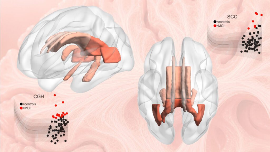
Investigators at the USC Mark and Mary Stevens Neuroimaging and Informatics Institute (Stevens INI) at the Keck School of Medicine say they have created one of the largest reference models ever developed for the human brain, using diffusion MRI scans from more than 54,000 people to chart how the brain’s communication pathways develop, mature, and decline across the lifespan.

Published in Nature Communications, the study “Lifespan normative modeling of brain microstructure” provides the equivalent of growth charts for the brain’s white matter, the vast network of neural wiring that allows brain regions to communicate, according to the Keck team, which adds that the novel tool offers researchers a new way to detect subtle patterns linked to aging, Alzheimer’s disease, schizophrenia risk, and other neurological and psychiatric conditions.

“Just as pediatric growth charts help clinicians determine whether a child’s height or weight is developing as expected, these brain charts provide a reference for how the brain’s neural pathways typically change over the lifespan,” said Julio E. Villalón-Reina, MD, PhD, a postdoctoral researcher at the Stevens INI and the study’s first author. “That gives us a powerful new way to identify when an individual’s brain wiring falls outside the expected range.”

To study white matter, the team used diffusion MRI, an imaging method that tracks how water moves through brain tissue. Because water movement is shaped by microscopic features such as nerve fibers and myelin, diffusion MRI can reveal subtle changes in tissue organization not visible on standard brain scans.

After compiling diffusion MRI data from 54,583 individuals across 19 international datasets, the researchers built statistical growth and decline charts for the brain’s neural pathways.

The researchers focused on four widely used measures of white matter microstructure across 21 major brain regions. By modeling how these measures vary by age and sex, they generated lifespan curves and percentile ranges that show what is typical at different stages of life.

Statistical charts compiled from a large population allow brain abnormalities to be detected in new individuals. [Stevens INI]

The results revealed that white matter follows distinct developmental and aging trajectories, with some measures reaching peak maturity in early adulthood and others later in midlife.

“Brain development and brain aging are not uniform processes,” continued Villalón-Reina. “The brain’s neural pathways mature on distinct timelines, and some are more vulnerable to decline than others. Our model reveals this structure by merging data on a truly global scale.”

The scientists also discovered evidence for a longstanding theory of brain aging, sometimes described as last in, first out. According to this theory, brain pathways that develop last in childhood and adolescence tend to be more susceptible to decline in older age. The researchers observed that white matter regions that mature later did indeed decline faster in old age, offering new insight linking brain development and aging.

To demonstrate the model’s practical value, the researchers applied it to clinical datasets from people with mild cognitive impairment, dementia, and 22q11.2 deletion syndrome, a genetic condition that increases risk of schizophrenia.

In each case, the model identified alterations in the brain’s circuitry that deviated from age-expected norms. Importantly, these deviations were not identical across individuals with the same diagnosis, highlighting the value of a person-specific approach.

Paul M. Thompson, PhD, associate director of the Stevens INI and senior author of the study

“This monumental study took seven years to complete,” explained Paul M. Thompson, PhD, associate director of the Stevens INI and senior author of the study. “The vast scale of the data and the fine scale of the brain features assessed means we can now evaluate your neural pathways relative to other people of the same age, sex, and demographics. We can see how your brain differs from what we would expect for a person of your age and sex, giving us a tool to use in clinical trials of treatments for dozens of brain diseases.”

When applied to people with dementia and mild cognitive impairment, the model detected atypical white matter patterns in brain regions involved in memory and interregional communication. In people with 22q11.2 deletion syndrome, it identified deviations in multiple key neural pathways, helping researchers discover which brain systems develop differently.

The reference charts may also help researchers evaluate treatments by tracking whether a person’s white matter measures move closer to the expected range, or whether a treatment slows the shift away from healthy patterns over time. The charts will now be used to compare more than 30 brain diseases and conditions, offering a common framework for studying how different disorders emerge, progress, and respond to intervention.

The models are also a publicly available resource that can be extended as additional brain imaging data become available. The methods are now being used to study neurological, psychiatric, and neurodevelopmental disorders by providing a common reference standard for white matter microstructure across the lifespan.

“This study demonstrates the power of large-scale, international data sharing to create tools the entire research community can use,” pointed out Arthur W. Toga, PhD, director of the Stevens INI and provost professor at USC. “By establishing a lifespan framework for the brain’s communication pathways, this work opens new opportunities to detect subtle disease-related changes, compare conditions more rigorously, and move toward a more individualized understanding of brain health.”


The post Disease Detection Gets Boost from Keck’s New Brain Reference Map appeared first on GEN – Genetic Engineering and Biotechnology News.

STAT+: Longevity startup NewLimit raises $435 million ahead of first clinical trial

Longevity startup NewLimit plans to launch its first clinical trial of a liver medicine after raising a staggering $435 million in new funding.

NewLimit announced its plans and Series C fundraising Tuesday. Founders Fund, the noted Silicon Valley venture capital firm co-founded by Peter Thiel, led the financing, which also included Thrive Capital, Lilly Ventures, and money from tech entrepreneurs Nat Friedman and Daniel Gross. The company is now valued at around $3.1 billion, according to co-founder and CEO Jacob Kimmel. 

NewLimit was founded in 2021 by Coinbase CEO and co-founder Brian Armstrong, former GV partner and bioengineer Blake Byers, and Kimmel, a stem cell biologist. The South San Francisco-based startup has been moving rapidly, and grabbing investors’ attention. This is the third time in the past year that the company has announced it has raised money from investors — first, a $130 million Series B round in May 2025, followed by another $45 million in October. 

Continue to STAT+ to read the full story…

STAT+: Radiopharmaceutical shows promise in post-Pluvicto setting

Want to stay on top of the science and politics driving biotech today? Sign up to get our biotech newsletter in your inbox.

Good morning. Today, we’re looking at mixed reactions to a closely watched immunology trial and growing scrutiny of a type of telehealth business model.

The need-to-know this morning

  • Vera Therapeutics and the FDA reached an agreement to allow Vera to accelerate the analysis of a confirmatory Phase 3 study involving atacicept, its treatment for the chronic kidney disease IgA nephropathy. The drug is already under review for accelerated approval, with a decision expected by July 7. The new FDA agreement will allow Vera to analyze its Phase 3 study, needed for full approval, in the third quarter, rather than wait for an additional year of data to accrue.

Abivax’s positive data weighed down by cancer concerns

Abivax said yesterday that its experimental treatment for ulcerative colitis showed significant efficacy in a closely watched maintenance trial.

Continue to STAT+ to read the full story…

Kinase Droplets Activate Growth Signals, Path for Cancer Therapy

A new study published in Cell Reports titled, “Kinase condensates enrich ATP and trigger autophosphorylation,” suggests that cellular phase separation, a mechanism that organizes biomolecules into dense, liquid-like condensates, may play a previously underappreciated role in regulating kinase activity. The findings suggest that aberrant condensate formation could contribute to oncogenic signaling while also offering new opportunities for drug targeting. 

“Many biological molecules have this propensity to spontaneously separate,” said Lindsay Case, PhD, assistant professor of biology at Massachusetts Institute of Technology (MIT) and corresponding author of the study. “We were really interested in asking, if we have these kinases forming droplets, what is the consequence of that in the context of signaling?” 

Phase separation occurs when proteins condense into highly concentrated liquid-like droplets within cells, analogous to oil droplets separating from vinegar. Although biomolecular condensates have emerged as important organizers of cellular processes, their impact on kinase signaling has remained incompletely understood. 

The researchers examined three kinases: focal adhesion kinase (FAK), Mst2, and Abl. Across all three systems, condensate formation increased kinase activity by concentrating enzymes and substrates, thereby promoting phosphorylation reactions. 

For FAK, the team found that elevated protein levels were sufficient to drive droplet formation and activate downstream growth signaling. The findings raise the possibility that FAK overexpression in tumors could promote constitutive signaling through condensate formation, potentially contributing to cancer progression and metastasis. 

“It was surprising that just by condensing this protein into a droplet, you can actually turn on a signaling pathway that should be turned off,” said Case. “If FAK concentration is too high, you’re always getting these droplets and you’re always signaling, regardless of what the receptors that are supposed to be controlling this are doing.” 

Mst2 and Abl also phase separated at high concentrations, which led to increased activity. For Mst2, phase separation is a strategy that healthy cells use to control the Hippo signaling pathway, which promotes cell growth and survival. Phase separation can also lead both enzymes to phosphorylate additional targets, and activate different signaling pathways. 

“It’s not just that you’re getting faster phosphorylation, but in those cases, the patterns of what is actually getting phosphorylated were very different inside of the droplet compared to what might be happening in a non-droplet context,” Case says. “The kinase is able to phosphorylate amino acid residues beyond the set of canonical sites that have been described before.” 

Mechanistically, the team found that kinase condensates selectively concentrate ATP, the phosphate donor required for kinase activity. Positively charged regions within kinases appear to recruit negatively charged ATP molecules to support phosphorylation. 

Using machine-learning analysis, the investigators predicted that approximately 45% of the roughly 500 human kinases possess the molecular features needed to form similar condensates. The findings suggest that phase separation may represent a widespread regulatory mechanism that could influence both normal cellular signaling and disease-associated kinase activity. 

In future work, Case hopes to explore designing drugs that could mimic ATP’s ability to be attracted into droplets within a cell, which could reduce side effects.

The post Kinase Droplets Activate Growth Signals, Path for Cancer Therapy appeared first on GEN – Genetic Engineering and Biotechnology News.

Circio and GenAssist Collaborate on Gene Therapy for Muscle Disease and In Vivo Cell Therapy

Oslo, Norway-based Circio and Suzhou, China-based GenAssist entered into a research collaboration to develop circVec-enhanced AAV vectors specifically engineered for in vivo cell therapy and targeted, low dose systemic gene therapy.

Genetic muscle disease is an area of major unmet medical need, where current gene therapy’s high dosing requirements are associated with severe toxicity. By integrating Circio’s and GenAssist’s complementary technologies, the parties aim to develop joint next generation of AAV gene therapy candidates, according to a Circio spokesperson. The focus is on addressing genetic muscle conditions where high and broad muscle-specific expression is required at substantially lower therapeutic AAV doses than can be achieved by conventional AAV gene therapy.

“Our second-generation AAV platform establishes a new benchmark for safety, utilizing highly tissue-specific, de-targeted capsids to dramatically lower systemic dosing while eliminating off-target toxicity,” said Chunyan He, PhD, CEO of GenAssist. “Through our collaboration with Circio, we integrate their unique circular RNA technology. This partnership directly addresses the core demands of next-generation genetic medicine, overcoming the traditional dose-expression trade-off to deliver safer and more effective therapies.”

In addition, Circio and GenAssist will explore the potential of generating joint in vivo CAR T candidates for oncology and autoimmune applications. The collaboration will involve production of novel AAVs combining GenAssist´s T-cell targeting with the circVec expression cassette from Circio. The combined AAVs will subsequently be tested in vitro and in vivo, and if successful, candidates for further development will be nominated for preclinical development.

“The targeted AAVs developed by GenAssist have the ability to specifically and efficiently transduce muscle or T-cells upon systemic delivery with near-complete liver de-targeting,” added Thomas Hansen, PhD, CTO of Circio. “The partnership between Circio and GenAssist will aim to evaluate whether the enhanced circVec expression acts synergistically with these targeted capsids and promoters.

“This fits perfectly into Circio’s strategy of testing circVec in multiple tissues using different AAV variants, both internally and externally. This will allow us to identify new therapeutic avenues where circVec delivers a benefit, and forge partnerships potentially enabling multiple future development opportunities. China is a particularly interesting geography, with cutting edge science and accelerated pathways to establish early clinical data.”

The post Circio and GenAssist Collaborate on Gene Therapy for Muscle Disease and <i>In Vivo</i> Cell Therapy appeared first on GEN – Genetic Engineering and Biotechnology News.
Low-Cost, Portable Biotech Tools Improve Access to Bioresearch and Diagnostics

A global research team headed by scientists at University of Toronto’s Leslie Dan Faculty of Pharmacy has demonstrated the effectiveness of a suite of low-cost, portable biotechnology tools that are designed to improve access to laboratory research and diagnostics in resource-limited settings.

The newly reported study highlights how decentralized biomanufacturing tools and freeze-dried reagents can help researchers produce high-value biological materials locally—reducing reliance on fragile international supply chains and expanding access to life sciences innovation globally.

“For labs in low- and middle-income countries [LMICs], access to high-quality supplies and equipment is a chronic problem,” says research lead Keith Pardee, PhD, associate professor at the Leslie Dan Faculty of Pharmacy Pardee. “Shipping can take a long time, it’s expensive, and products often require a cold chain to retain their effectiveness. This research is in response to those challenges to develop tools that are more accessible for labs in lower-resource settings and improve research equity.”

Pardee, alongside collaborators including Camila González, PhD, at the Universidad de los Andes, Bogotá, Fernán Federici, PhD, at Millennium Institute for Integrative Biology (iBio), Santiago, and Lindomar Pena, PhD, at Aggeu Magalhães Institute (IAM), Oswaldo Cruz Foundation (Fiocruz), Recife, reported on the study in Science Advances. In their paper, “International multisite implementation of distributed cell-free protein biomanufacturing to advance health and research equity,” the authors concluded, “This study lays the foundation for fundamental shifts in biotechnology manufacturing practices in LMICs and developing nations, moving from reliance on centralized and outsourced production facilities to adopting decentralized, local production platforms.”

“Emerging biotechnologies hold transformative potential to strengthen economic and health security, while benefiting the planet,” the authors wrote. However, they pointed out, “Access to advanced tools, such as molecular diagnostics, life-saving treatments, and biomanufacturing infrastructure, remains largely concentrated in wealthier regions, thereby restricting access to transformative solutions for communities that need them most.”

A key factor is the reliance on centralized bioproduction systems, “… which require sophisticated, capital-intensive infrastructure and cold supply chains that are often unavailable in resource-limited settings.” Access to healthcare is similarly affected by the availability of biomanufacturing capacity and biologistics, which can slow delivery of diagnostics, delay disease control programs, and limit the ability to carry out life sciences research.

For their newly reported work the team focused on synthetic biology and cell-free systems—technologies that isolate and freeze-dry the molecular machinery needed to produce proteins commonly used in life sciences research. Because the reagents are freeze-dried, they can be shipped and stored without refrigeration, then reactivated simply by adding water. “One promising avenue to improving access is cell-free protein synthesis (CFPS), which offers the potential to empower communities through affordable, low-burden, on-site production of critical bioreagents, diagnostic tools, and therapeutic agents,” the researchers stated.

They paired these systems with low-cost, adaptable hardware, including a 3D-printed hand-powered centrifuge developed by postdoctoral fellow Mohammad Simchi, PhD, at the Leslie Dan Faculty of Pharmacy. Together, the technologies enabled teams to produce a range of research proteins and diagnostic tools in diverse settings, from conventional laboratories to remote field locations.

“With efficient, low-cost systems in place, rapid on-site production of high-value bioproducts for research, including growth factors, vaccines, and diagnostic enzymes, became achievable within a single day and at a fraction of the typical cost,” they commented.

Using the platform, researchers successfully produced growth factors used in life sciences research and therapeutics, as well as a SARS-CoV-2 vaccine candidate tested in mice and diagnostic tools targeting several clinically relevant pathogens. Using molecular, cell-based, animal model, and clinical sample testing, the bioproducts were validated through proof-of-concept studies and multisite clinical trials. “Direct comparisons with high-cost commercial reagents, the current gold standards, demonstrated similar performance, efficiency, precision, and reproducibility,” the investigators further noted.

First author Severino Jefferson Ribeiro da Silva, PhD, a postdoctoral fellow in Pardee’s lab, said, “Our work shows that it is possible to produce high-value bioreagents on site, essentially anywhere. Through this work, we demonstrated our tools across diverse international settings while maintaining performance comparable to commercial products.”

The authors say that, to their knowledge, the study is the first to translate cell-free biomanufacturing from laboratory to real-world use, across multiple geographic settings, including those that historically have had limited access to the bioeconomy. “By prioritizing accessibility, affordability, and reproducibility, we show that cell-free biomanufacturing is a transformative tool for expanding global research capacity and, ultimately, health equity and participation in the bioeconomy.”

A key component of the project involved testing the systems in a variety of environments across Canada and internationally. Da Silva travelled to the Algonquin Highlands to evaluate diagnostic tools for tick-borne pathogens and tuberculosis, while graduate student Quinn Matthews travelled to the Yukon where he produced and purified proteins using the portable system on a mountain outside Whitehorse.

Collaborators in Chile, Brazil, Colombia, and India also tested the systems, helping ensure the technologies addressed the practical realities faced by researchers in different regions. The project involved extensive international collaboration, including regular meetings, student exchanges and knowledge sharing among participating teams.

Da Silva says the research team experienced first-hand many of the logistical challenges their collaborators routinely face, including lengthy customs delays and damaged shipments containing critical reagents.

“Those experiences highlighted how dependent many researchers and labs still are on fragile international supply chains. If a shipment is delayed, an entire project can stop,” says da Silva. “This work makes it possible to reduce that dependency by enabling local production of key proteins directly at the point of need.”

The researchers say the long-term goal is to help research labs in remote and underserved regions gain access to high-quality diagnostics, research reagents and biomanufacturing capabilities produced closer to home, strengthening resilience against future supply chain disruptions while empowering their research capacity and address local healthcare needs. “With their low cost and operational simplicity, we see these platforms and similar disruptive technologies … as part of a new generation of tools that will help shape a future in which bioreagents, advanced diagnostics, and life-saving therapeutics are accessible to all.”

Da Silva added, “This work is really about access and scientific empowerment. Many labs worldwide have the expertise and ideas to conduct life sciences and applied science research, but they face major challenges accessing key bioreagents and essential materials. Decentralized biomanufacturing could help reduce those barriers and make research and diagnostics more accessible globally.”

The post Low-Cost, Portable Biotech Tools Improve Access to Bioresearch and Diagnostics appeared first on GEN – Genetic Engineering and Biotechnology News.