Officials at Cenevo say the company has launched the Labguru Customer Portal to help streamline client communication for CROs, CDMOs, and others.
CROs and CDMOs operate in high-throughput, service-driven environments where speed, accuracy, and transparency are critical, explains Eran Sandman, product manager, Cenevo. Labguru allows CROs and CDMOs to centralize operational visibility across departments and projects, standardize workflows, and manage multiple client programs at scale, he adds.
Integrated with Labguru’s ELN and LIMS platforms, the new portal was designed to extend internal workflows to support external collaborators. With this structured approach, labs experience increased efficiency while minimizing version conflicts, human error, and compliance risks, maintains Sandman, noting that clients can submit requests, monitor progress and access results, while labs retain full control and visibility over shared information.
Lab managers can use the portal to gain actionable insights across every client individually and all clients in aggregate, as they relate to equipment and consumables usage, client activity trends, regulatory compliance, and other critical information, points out Sandman.
“Our goal is to make our clients’ operations run more smoothly,” he says. “High-performing CROs are moving toward shared digital environments. With the Customer Portal, CROs and their clients are able to see the same information and collaboration becomes faster and more strategic. Lab managers don’t have to spend all their time on back-and-forth communications.”
Cholera, caused by the bacterium Vibrio cholerae, remains a major global health threat. Like most bacteria, Vibrio cholerae lives under constant attack from viruses. To survive, bacteria equip themselves with antiviral immune systems. Previous work has shown that V. cholerae carries a large genetic element called a sedentary chromosomal integron (SCI). This structure contains hundreds of small mobile DNA units known as “gene cassettes” arranged in a long array, like a chain of pearls. A new study by researchers at the School of Life Sciences, Ecole Polytechnique Fédérale de Lausanne (EPFL), has now shown that V. cholerae can efficiently acquire new SCI gene cassettes from extracellular DNA released by other V. cholerae strains, and non-cholera vibrio species, potentially adding to their own defense arsenals.
Research lead Melanie Blokesch, PhD, and colleagues reported on their findings in Science, in a paper titled “Competence-mediated DNA uptake diversifies Vibrio cholerae sedentary chromosomal integrons,” in which they concluded “Given the widespread presence of SCIs and the conservation of natural competence across the genus, we propose that SCIs function as genus-wide reservoirs of exchangeable protective genes.”
Bacteria often survive viral attack and environmental stress by sharing genes that enhance their defenses, the authors wrote. A “defining genomic feature” of V. cholerae is its sedentary chromosomal integron (SCI), a genetic element containing hundreds of mostly promoterless gene cassettes. While the function of many cassettes remains unknown, some do encode antiviral immune systems. “Although most cassettes encode proteins of unknown function, ~10% encode phage defense systems, suggesting that SCIs as well as mobile integrons function as reservoirs, or “biobanks,” of defense genes,” the authors continued.
However, most of these genes are located far from the start of the array and remain silent. Prevailing models proposed that cassettes could be internally reshuffled to activate them, yet no such rearrangements have been observed in the pandemic lineage of V. cholerae for decades. “Cassettes are thought to reshuffle under stress to the favorable first array position, yet the SCI in pandemic V. cholerae has remained static for more than 60 years.”
This raises a key question: if internal reshuffling is rare, how are cassette-encoded immune systems activated, and how do new cassettes enter the array at all? To address this question, a team led by Blokesch at the Laboratory of Molecular Microbiology at EPFL investigated whether the SCI might capture gene cassettes from genetic material entering the cell from the outside. “We asked whether SCI cassettes move horizontally rather than by intracellular reshuffling.”
A key feature of this process is natural competence, the ability of bacteria to take up free DNA from their surroundings. V. cholerae becomes naturally competent when it grows on chitinous surfaces, a polymer found in the shells of crustaceans that is abundant in aquatic environments.
In the laboratory, the team mimicked these conditions by growing bacteria on chitin and supplying DNA from different Vibrio cholerae strains or from other Vibrio species. They then tracked whether newly acquired gene cassettes were inserted into the first position of the SCI array.
Through their studies the team confirmed that that V. cholerae can acquire new SCI gene cassettes from extracellular DNA. Collective experimental results, the authors stated, “We show that SCI cassettes are efficiently acquired by naturally competent V. cholerae and inserted at the first SCI array position in an integrase-dependent manner. This process incorporates cassettes not only from other V. cholerae strains but also from diverse Vibrio species.”
In aquatic habitats, DNA is released when bacterial cells are killed by viruses, antimicrobial compounds, or bacterial weapons. Nearby competent bacteria can take up this DNA and incorporate selected fragments into their own SCI. “A loose comparison would be the following,” said Blokesch. “Imagine your grandmother passes away and, as a farewell gift, hands over the immunity she built up against the Spanish flu a century ago, immediately protecting you from that same virus. Wouldn’t that be amazing? This is essentially what we show that V. cholerae can do.”
The team also showed that cassettes inserted in this position are functional. Several defense systems provided protection against viruses that infect Vibrio species, known as vibriophages. They stated, “In this study, we show that SCI diversification efficiently occurs by horizontal transfer linked to the genus’s aquatic lifestyle: DNA released from lysed cells is taken up by naturally competent vibrios and integrated into the first position of the SCI array, the primary site of strong expression, where it confers resistance to phage and potentially other threat,” the wrote in summary. “Together, these results demonstrate that SCI cassettes can cross species boundaries, supporting a model in which SCIs may function as genus-wide reservoirs of exchangeable genes, including defense genes, that confer selective advantages under certain conditions.”
An important exception emerged. In the pandemic 7PET lineage of V. cholerae, the SCI appears largely static. “The SCI of 7PET V. cholerae is large but remarkably stable,” the authors noted. They propose that this reflects adaptation to a human-associated niche. “We propose that this reflects adaptation to a human-associated niche, where chitin is less abundant and competence induction—requiring growth on chitin to high cell density plus relief from catabolite repression is unlikely to occur,” they suggested. “As a result, SCI-mediated diversification may be largely inactive in pandemic strains.”
However, if pandemic strains were to encounter environmental conditions that enable SCI cassette acquisition, they could expand their antiviral defenses. Blokesch commented. “This possibility matters because vibriophage-based approaches are currently being explored to prevent cholera in endemic regions, and such evolutionary flexibility could ultimately affect how effective these strategies remain.”
In their paper the team concluded that since onset of the seventh pandemic, “… acquisition of novel and diverse, large defense-related genomic regions by 7PET strains appears to have been relatively limited. Consequently, reduced SCI-mediated diversification may lower the capacity of this lineage to rapidly evolve new defenses—an important consideration for ongoing efforts to deploy phage-based prophylaxis against cholera in endemic settings such as Bangladesh.”
For decades, venture capitalists have relied on a tried-and-true recipe to make money in biotech: Start with compelling scientific research — usually out of a U.S. university lab — add a dash of veteran executives from a pharmaceutical goliath or big biotech, and finish with tens of millions of dollars. The approach has led to scores of new medicines, successful companies, and financial returns for VC firms and their investors.
Now, it’s being disrupted.
In the aftermath of biotech’s Covid-era rally and subsequent decline, the industry has been startled to find that Chinese scientists are conducting innovative research, faster and at lower costs, than their U.S. counterparts. In the United States and elsewhere, meanwhile, investors are being wooed by artificial intelligence, drawing attention and dollars away from biotech.
The biological connection between a pregnant woman and her developing baby—the human maternal–fetal interface—is a specialized, transient organ composed of uterine cells from the mother and fetal cells that acts as a barrier, supports fetal growth, and maintains the mother’s health. The cellular complexity of the maternal-fetal interface has limited scientists’ ability to study how healthy pregnancies develop and why complications arise. The underlying cellular, molecular, and spatial programs of the interface—which forms about a week after fertilization and lasts until birth—has remain incompletely defined.
Now, the human maternal–fetal interface has been mapped in unprecedented detail by scientists at the University of California, San Francisco (UCSF), revealing new cell types and providing insights into conditions such as preeclampsia, preterm birth, and miscarriage.
“By examining this tissue cell by cell across pregnancy, we can begin to understand both normal development and what may go wrong,” said Susan J. Fisher, PhD, professor of obstetrics, gynecology, and reproductive sciences at UCSF.
The team generated a comprehensive atlas of the human maternal–fetal interface across normal pregnancies, from early gestation to term. The researchers did this by “integrating large-scale paired single-nucleus transcriptomic and chromatin accessibility profiling with submicrometer-resolution spatial transcriptomics and CODEX multiplex protein imaging.”
Using these tools, the researchers analyzed about 200,000 individual cells and compared them with nearly one million cells in their original positions within the uterine and placental tissue. This enabled them to identify different cell types, track how they develop, and see how they are linked to pregnancy complications.
“This work gives us a much clearer picture of this critical region than ever before,” said Jingjing Li, PhD, associate professor in UCSF’s Department of Neurology and the Eli and Edythe Broad Center of Regenerative Medicine and Stem Cell Research.
The atlas revealed a previously unknown maternal cell type located where fetal placental cells first enter the uterus. These cells appear to regulate how deeply placental cells invade uterine tissue, a process that is essential for establishing blood flow to the fetus. The researchers found that these cells carry a cannabinoid receptor, and exposure to cannabinoid molecules caused them to further restrict placental cell invasion.
“Population studies have linked cannabis use during pregnancy to poorer outcomes,” said Cheng Wang, PhD, a postdoctoral fellow at UCSF. “This cell type may help explain the biological basis of that association.”
To understand how complications arise, the team integrated genetic data from more than 10,000 patients. They mapped genetic risk signals for conditions including preterm birth, preeclampsia, and miscarriage onto regulatory regions of DNA that control gene activity. This approach allowed the researchers to identify the specific cell types and states most strongly associated with each condition.
The team then focused on preeclampsia, a potentially life-threatening disorder marked by sudden high blood pressure. They found that the most affected cell types are involved in remodeling the mother’s uterine blood vessels, a process required to supply adequate blood to the placenta. The findings suggest that preeclampsia may result from disrupted communication between maternal and fetal cells that normally coordinate this process.
Having established a detailed map of healthy pregnancies, the researchers plan to study complicated pregnancies to identify potential targets for treatment.
Dynamic42 and EPO (Experimental Pharmacology and Oncology), both based in Germany, report that they are addressing the limited availability of preclinical models in brain cancer research by forming a strategic collaboration that focuses on bringing organ-on-chip technologies “closer to the core of preclinical drug development.”
The partnership combines Dynamic42’s organ-on-chip platforms with EPO’s expertise in translational oncology and access to well-characterized tumor models and patient-derived material. Together, the teams are developing experimental setups designed to reflect human tumor biology more closely and generate data that translates more reliably into clinical outcomes.
The first joint projects target glioblastoma and the blood–brain barrier (BBB). Using Dynamic42’s human-based BBB-on-chip model, the partners will explore how differences between human and non-human BBB-biology can influence therapeutic responses, which is a major factor for the limited activity of brain cancer drugs.
“Too often, critical decisions in drug development rely on data that do not fully reflect human biology,” said Thomas Sommermann, PhD, head of cancer research at Dynamic42. “We want to change that. By bringing human-based models earlier into the process, we can sharpen decision-making and reduce late-stage failure risks.”
“For us, this collaboration is about strengthening the translational link,” added Jens Hoffmann, CEO at EPO. “Integrating advanced in vitro systems allows us to look at tumor biology from a different angle and to build robust experimental in vivo strategies.”
The collaboration is designed as a complementary approach that connects established preclinical in vivo expertise with emerging human-based in vitro technologies. It supports more targeted, biology-driven research strategies and the principles of the 3Rs (Replace, Reduce, Refine), contributing to the ongoing shift toward more human-relevant experimental systems.
Beyond joint research, the partnership includes model development activities, elaboration of commercialization strategies, and close scientific exchange, including collaboration between early-career researchers from both organizations.
Dynamic42 and EPO will jointly present the first results of their collaboration at the American Association for Cancer Research® Annual Meeting 2026. Both companies plan to expand the collaboration further, exploring additional indications and extending the use of organ-on-chip technologies across different areas of drug development.
There is no doubt that autologous chimeric antigen receptor (CAR) T-cell therapy has revolutionized the treatment of serious blood cancers. A significant proportion of advanced-stage blood cancer patients who failed to respond to previous therapies now go into remission with this treatment, with some remaining cancer-free in the long term.
However, despite their success, these immunotherapies have significant disadvantages. Although CAR T-cell therapies have essentially rescued advanced-stage patients who previously would only have been offered palliative care, serious side effects such as cytokine release syndrome and immune effector cell-associated neurotoxicity syndrome (ICANS) are associated with the treatment.
All seven CAR T-cell therapies approved by the U.S. Food and Drug Administration (FDA) since August 2017 are autologous cell therapies, wherein the patient’s own T cells must be extracted and genetically engineered in the lab to produce cancer-targeting CAR T cells that are reinfused into the patient to fight the cancer.
Unless they live near a major cancer center or company with the relevant expertise and lab capacity in-house, many eligible patients miss out on the therapy because the wait time is too long or the whole process is too expensive. Patients must also be admitted to the hospital to undergo lymphodepletion chemotherapy before receiving the final infusion to allow the infused cells to expand, persist, and work better.
“It’s just simply too expensive, too complex to manufacture, and has all kinds of logistical issues that translate to limited patient access,” explained Maurits Geerlings, MD, co-founder, CEO, and president of in vivo CAR T-cell therapy biotech NanoCell Therapeutics, which has offices in Pennsylvania and Utrecht.
“Also, importantly, the batch capacity in highly specialized hospitals is so limited that altogether maybe 10% of patients that are eligible effectively get access to CAR T-cell therapy in the Western world.”
Maurits Geerlings, MD Co-founder, CEO NanoCell Therapeutics
Initially, after the first ex vivo, autologous CAR T-cell therapies like Novartis’s Kymriah and Kite’s Yescarta were approved in 2017, the field looked to develop “off-the-shelf” allogeneic therapies made from donor cells that would overcome some of the issues with autologous CAR T-cell therapies.
Despite the best efforts of a number of companies and researchers, no allogeneic CAR T-cell therapies have yet reached the market, although some companies like Allogene have reached Phase II trials. This is likely due to a few factors, such as adverse events linked to the rejection of donor cells, complex engineering problems, and the small margin of benefits of allogeneic over autologous CAR T-cell therapies.
Instead, over the last couple of years, the focus of the field has moved towards developing next-generation in vivo CAR T-cell therapies. Until recently, vectors or nanoparticles that could hit T cells precisely, safely, and predictably enough in humans to justify skipping ex vivo engineering were simply not available, but this is now changing.
In vivo CAR T-cell therapy uses the patient as a bioreactor. Upon injecting an engineered treatment carried by a vector such as a lentivirus or a lipid nanoparticle (LNP), it programs the patient’s T cells to attack either the cancer or autoreactive B cells in the case of autoimmune disease.
The field is still young, but initial clinical results reported last year in multiple myeloma blood cancer by Kelonia Therapeutics and in the B cell-driven autoimmune disease systemic lupus erythematosus by MagicRNA, as well as from EsoBiotec and academic labs, are promising.
Kevin Friedman, PhD Co-founder, CEO Kelonia Therapeutics
“It’s early days, so I don’t want to overinterpret the data. It’s also only in four patients, but what we are seeing is substantially better than what ex vivo CAR T cells have shown from an efficacy perspective,” emphasized Kelonia CEO and co-founder Kevin Friedman, PhD.
Indeed, this early success seems to have prompted intense investor and big pharma interest in the field. Since March 2025, when EsoBiotec was acquired by AstraZeneca, at least four other in vivo CAR T cell biotechs have been acquired, including Capstan Therapeutics by AbbVie and Interius BioTherapeutics by Kite/Gilead.
Whether in vivo CAR T-cell therapy will truly be the future of the field remains to be seen, but its convenience, economic viability, and the fact that it is effectively an “off-the-shelf” therapy that does not require lymphodepletion make it an attractive prospect for many.
Credit: Kelonia
Lentiviral vectors: Sticking with a known quantity
Five of the seven FDA-approved autologous CAR T-cell therapies, including Kymriah, use lentiviral vectors in the lab to engineer a patient’s T cells and transform them into CAR T cells.
Many of the most advanced companies in the in vivo CAR T- cell therapy space are applying similar technologies and using lentiviral vectors to target and transform T cells, but inside the body rather than in the lab.
Kelonia, which is based in Boston, is a leader in the in vivo CAR T-cell space and has already started clinical trials with its lead candidate KLN-1010 for the treatment of patients with relapsed and refractory multiple myeloma.
“It’s essentially delivering a fully human anti-BCMA (B cell maturation antigen) CAR to T cells, to reeducate them by expressing this anti-BCMA CAR inside the body to fight their tumor cells. Just like Abecma, or Carvykti, but it’s all done inside the body,” explained Friedman.
At the American Society of Hematology Annual Meeting in December last year, the company presented early Phase I results from four patients with relapsed and treatment-resistant myeloma who were treated with KLN-1010.
Although the study was small, the results were promising, with all four patients showing 100% minimal residual disease-negative response rate at follow-up and a lower rate of side effects than approved autologous CAR T-cell therapies.
“With our data and the efficacy and the safety profile that we’re seeing, this has a real shot at getting out of the major medical centers and into the community hospitals where the patients live, so they don’t have to travel to major medical centers,” said Friedman.
“Doctors can potentially treat patients in their own community and get access to the 90% of myeloma patients who, right now, despite the profound clinical benefit that CAR T cells provide, cannot be treated.”
Ryan Larson, PhD Senior Vice President Umoja Biopharma
Umoja Biopharma is another biotech using lentiviral vectors to develop in vivo CAR T-cell therapies. “We have three different products in the clinic currently. Two of those products are in B-cell malignancies, and one of the products is in autoimmune disease. And we’re making really great progress in enrolling patients across those studies,” said Ryan Larson, PhD, senior vice president and head of research at Umoja, although the Seattle-based company has not yet released any results from its Phase I studies.
Although Umoja is using lentiviral vectors, it has built in a rapamycin-activated cytokine receptor, which essentially acts as a booster switch for the engineered T cells in cancer patients while slightly dampening the rest of the immune system.
“It allows us to deliver a controlled pro-survival signal selectively to our CAR T cells in vivo,” explained Larson. “We’re able to potentiate persistence in a controlled manner in our in vivo generated CAR T cells with this rapamycin-activated cytokine receptor to drive persistence and ongoing immune surveillance, thus driving the key durable outcomes in oncology specifically.”
The requirements for autoimmune disease patients are different from those of advanced cancer patients, with a greater focus on safety. Long-term depletion of B cells is also not ideal, with the aim being to reset the immune system by getting rid of autoreactive B cells and replacing them with healthy ones.
“You’re eliminating all of the autoreactive repertoire and replacing it with a normal B cell repertoire, thus driving, ideally, a durable response wherein those autoimmune disease patients are no longer reliant on all the various immunosuppressants that are typically used to treat autoimmune disease,” said Larson.
Kite Pharma, now owned by Gilead and headquartered in California, was a pioneer in the autologous CAR T-cell therapy space. It developed Yescarta, one of the first two autologous CAR T-cell therapies approved by the FDA to treat blood cancers in 2017. Kite recently acquired Interius BioTherapeutics, a biotech in the lentiviral in vivo CAR T-cell space, for $350 million.
Priti Hegde, PhD Senior Vice President Kite Pharma, a Gilead Company
“The reason why we moved forward with Interius was that the clinical proof of concept for lentiviral-based delivery systems is far more advanced than for LNP-based systems,” said Priti Hegde, PhD, senior vice president and global head of research at Kite. “We were really excited to see that translation of the pharmacokinetics from an ex vivo platform to an in vivo platform.”
While lentiviral vectors are arguably “tried and tested” in the CAR T-cell space, there are some disadvantages associated with using them. For example, they can be hard to produce, implying that it is expensive and challenging to scale up manufacturing.
This is something both Umoja and Kelonia seem to have addressed, however. “We actually are quite unique from a biotech perspective in that we have our own, wholly owned manufacturing facility … It’s really allowed us to have a true pipeline from an in vivo cell therapy development perspective,” said Larson. “We’re actively working in our early phase clinical trials in a manufacturing setting that we know is scalable to commercial readiness.”
Kelonia does not do all its manufacturing in-house, but Friedman said that they have worked hard to develop a system that can be scaled. “Manufacturing is complicated. We like to think that we were thoughtful about our manufacturing approach, but it’s challenging generating these particles, these complicated medicines for Phase I use. We did it, though, and we now have a very reliable and scalable manufacturing process.”
Another potential risk linked to lentiviral and other viral vectors is that there is a small but significant risk of the vector inducing unwanted mutations in the DNA of target cells.
“Viral vectors have a propensity to integrate in transcriptionally active gene regions where you don’t want to go, because that enhances the mutagenesis risk,” noted NanoCell’s Geerlings.
Taking the non-viral route
Not everyone working to develop in vivo CAR T-cell therapies is using viral vectors. The second main route that companies and researchers are following to develop these cell and gene therapies is to use mRNA encapsulated in an LNP.
Last September, Shenzhen-based Chinese biotech MagicRNA published data from a Phase I trial of its in vivo mRNA and LNP-based CAR T-cell therapy in five patients with systemic lupus erythematosus. Similar to in Kelonia’s cancer trial, the results were promising. However, larger studies are needed for more conclusive results, as the sample size was small. But rapid, near-complete B cell depletion was seen for up to 10 days with no significant side effects like serious cytokine release syndrome or ICANS.
Since the pandemic, the use of mRNA therapeutics has become much more mainstream. For example, both of the prevalent vaccines against COVID-19 use a combined mRNA–LNP approach. In in vivo CAR T-cell therapy, the LNPs are used to take CAR-encoding mRNA to the right target cells in the body. Once inside a T cell, the LNP breaks apart and releases the mRNA into the cytoplasm. The cell’s protein synthesis machinery reads the mRNA and makes the correct CAR protein, which is then added to the surface of that T cell.
Aera Therapeutics, founded by CRISPR pioneer Feng Zhang, PhD, and based in Cambridge, Massachusetts, takes a combined mRNA–LNP approach to in vivo CAR T-cell therapy development, with a focus on treating B cell-mediated autoimmune disease.
Akin Akinc, PhD CEO, Aera Therapeutics
“We were really focused on autoimmune indications, so we said, ‘Let’s try to build a product profile that’s a great fit for that,’” explained Akin Akinc, PhD, who is CEO at Aera.
“You have the risk of insertional mutagenesis with lentiviral vectors. Even if those rates are small, they’re not zero … So that’s why we thought an mRNA–LNP approach, where there’s no chance of insertion, is theoretically a more attractive approach.”
Aera has not yet moved into clinical studies but reported preclinical data for its therapy candidate AERA-109 in non-human primates at the American Society of Hematology Annual Meeting at the end of last year. They showed potent and durable B cell depletion across different tissues in the body.
One potential disadvantage of using a combined mRNA–LNP approach, particularly for treating cancer, is that it is unlikely to last as long as a lentiviral approach. As this could be potentially advantageous in people with autoimmune disease, where B cell depletion does not need to occur over such a long period of time, it seems to be the most common method followed by companies designing in vivo CAR T-cell therapies for autoimmune conditions.
“Our therapeutic goal is to go in and clear out the B cells that exist in the body, both in the periphery and the tissues, and then allow them to repopulate. If we achieve that immune reset, then that’s all that we can do,” said Akinc.
Scott Barros, Head of Early Development, Akin Akinc, Chief Executive Officer, Bill Querbes, Chief Scientific Officer.
“Then the question is, is there going to be a relapse 12–18 months later? But so long as we clear out all the B cells, which happens pretty quickly, I think there’s no benefit to having the CAR T cells hanging around for longer, because at that point you’ve done the job. Then it’s about whether or not that remission is going to persist.”
NanoCell Therapeutics is also taking a non-viral approach to developing in vivo CAR T-cell therapy for treating B-cell malignancies, but is using DNA instead of RNA. The candidate has not yet reached the clinic, but it has achieved good preclinical results and will soon be tested in non-human primates.
Similar to Aera, NanoCell packages its therapy in targeted LNPs. However, these carry a minicircle DNA that encodes the CAR information and an mRNA transposase that allows the DNA to integrate into the target cell genome.
“We still remain, I think, pretty much in the lead as a company delivering non-viral DNA, because it is very difficult … We see an opportunity for us to actually make a breakthrough there,” said Geerlings.
“The nuclear membrane of the cell is such a barrier. You need to find an opportunity to open it up and to be just in time with your DNA in a way that is not triggering an innate immune response. You also need to have a mechanism by which that DNA can integrate, because otherwise, you will end up having an episomal expression of your DNA.”
The approach taken by NanoCell is definitely at an earlier stage than the lentiviral and mRNA–LNP approaches that are already generating clinical data, but there are a couple of other companies working on similar products, like Stylus Medicine and CPTx. If it works, then this approach has the promise of ruling out problems with viral vectors, such as manufacturing difficulties. It would also theoretically generate longer-lasting and more durable treatment effects than could be achieved with mRNA.
What’s next for CAR T-cell therapy?
It seems that we are on the cusp of next-generation in vivo CAR T-cell therapies, although the studies published so far have all been small and it remains to be seen if the current buzz in the space is based on hype or reality.
“I do think that in vivo will go from a platform with initial proof-of-concept to broad applicability faster than perhaps ex vivo platforms did,” said Hegde. “But we have a lot of scientific questions. For example, in the absence of lymphodepletion, can an in vivo platform give you the depth and durability of response that an ex vivo platform does?”
There is a lot of interest in whether safer and more accessible in vivo CAR T-cell therapy can make this treatment approach more appealing to people with B-cell-mediated autoimmune conditions than autologous CAR T-cell approaches. The initial clinical results are good, but questions remain about how long the results will last.
“I think these are going to work, but we’re going to learn things that allow us to make second-generation products that are even better and even more potent,” said Akinc.
On the cancer side of things, most companies developing in vivo CAR T-cell therapies for oncology indications are sticking with blood cancers against which autologous CAR T-cell therapies have already been shown to be efficacious.
“I think we’ll continue to see strong proof of concept in de-risked indications like the hematologic malignancies over the next year,” said Larson. “Over the next two years, I think we’re going to be closely watching the field for durable outcomes in oncology and the ability to drive immune reset in autoimmune disease that then translates to durable remissions in autoimmune disease patients.”
A big question on everyone’s mind is whether this new technology could help overcome some of the hurdles that prevent CAR T-cell therapy from being successful at treating “solid” tumors, such as working out how to overcome diverse tumor microenvironments.
“I think there’s great potential in solid tumors. One reason why we went with Interius was [that] we think that the application of the in vivo platforms could really break open the problems that we perhaps had in solid tumors with ex vivo CAR T cells,” said Hegde.
“The nice thing about the in vivo space is you can put whatever targeting antigen you want on the virus to go to a specific cell type. So it’s really up to your imagination, how you want to design an in vivo CAR T cell.”
Dispatch Bio is also in the CAR T-cell therapy space, but is targeting solid tumors rather than developing in vivo CAR T cells. The company is based in Philadelphia and was co-founded by Carl June, MD, one of the pioneers of CAR T-cell therapy.
The technology they are developing is a two-component system, in which a human-specific adenovirus designed to infect cancer cells, but not healthy tissue, is used to “paint” the tumor cells so that a CAR T cell can more easily home in on the cancer and destroy it.
Barbra Sasu, PhD Chief Scientific Officer Dispatch Bio
“The virus gets into the tumor microenvironment and then, because it’s a virus, creates an inflammatory condition. When it does that, it’s immediately more supportive for T cells,” explained Dispatch chief scientific officer Barbra Sasu, PhD.
“We’re doing what T cells can’t do for themselves. We’re expressing a target and directing them to kill what we want them to kill. We’re also adding a cytokine to support them and, actually, the endogenous immune system too.”
The two-part approach is very new, so Sasu and colleagues are testing the system using a known autologous CAR T-cell therapy approach. But she says that the system is potentially very flexible and could allow a wide range of therapies, including in vivo CAR T-cell therapies, to be combined with the viral targeting approach if they prove effective.
“What we wanted to do was to start with something that we felt we understood very well. We also had the benefit in our first program of being able to work with people who’ve already developed CAR T-cell therapies,” said Sasu. “That’s a big advantage because [when] coming in with a two-component system, it’s good if you don’t have to refine both parts at once.”
Another CAR T-based approach being developed by Kite and others in this space is logic gating, which is the development of IF, AND, and NOT switches to allow much more refined control of CAR T-cell therapies by clinicians and potentially increase effectiveness in complex solid tumors.
“We’re really interested in exploring the logic gating space and what it can do to deliver CAR T cells more safely, especially in solid tumors, where the antigens aren’t as broadly homogeneously expressed,” said Hegde.
Helen Albert is senior editor at Inside Precision Medicine and a freelance science journalist. Prior to going freelance, she was editor-in-chief at Labiotech, an English-language, digital publication based in Berlin focusing on the European biotech industry. Before moving to Germany, she worked at a range of different science and health-focused publications in London. She was editor of The Biochemist magazine and blog, but also worked as a senior reporter at Springer Nature’s medwireNews for a number of years, as well as freelancing for various international publications. She has written for New Scientist, Chemistry World, Biodesigned, The BMJ, Forbes, Science Business, Cosmos magazine, and GEN. Helen has academic degrees in genetics and anthropology, and also spent some time early in her career working at the Sanger Institute in Cambridge before deciding to move into journalism.
Salk Institute scientists have developed a biological platform for studying mitochondrial DNA in physiology, adaptation, disease mechanisms, and therapeutic development. Headed by Ronald Evans, PhD, professor and director of the Gene Expression Laboratory and holder of the March of Dimes Chair in Molecular and Developmental Biology at Salk, the team has already used the platform to generate a library of 155 mitochondrial DNA mutant cell lines and reveal correlations between mouse development and mitochondrial function. They suggest that the platform, library, and findings will accelerate therapeutic development for mitochondrial disorders, as well as help scientists treat mitochondrial dysfunction in other diseases and conditions like cancer or aging.
“Mitochondrial DNA accumulates mutations at a high rate, and more than 260 inherited disease-causing mtDNA mutations have been identified in humans,” said Evans. “Until now, a lack of models representing this diversity has limited mechanistic insight and therapeutic development. Our new platform will allow scientists to investigate mitochondrial DNA variation in health, disease, and evolution, which will enable therapeutic innovation for mitochondrial disorders.”
Evans is co-corresponding author of the team’s published paper in PNAS, titled “A scalable embryonic stem cell–based platform for efficient generation of mitochondrial DNA mutant mice,” in which they concluded that their new platform, “… opens the door to mechanistic dissection of how mtDNA variation influences metabolism, adaptation, and disease, and provides a strategically valuable foundation for accelerating therapeutic development through genetically precise mitochondrial disease models.”
Some of your most important life partners are the mitochondria that power all your cells. You and these little cellular powerhouses are in a 1.5-billion-year-old evolutionary relationship—but mitochondria brought some baggage. Mitochondria brought their own DNA with them when they joined with the bigger, more complex cells so long ago, and today that mitochondrial DNA influences human health. Mitochondrial DNA does the extremely important job of creating the proteins needed for energy production—but it also has an especially high rate of mutation, and those mutations can accumulate thanks to inefficient repair mechanisms. Because mitochondria are essential parts of every cell, their dysfunction can lead to body-wide dysfunction, with especially devastating impact on high-energy organs like the brain and heart. Without enough power in your cells, symptoms like migraines, muscle weakness, and loss of hearing or sight can begin to manifest.
“Mitochondria are central to energy metabolism and cellular signaling, and mutations in mitochondrial DNA (mtDNA) can disrupt these processes and contribute to human disease,” the authors wrote. “Mitochondrial DNA (mtDNA) accumulates mutations at a high rate, and more than 260 pathogenic germline mtDNA mutations have been identified in humans, producing diverse and often tissue-specific disorders.”
The chronic and broad impact of mitochondrial dysfunction makes it especially important to study. However, trying to pinpoint the outcome of specific mitochondrial DNA mutations has for many years been a slow, arduous process for many years. “… progress in defining how mtDNA variation influences adaptation, pathophysiology, and disease susceptibility has been limited by the lack of suitable animal models,” the team continued. “Researchers would create mouse models one-by-one with different mitochondrial DNA mutations, with just one model sometimes taking years,” said Salk staff scientist Weiwei Fan, PhD. This was a problem that Fan had noted early in his scientific career and set his mind to as a PhD student.
The new Salk model is a scalable, embryonic stem-cell (ES)-based platform creating mice with mutations to their mitochondrial DNA. “This new work is all building off an original platform I generated during my PhD,” says Fan, first and co-corresponding author of the study. “That platform was inefficient—it took a long time to generate just one mitochondrial DNA mutant. With some technological improvements and modifications, this new platform is much more efficient and can create dozens of mutants with far greater ease.”
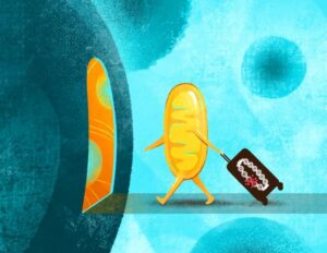
A playful representation of a mitochondrion moving into a larger cell, bringing with it the “baggage” of mutated mitochondrial DNA. Researchers at the Salk Institute developed a new platform for studying that mutated mitochondrial DNA, helping explain the ways it influences human health. [Salk Institute]
The authors explained, “… we developed a scalable ES cell–based platform that integrates mtDNA mutagenesis, cybrid technology, high-sensitivity mutation detection, and optimized mouse transgenesis.” The platform starts with a protein, called mitochondrial DNA polymerase, generating randomly mutated mitochondrial DNA. That mutated mitochondrial DNA is then transferred into stem cells, which can be integrated with mouse embryos to create mice for study. Once one of these mice is established, researchers can investigate the specific symptoms of their specific mitochondrial DNA mutation and the mechanisms by which those symptoms arise—insight that can be used to design targeted therapies down the line. “Optimized ES cell–embryo aggregation enables robust contribution of mtDNA mutant ES cells to host embryos, producing chimeric mice with germline transmission,” the investigators noted.
Using this platform, the Salk team generated a library of 155 mitochondrial DNA mutation cell lines, each with its own distinct impact on mitochondrial performance. “Using this platform, we generate a library of 155 donor fibroblast lines carrying distinct homoplasmic single-nucleotide mtDNA mutations that produce diverse mitochondrial phenotypes, including impaired oxidative phosphorylation, increased reactive oxygen species, and altered mitochondrial membrane potential,” they stated. They then used that library to validate that the cells could be used to generate mice with single mitochondrial DNA mutations. These mice allowed them to find a strong correlation between mitochondrial function and early embryonic development, suggesting a baseline energy level is required for normal development.
“Our library is a huge milestone and is very diverse, with a scale of diversity similar to the known human disease-causing mutation diversity of around 260,” said Fan. “And with this collection of mutant cells, we can not only look at inherited mutations but also at ones that occur based on other stresses like environmental cues or aging.” The authors added, “Together, the advances outlined in this study establish a powerful and generalizable platform for systematically modeling the functional diversity of human mtDNA mutations and polymorphisms in vivo.”
The new platform and library are cracking open the world of mitochondrial DNA. With the ability to generate mitochondrial DNA mutants more rapidly, therapeutic development for mitochondrial disease and dysfunction will come more rapidly, too. The mouse models are already a huge step forward for the field, but the researchers are also eager to move into human models in a more human-relevant context.
“The majority of human diseases come with or cause mitochondrial dysfunction,” said Evans. “Progress in this field has been limited, but this new platform is going to fuel so much important research that points to therapeutic approaches to combat mitochondrial diseases, as well as diseases or conditions associated with mitochondrial dysfunction like cancer or aging.”
In their paper the team concluded, “The library provides a unique and comprehensive resource for modeling the diversity of human mtDNA variation in vitro and can also be used to generate in vivo models through ES-cybrid technology … By enabling the generation of both pathogenic and physiologically relevant mtDNA variants—including those resembling somatic mutations associated with aging and cancer—this platform substantially expands the toolkit available to mitochondrial researchers.”
The Chinese biotech CorrectSequence Therapeutics, also known as Correctseq, reports good results from a Phase I study of its technology involving editing a person’s hematopoietic stem cells to treat beta thalassemia.
The trial, published in Nature, included five patients with transfusion dependent beta thalassemia who were able to stop red blood cell transfusions, the standard treatment for the condition, after receiving the base-edited treatment CS-101. The participants continued to have good levels of hemoglobin with no serious side effects during follow-up.
Beta thalassemia is a rare inherited condition affecting around one in 100,000 people in the U.S. Mutations in the beta‑globin gene HBB reduce or stop production of the beta chains of hemoglobin, leading to chronic anemia that varies in its severity.
There are already several therapies on the market for beta thalassemia. The most common treatment is still regular blood transfusions to treat the anemia, but recently the genetic therapies Zynteglo, a lentiviral gene therapy developed by Bluebird Bio, and Casgevy, a CRISPR edited therapy developed by Vertex Pharmaceuticals and CRISPR Therapeutics were approved by the FDA.
Casgevy works by boosting fetal hemoglobin levels to treat the anemia seen in thalassemia patients. It uses CRISPR–Cas9 to cut both strands of DNA at the BCL11A enhancer site, which relies on error‑prone repair and can theoretically generate insertions, deletions, and larger rearrangements.
Correctseq is also aiming to raise fetal hemoglobin levels with CS-101, targeting the same site, but is only changing individual bases without making a full cut, which should reduce risks linked to double‑strand breaks, such as large deletions or chromosomal translocations.
In this study, CS-101 was given to five patients with beta thalassemia, previously treated with blood transfusions. The process involves extracting their stem cells, reactivating fetal hemoglobin production using base editing, giving the patients chemotherapy to clear existing stem cells and make way for the newly edited population, and finally injecting the patients with the edited stem cells.
All five patients were able to stop red blood cell transfusions and had maintained good levels of hemoglobin at three months. These levels stayed at a similar level through a median follow up period of 23 months. No deaths or reported cancers due to the chemotherapy treatment were observed and the safety profile so far is acceptable.
Although these results are promising, this trial is just a small initial study and further work is needed to confirm safety and efficacy of CS-101.“The planned Phase II/III trial will be crucial for evaluating a larger and more genetically diverse patient population across multiple centers,” write the authors.
“Extended follow-up will be required to enable comprehensive analyses of chimerism and clonality, which will facilitate more definitive assessment of long-term safety, engraftment dynamics and clinical benefit.”
One Correctseq’s main competitors is U.S.-based Beam Therapeutics, which is developing a similar base edited treatment. Beam is behind Correctseq in developing its edited therapy for beta thalassemia, but ahead with its therapy for sickle cell disease, something Correctseq are also targeting using a similar pathway.
The Chinese biotech industry is currently on an upward trajectory. Correctseq is one of many Chinese biotech companies currently working to produce competitors for gene therapies like Casgevy and Zynteglo at a more affordable price than those seen in the U.S.