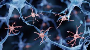
Scientists at the Salk Institute and the Arc Institute, along with their collaborators, unveiled the first body-wide single-cell atlas of two major epigenetic systems: three-dimensional genome folding and DNA methylation, measured simultaneously in the same cells.
The atlas spans 86,689 cells from 16 human tissues, revealing 35 major cell types and 206 subtypes, and is freely available online. The work is part of the National Institutes of Health’s 4D Nucleome (NIH 4DN) program, which aims to understand how the genome is organized in space and time to regulate gene expression in health and disease.
Because the two epigenetic layers were measured together, the researchers could compare what each layer says about a cell’s identity. And while often the two pictures agree, they found that sometimes they do not.
The Human Genome Project, completed in 2003, produced a linear read of the three billion DNA letters in the human body. But the letters alone don’t explain how a single genome produces hundreds of different cell types. That information lives in the epigenome in the form of chemical modifications and structural folds layered on top of the DNA sequence, where they can switch genes “on” and “off” in patterns specific to each cell type.
Caption: Salk scientists Jingtian Zhou (left), Jesse Dixon (center), and Joseph Ecker (right) profiled 86,689 cells across 16 human tissues, linking cell-type-specific epigenetic features to disease risk and revealing that a cell’s 3D genome and DNA methylation don’t always tell the same story. [Salk Institute]
Two of the most consequential epigenetic features are 1) DNA methylation, where small chemical groups called methyl groups are attached to specific DNA bases, and 2) 3D genome organization, where intricate loops, folds, and compartments bring distant stretches of DNA into contact. Both influence gene expression, but they had never been measured together in single cells across the human body.
“There has been an appreciation for trying to understand, at the individual cell level, how the genome is organized, so that we can get a better idea of how genetic variants impact disease,” said co-corresponding author Joseph Ecker, PhD, a professor and Salk International Council Chair in Genetics at Salk and a Howard Hughes Medical Institute investigator. “Some cell types may be more vulnerable than others to genetic variants, because the genome is organized differently in different cell types—and whether a variant matters can depend on that organization.”
Why is noncoding DNA relevant in disease?
Most disease-associated genetic variants fall in the noncoding regions of the genome. That has made it difficult to figure out how a variant contributes to disease, which cell type it acts in, and what gene it ultimately affects.
The new atlas identifies more than 1.36 million differentially methylated regions and 283,606 differential chromatin loops across the human body’s cell types, using tissues from the heart, brain, lungs, stomach, skin, and more. When the researchers overlaid genetic variants known to raise disease risk, specific pairings emerged like variants for blood-glucose regulation concentrated in endocrine cells, atrial fibrillation variants in heart muscle cells, balding variants in skin fibroblasts, and bipolar disorder and schizophrenia variants in excitatory and inhibitory neurons.
“A lot of the genetic variation that predisposes someone to disease is in noncoding parts of the genome,” said co-corresponding author Jesse Dixon, MD, PhD, associate professor and Helen McLoraine Developmental Chair at Salk. “By adding in the 3D genome aspect, we can potentially bridge that gap—connecting noncoding variations with the genes they affect in specific cells and tissues.”
Microglia, illustration. Researchers from the New York Genome Center and Columbia University used the atlas’ cross-tissue methylation data to show that a substantial fraction of the brain’s resident immune cells (microglia) are replaced by cells resembling blood monocytes between roughly ages 50 and 75. The finding challenges the long-held view that microglia persist from embryonic development throughout the life span. [Artur Plawgo/Getty Images]
What happens when two epigenetic lenses disagree?
One of the study’s most surprising findings is that DNA methylation and 3D genome structure don’t always tell the same story about a cell. In skeletal muscle, the team found fibers that look like mature, differentiated muscle cells by their 3D genome folding, but still carry the methylation signature of muscle stem cells. The reverse almost never happens. The most plausible explanation, they explained, is that these cells are caught mid-differentiation, with 3D architecture updating first and methylation catching up.
Similar mismatches appeared in Schwann cells of the peripheral nervous system and in placental trophoblasts. The pattern suggests that different epigenetic features update on different time scales during cell state transitions—a finding that could reshape how researchers define “cell type” in adult tissues and how they track cells moving between states in disease.
The atlas also revises a long-standing assumption about “non-CG methylation,” an unusual form of methylation previously thought to be largely confined to brain cells and stem cells. The study shows that it carries cell-identity information across many human tissues, including muscle, pancreas, and immune cell types, at lower but biologically meaningful levels.
“The inconsistency between modalities may be further used to determine what cell populations are switching between each other in adult tissues and diseases, which could, for example, expand our understanding of cancer cell dynamics,” said co-first and co-corresponding author Jingtian Zhou, PhD, a former graduate researcher in Ecker’s lab who now leads his own lab at the Arc Institute.
A public resource for scientists and artificial intelligence
To make the atlas broadly usable, the team built an interactive web browser that lets researchers visualize DNA methylation and 3D chromatin contacts across every tissue, cell type, and subtype in the study. The underlying data, including 195 billion methylation measurements and 18 billion chromatin contacts, are freely available.
The resource arrives as artificial intelligence tools are increasingly used to predict the functional impact of genetic variants. Atlases like this one can provide the labeled, cell-type-resolved training data that models need to make accurate predictions—a bottleneck that has historically limited the field.
For example, in a companion paper in the same issue of Science, a study led by Bing Ren, PhD, from the New York Genome Center and Columbia University used the atlas’ cross-tissue methylation data to show that a substantial fraction of the brain’s resident immune cells, called microglia, are replaced by cells resembling blood monocytes between roughly ages 50 and 75. The finding challenges the long-held view that microglia persist from embryonic development throughout the life span.
“DNA methylation patterns are specific to each cell type and analogous to a cellular barcode,” said Ren, who also co-authored the Salk-led study. “The comprehensive cross-tissue DNA methylation atlases show that the aging microglia in the human hippocampus more closely match the monocytes from peripheral blood than microglia from young adults, providing a crucial clue for the biological identity of these cells.”
The NIH 4D Nucleome consortium, of which this study is a part, aims to extend this kind of mapping into the fourth dimension: time. A 4D understanding of the genome—how its structure and chemistry change as cells develop, age, and respond to disease—remains a major goal, and the cross-tissue atlas provides reference scaffolding that future time-course studies will build on.
Along with scientists from the Salk Institute and Arc Institute, investigators from the University of California, San Diego, Columbia University, New York Genome Center, University of California, Los Angeles, Harvard, Henan University in China, Vanderbilt University, Stanford University, Broad Institute, University of Sheffield in the U.K., Yale, University of Florida, University of Freiburg in Germany, University of Graz in Austria, and Nanchang University in China; and Chongyuan Luo also contributed to the Science paper.
While Alzheimer’s disease is the most common cause of dementia, many of the molecular mechanisms that drive its progression remain poorly understood. While researchers have cataloged changes in gene activity across different brain cell types, a key unanswered question has been how the genome’s 3D organization influences those changes. Now, researchers have linked alterations in genome folding to disrupted gene regulation in Alzheimer’s disease, providing a new layer of insight into the biology of neurodegeneration.
The findings, published in Science in the paper “Single-cell multiomics connects 3D genome and transcriptome alterations in Alzheimer’s disease,” were reported by researchers from Carnegie Mellon University’s School of Computer Science, the University of Pittsburgh School of Medicine, the University of Washington, and collaborating institutions. Using single-cell multiomics, spatial transcriptomics, and artificial intelligence (AI), the team generated a multiscale view connecting genome structure, gene expression, and tissue organization in Alzheimer’s disease.
To investigate the role of genome architecture in Alzheimer’s disease, the researchers analyzed postmortem prefrontal cortex tissue from individuals with and without the disease. They used GAGE-seq (genome architecture and gene expression by sequencing), a technique that measures both gene expression and physical genome contacts in the same single cell. The team combined those data with chromatin accessibility data, spatial transcriptomic maps, and a transformer-based AI model called Hicformer, which integrates DNA sequence and 3D genome features to predict cell-type-specific gene activity.
The study revealed widespread changes in chromatin organization across major brain cell types. According to the paper, Alzheimer’s disease was associated with “reduced short-range interactions and increased longer-range interactions” within the genome. Active and inactive genomic regions also exhibited increased mixing, consistent with weaker compartment segregation. The researchers linked these structural changes to cell type–specific alterations in gene expression programs involved in disease-relevant pathways.
Researchers also observed weakening of promoter-proximal interactions and changes in regulatory elements, alongside evidence of senescence-related activation in microglia and sex-dependent dysregulation of X-linked genes in females. Integrating the molecular data with spatial transcriptomics revealed altered cellular neighborhoods and disrupted coordination of gene programs within diseased brain tissue. The authors wrote that the results connect “genome structure, gene regulation, and tissue organization through a unified multimodal analysis.”
Their predictive model Hicformer also demonstrated that “3D genome features provide information beyond DNA sequence alone for explaining AD-relevant gene expression, enabling prioritization of distal regulatory elements whose effects are mediated through chromatin contacts,” the authors wrote.
“Measuring gene activity and genome folding in the same cell allows us to directly connect chromosome structure with disease-related gene programs,” said Yang Zhang, PhD, a project scientist in Carnegie Mellon’s Computational Biology Department and co-lead author. “Across several kinds of brain cells, this paired view revealed a consistent signature of 3D genome reorganization in Alzheimer’s disease and helped us prioritize regulatory regions for future mechanistic and therapeutic investigation.”
The researchers concluded that genome folding represents a previously underappreciated regulatory layer associated with Alzheimer’s pathology. By creating a detailed map linking 3D genome remodeling to gene expression and tissue organization, the study provides a framework for future experiments aimed at determining which structural changes contribute directly to disease progression. This may also provide clues to future therapeutic focuses.
“Alzheimer’s disease cannot be understood one layer at a time,” said senior author Jian Ma, PhD, the Ray and Stephanie Lane Professor of Computational Biology at Carnegie Mellon University. “The genome’s 3D structure is a fundamental regulatory layer that helps to connect DNA sequence to gene activity. By integrating genome folding, cell state, and tissue context, we can move beyond cataloging disease-associated changes toward understanding how they fit together and which mechanisms to test next,” said Ma. “Alzheimer’s disease cannot be understood one layer at a time.”
The results of a preclinical study by researchers at the University of Texas MD Anderson Cancer Center have found that an investigational epigenetic therapy called NTX-301 remained effective in treatment-resistant acute myeloid leukemia (AML) by activating the Hippo pathway, a tumor-suppressing pathway linked to cancer growth and drug resistance.
In preclinical models, the hypomethylating agent (HMA) NTX-301 was more effective than standard hypomethylating agent therapy and retained anti-leukemia activity in treatment-resistant and TP53-mutant AML. The team also found that the therapy activated the Hippo pathway through targeted epigenetic changes, revealing a previously unrecognized mechanism that may contribute to its anti-leukemia effects.
“Leukemia cells are remarkably adaptable and often find new pathways to survive after treatment,” said Michael Andreeff, MD, PhD, professor of medicine at the University of Texas MD Anderson Cancer Center and research co-lead. “These findings suggest NTX-301 may disrupt several of those survival mechanisms simultaneously while reactivating pathways that normally restrain cell growth. That dual effect could help explain why NTX-301 remained active in some of the most therapy-resistant forms of AML.”
First-generation hypomethylating agents, including 5-azacytidine (5-AZA) and decitabine (DAC), are used as standard clinical care for patients with AML and myeloid dysplastic syndromes (MDS), the authors wrote. Combining HMAs with the BCL-2 inhibitor venetoclax has further improved outcomes for patients.
“Hypomethylating agent (HMA) and the BCL-2 inhibitor venetoclax (VEN) combinations have evolved into frontline therapies for patients with acute myeloid leukemia (AML), yielding high response rates,” they stated. However, while such combination therapy works well initially, resistance and relapse remain common.
The challenge is particularly significant in AML with mutations in the TP53 gene, which normally helps cells respond to damage and prevent uncontrolled growth. When that gene is mutated, leukemia cells can become resistant to therapy and more difficult to eliminate. “… most patients ultimately relapse, particularly those with TP53 mutations,” the researchers continued.
Efforts have been made to develop improved and more effective HMAs, they noted, and NTX-301 is such a next-generation HMA. But as they pointed out, “… previous reports of NTX-301 preclinical studies in leukemia were conducted primarily in cell lines and xenograft models … its activities in therapy-resistant settings have not been investigated.” And while a Phase I study (NCT04167917) of the oral agent NTX-301in patients with AML and MDS has been completed, the team noted in their paper that the study has not yet been reported.
For their newly reported preclinical study, the researchers evaluated NTX-301 across multiple preclinical models of treatment-resistant AML, including patient-derived xenograft (PDX) models of AML with acquired resistance. Their results showed that NTX-301 therapy consistently reduced leukemia cell survival more effectively than azacitidine (AZA), a commonly used hypomethylating agent.
Importantly, NTX-301 remained active in leukemia cells that had already developed resistance to both hypomethylating therapy and venetoclax, and demonstrated anti-leukemia activity in TP53-mutant AML models. When combined with venetoclax in resistant leukemia samples, NTX-301 produced stronger anti-leukemia effects than either treatment alone. The combination was effective not only against leukemia blasts but also against leukemia stem and progenitor cells, which are believed to contribute to disease persistence and relapse.
In summary, they wrote, “Therapeutically, NTX-301 is more potent than 5-AZA in AML cells with various genetic backgrounds, is active in AML cells with acquired resistance to HMA or VEN, overexpressing VEN-resistant factors MCL-1 or BCL-2A1, and in isogenic AML cells with TP53 deletions/mutations in vitro and in vivo in xenograft models, exhibits activities against AML blasts and stem/progenitor cells from patients resistant to/relapsed from VEN-based therapies and with TP53 mutations in vitro and in vivo VEN/DAC-resistant PDX models, and enhances VEN activity.”
To understand why NTX-301 appeared more effective than existing drugs, researchers analyzed changes in DNA methylation, a process that can switch genes on or off without altering the underlying genetic code. Unlike current hypomethylating therapies, which broadly affect DNA methylation, NTX-301 focused on a more selective set of genes and pathways, including the Hippo pathway, which functions as a natural cell growth regulator.
NTX-301 increased activity of key Hippo pathway genes while reducing activity of YAP, a protein frequently linked to cancer cell survival, treatment resistance, and stemness. These findings suggest Hippo pathway reactivation may be an important reason the therapy remained effective in resistant leukemia models and could represent a new strategy for overcoming treatment resistance in AML. “Collectively, our data suggest that NTX-301 exhibits more potent anti-leukemia activities compared to current HMAs and synergizes with VEN in VEN-resistant and TP53-mutant AML and AML stem/progenitor cells,” the team concluded.
Additional studies are needed to determine whether these results translate to patients and to identify which populations may benefit most. The findings suggest that patients with relapsed AML, venetoclax-resistant disease, and TP53 mutations may be important groups for future clinical evaluation. “Taken together, the numerous NTX-301 targets identified here, its novel mechanism of action, and its superior activity against VEN-resistant and TP53-mutant AML compared to 5-AZA, warrant the future clinical development,” the investigators noted. “Given the strong preclinical data in TP53-mutant AML and the unmet clinical need, this should be a primary target group in the next clinical trial.”
Carter said, “An encouraging aspect of this study is that it identified both a potential therapeutic opportunity and a biological explanation for why it may be effective. The results provide a rationale for continued clinical development and suggest that targeting Hippo signaling may help address treatment resistance in AML.”
As one of the most aggressive blood cancers, the way acute myeloid leukemia (AML) is classified continues to shape every major clinical decision—from risk stratification to the choice of targeted therapies. For decades, that classification has rested almost entirely on the gene mutations found in leukemic cells. But mutations alone have never fully explained why AML behaves so differently from patient to patient. A new study published in Nature now provides the missing layer: the epigenome.
In the largest chromatin‑profiling effort ever conducted for any cancer, a research team led by Seishi Ogawa, MD, PhD, and Yotaro Ochi, MD, PhD, of Kyoto University, together with Sören Lehmann, MD, PhD, of the Karolinska Institute, mapped the chromatin accessibility landscape of 1,563 AML patient samples. Their analysis—built on ATAC‑seq, RNA‑seq, DNA methylation, ChIP‑seq, whole‑genome sequencing, and single‑cell multiomics—reveals that AML can be classified into 16 distinct epigenomic subgroups, each defined by a characteristic chromatin state and its own regulatory wiring.
As the authors wrote, “ATAC-seq…show[s] that AML can be classified into 16 subgroups on the basis of chromatin accessibility profiles.” This chromatin‑based structure was remarkably stable: single‑cell ATAC‑seq across more than 280,000 cells confirmed that each patient’s leukemic population shares a conserved accessibility fingerprint.
Each subgroup carries a unique combination of driver mutations, differentiation states, transcription‑factor networks, DNA methylation patterns, and super‑enhancer architecture. Many do not align cleanly with existing genomic classifications such as WHO or ICC, according to the researchers. In fact, the team found that even exhaustive decision‑tree analyses of known driver mutations could not explain most subgroup identities. As the paper noted, “Evidence suggests that genetic alterations do not fully explain AML pathophysiology and heterogeneity.”
Clinically, chromatin information sharpened prognostic assessment in both Swedish and Japanese cohorts. Several subgroups also showed unexpected drug sensitivities. Three subgroups responded to MEK inhibitors despite lacking RAS‑pathway mutations. Another subgroup, enriched for RUNX1 mutations and marked by a chromatin profile resembling early B‑cell precursors, proved highly sensitive to ABL inhibitors.
The study, “Chromatin landscape and epigenetic heterogeneity of acute myeloid leukemia,” positions chromatin architecture as a foundational dimension of AML biology. It also provides a practical path toward clinical adoption: the team distilled a 30‑gene expression signature capable of identifying high‑risk chromatin subgroups using standard sequencing workflows.
Looking ahead, the group aims to develop lower‑cost diagnostic approaches and refine treatment strategies tailored to each epigenomic subgroup. The newly generated eCHROMA AML atlas is expected to serve as a resource for cancer epigenomics broadly, enabling discovery of new therapeutic targets and mechanistic insights.
Having higher levels of vitamins A and D in the body could help improve lung function in adults with asthma, suggests research led by Brigham and Women’s Hospital in Boston.
The study, published in the journal Thorax, also showed that higher vitamin A levels could also benefit lung health in children with asthma.
“Vitamins A and D are key regulators of gene expression involved in lung development and immune function…Both vitamins have complex roles in asthma and lung function,” write lead author Michael McGeachie, PhD, assistant professor at Brigham and Women’s Hospital, and colleagues.
“Vitamin A deficiency is more common in people with asthma and is linked to airway hyperresponsiveness. Moderate intake in childhood may improve lung function and reduce asthma risk, whereas excessive intake may increase adult-onset asthma risk. Vitamin D deficiency is associated with poorer lung function, increased exacerbations and worse disease control, although results are variable due to diet, age and sun exposure.”
In this study, the researchers evaluated data from two asthma study cohorts—the Genetic Epidemiology of Asthma in Costa Rica Study including 1,165 children with asthma and the adult Omic Determinants of Longitudinal Lung Function in Asthma cohort including 1,041 adults with the condition.
The researchers looked at both circulating vitamin A and D in both groups and also looked at links with epigenetic regulation.
In children, there were no significant links between lung function and vitamin D levels. Higher vitamin A levels were linked to better breathing capacity though. For each step up in vitamin A levels, the amount of air the children could blow out in the first second was about 2.5 percentage points higher, and the total amount of air they could breathe out was about 7.6 percentage points higher.
In the adults, one step up in vitamin A levels was linked to a 4.7 unit increase in the amount of air they could blow out in the first second and a 3.4 unit increase in the total amount of air they could breathe out. The effect of vitamin D levels in this group was smaller, but statistically significant, with lung function improvements between 0.16-0.18 units per step up in vitamin D levels.
The researchers also looked at DNA methylation and at several epigenetic clocks that estimate a person’s biological age from methylation patterns in the adult cohort. They found that adults with higher vitamin A and vitamin D levels had fewer methylation tags at key control sites in the IRF5 gene than those with lower levels and showed changes in small regulatory micro RNAs that respond to vitamin levels. These epigenetic changes were linked to better lung function and slower biological aging.
The epigenetic tests suggest that vitamins influence lung function and aging partly by producing these epigenetic changes, rather than only through a direct effect, according to the researchers.
“While future studies to replicate these findings in independent populations are needed to confirm the generalizability and robustness of these observations,” write Sze Man Tse, MD, and Geneviève Mailhot, PhD, of the CHU Sainte-Justine Research Center, Montreal, and the University of Montreal in an accompanying editorial in the same journal, “subsequent interventional studies examining the impact of vitamin supplementation on biological ageing and on IRF5 function will be particularly relevant.”
A first-in-human clinical trial of an experimental epigenetic therapy has produced an outcome long considered elusive in the progressive genetic muscle degeneration disease facioscapulohumeral muscular dystrophy (FSHD): measurable gains in lean muscle volume.
Epicrispr Biotechnologies announced updated interim results from its ongoing Phase I/II study of EPI-321, reporting that all three FSHD patients who reached the six-month evaluation point experienced increases in lean muscle mass following a single intravenous (IV) infusion of the therapy. The findings represent what the company says is the first clinical evidence that a treatment may be capable of increasing muscle volume in FSHD, which affects an estimated 870,000 people worldwide and is one of the most common forms of muscular dystrophy.
Although the study remains in its early stages with only nine total patients across two dosing cohorts, the promising findings were supported by imaging data, functional measurements, and biomarker changes that together suggest the therapy may be altering the biological drivers of the disease.
FSHD lacks disease-modifying treatments to intervene in the characteristic loss of skeletal muscle function, starting in the face, shoulders, and upper arms and spreading throughout the body. “We were pretty blown away,” Epicrispr CEO Amber Salzman told Inside Precision Medicine. “Every single patient gained lean muscle volume. No one has seen that.”
A silencing GEM
FSHD is caused by hypomethylation of the D4Z4 region, which is a polymorphic variable number tandem repeat (VNTR) array made up of 3.3 kilobase units, with each unit encoding the DUX4 gene. This hypomethylation leads to abnormal activation of the DUX4 gene, triggering muscle cell death and tissue degeneration, resulting in progressive muscle weakness, asymmetry, and fat infiltration.
Unlike conventional gene-editing therapies that permanently alter DNA sequences, Epicrispr’s therapy uses their Gene Expression Modulation System (GEMS). Epicrispr’s EPI-321 is an adeno-associated virus (AAV)-delivered epigenetic gene therapy that restores D4Z4 methylation and suppresses DUX4 expression, and preclinical studies show improved muscle function and reduced muscle cell death. “We don’t cut DNA,” Salzman said. “We’re not using Cas9 in the traditional editing sense. We use a dead Cas protein and an epigenetic mechanism to silence the gene.”
According to Epicrispr, preclinical studies demonstrated that the epigenetic modifications persist through many rounds of cell division, suggesting long-term suppression may be possible after a single treatment.
In addition to the therapeutic innovation, Epicrispr has also made headway into the long-standing challenge of identifying reliable molecular biomarkers for FSHD to evaluate activity downstream of DUX4. Traditional muscle biopsies provide information from only a small tissue sample and can be difficult to interpret when substantial fat replacement has already occurred.
Instead, Epicrispr collaborated with researchers at the University of Colorado investigating circulating cell-free DNA signatures associated with DUX4-driven disease activity. The biomarker, known as CBT17, emerged from studies comparing blood samples from approximately 50 healthy individuals and 50 FSHD patients. The biomarker may provide a whole-body readout of disease activity, offering evidence that DUX4 suppression is occurring beyond individual muscles examined through biopsy.
As of the May 12, 2026 data cutoff, EPI-321 has demonstrated a manageable safety profile, with nine patients treated across two dose cohorts. One IV infusion was administered to six patients in the first cohort at a target dose of 2×10¹³ vg/kg and to three patients in the second cohort at a target dose of 4×10¹³ vg/kg.
The company reported a “manageable” safety profile, with no severe dose-limiting toxicities disclosed to date. Salzman was careful not to overstate the findings. “I never want to be hubristic when it comes to AAV,” she said. “You’re giving people a lot of virus.”
She described the treatment experience as somewhat analogous to vaccine-related immune responses, noting that prophylactic immunosuppression is used as part of the protocol. “It’s manageable,” she said. “I’m not going to say it’s favorable. It’s manageable.” No serious grade 3 or grade 4 treatment-related safety concerns have been reported so far.
Baseline levels of CBT17 in treated patients fell substantially after therapy. “When we looked at baseline and three months, our patients’ levels came down so that it was just about in the healthy range,” she said.
Functional measures match MRI
The headline result comes from whole-body MRI analyses conducted six months after treatment. Among the first three evaluable patients, lean muscle volume increased in every individual compared with baseline measurements. On average, patients gained approximately 370 milliliters of lean muscle tissue, equivalent to roughly 0.8 pounds of muscle mass. Individual gains ranged from about 0.5 to 1.3 pounds. For a disease characterized by chronic muscle loss, even stabilizing muscle mass would be considered a meaningful achievement. Actual gains are potentially more significant.
The MRI analyses were performed in collaboration with Springbok Analytics, a company specializing in AI-powered muscle imaging. In addition to being widely used in professional sports to monitor injury recovery and rehabilitation, as well as a growing number of clinical settings, FSHD is one of Springbok’s primary clinical focus areas, along with disorders such as Charcot-Marie-Tooth disease and Duchenne muscular dystrophy. Using a specialized MRI protocol that runs on standard scanners, the company can quantify changes across as many as 140 individual muscles throughout the body, measuring muscle volume, muscle composition, and fat infiltration, producing objective data on muscle health.
According to Salzman, Springbok compared patients’ baseline scans against machine-learning models trained on longitudinal imaging datasets from more than 100 FSHD patients. “They can predict what it’s going to look like in six months—how much muscle patients will lose and where,” she said. “Then we sent them the six-month scans, and every patient gained lean muscle volume.”
The gains were not evenly distributed throughout the body. Patients appeared to benefit most in muscles that still retained substantial healthy tissue before treatment. “If you have too much fat in that muscle, there’s not that much we can do,” Salzman explained. “But if you have some residual muscle, it can not only stop getting worse because we’re cutting off the poison, but you can also regenerate muscle.”
One participant, a post-menopausal woman, gained approximately 1.3 pounds of lean muscle mass according to the MRI analysis. The increase was particularly notable because age-related muscle loss would normally be expected in that population. Earlier data released by Epicrispr showed favorable trends across several functional and strength assessments at the three-month mark. These measures included tests commonly used in neuromuscular disease studies, such as the Timed Up and Go test, 10-meter walk/run assessments, and quantitative muscle testing.
Because many functional endpoints depend on patient effort, interpreting changes in small open-label studies can be challenging. Investigators and investors alike often question whether participants simply perform better because they know they received treatment. Salzman acknowledged those concerns. “You could say maybe they had a good day, and that’s why the functional measures were better,” she said.
MRI measurements, however, are not subject to effort-dependent variability. “You can’t fake MRI,” she added. Importantly, the regions showing muscle growth on imaging appeared to correspond with areas where patients demonstrated improved performance.
One participant who gained substantial upper-body muscle volume showed corresponding gains on upper-body strength assessments, while lower-body improvements were more limited in areas where muscle loss had already become severe. “The whole story is holding together,” Salzman said. “Their functional measures are getting better; their lean muscle is increasing.”
Taken together, the MRI, biomarker, and functional data provide multiple independent signals supporting biological activity. “You can’t fake blood; you can’t fake MRIs,” Salzman said. “Those are totally supportive.”
Epitome of epigenetic editing?
Beyond FSHD, researchers are closely watching EPI-321 because it represents one of the first clinical tests of an epigenetic CRISPR-based therapeutic strategy. “This is an important milestone not just for FSHD but for epigenetic medicine,” Salzman said. The approach could potentially be adapted to other diseases caused by harmful gene activation or insufficient gene silencing.
Still, significant hurdles remain before EPI-321 can be considered a proven therapy. The current dataset includes only three patients with six months of follow-up. Epicrispr plans to present additional data from six patients at the World Muscle Society Annual Congress in September 2026.
The company expects the initial Phase I/II study to complete enrollment and generate a full readout in mid-2027. Yet, larger studies will be needed to determine whether muscle gains persist, whether they translate into meaningful long-term functional benefits, and whether safety remains acceptable over time.
Regulators will also require evidence linking increases in lean muscle volume to tangible improvements in patient function and quality of life. To that end, Epicrispr is preparing discussions with the U.S. Food and Drug Administration regarding potential future endpoints and whether MRI-based muscle measurements could eventually serve as surrogate markers of clinical benefit.
Whether those early signals hold up in larger populations remains to be seen. But for a disease in which muscle loss has long been viewed as inevitable, the possibility of reversing that trend, even modestly, marks a notable moment for the FSHD field.
Many DNA variants linked with neuropsychiatric disorders (NPD) that do not code for proteins depend on neuronal activation, a study suggests.
The findings, in Science, highlight the power of cell stimulation to reveal context-specific “hidden” genetic effects in conditions such as schizophrenia.
They suggest that genetic regulation is not fully revealed by measuring gene expression alone.
Instead, gene activity—at least in the brain—may depend on context and the physiological state of neurons.
“Liang et al. demonstrate that the genetic processes that underlie neuropsychiatric disease are heavily determined by a dynamic physiological environment rather than by fixed cellular conditions,” said Biao Zheng, PhD, and Panos Roussos, PhD, from Icahn School of Medicine at Mount Sinai in New York, in a Perspective article accompanying the study.
They added: “To understand disease genetics, we might need to study the genome in motion and not at rest.”
Genome-wide association studies have revealed hundreds of genetic loci associated with mental illness, with more than 280 identified for schizophrenia alone.
But many of these DNA regions do not encode proteins and their impact is often subtle and difficult to detect.
To investigate further, Lifan Liang, PhD, from the University of Chicago, and co-workers studied gene expression and chromatin accessibility in single neurons derived from induced pluripotent stem cells collected from a hundred human donors.
The single-cell multi-omics study involved assessing transcriptional and epigenomic profiles before and after neurons were activated through potassium-induced depolarization.
The team found that much of the activity in regulatory DNA regions only became apparent with neuronal stimulation.
Both the number of detectable expression quantitative trait loci (eQTLs)—genetic variants associated with differences in gene expression—and chromatin accessibility QTLs (caQTLs)—DNA variants associated with differences in chromatin accessibility—rose after neuronal stimulation.
Shared and cell type-specific transcription factors worked together, possibly through regulatory cascades, to drive cell type-specific neuronal responses to stimuli.
eQTLs after stimulation had substantially weaker overlap with brain eQTL catalogs derived from postmortem tissue compared with eQTLs before stimulation.
This suggested that many relationships between regulatory DNA activity and gene expression become detectable only during neuronal activation and could be missed by traditional tissue-based studies.
A higher number of caQTLs were associated with neuropsychiatric disease compared with eQTLs, suggesting that disease-associated genetic variants could have detectable effects on regulatory DNA even when downstream changes in gene expression were not obvious.
Supporting this, chromatin accessibility and transcriptional responses to neuronal activation often occurred at different times.
Regulatory regions associated with genes that respond rapidly to neuronal stimulation often remained accessible after transcription subsided. By contrast, some late response genes exhibited accessible chromatin before their expression was induced.
When taken together, these observations implied that chromatin accessibility can be an indication of both prior and future transcriptional potential.
“We identified thousands of cell type–specific and activity-dependent quantitative trait loci for gene expression (eQTLs) and chromatin accessibility (caQTLs), helping prioritize NPD risk variants and genes that manifested functional effects only upon neuronal stimulation,” the researchers asserted.
They added: “Our work provides mechanistic insights on neuron subtype–specific activity-dependent gene regulation, substantially expanding the repertoire of context-specific causal variants and genes for NPD and other brain traits.”
BackgroundThe relationship between adverse childhood experiences (ACEs) and personality disorders (PDs) has attracted sustained attention in psychiatry, psychology, and public health. Existing studies have mainly examined epidemiological associations, specific PDs diagnoses, or mechanisms, whereas bibliometric evidence mapping the field’s knowledge structure and thematic evolution remains limited. This study aimed to characterize trends, contributors, collaboration networks, core themes, and frontiers in ACEs–PDs research.MethodsEnglish-language publications on ACEs and PDs were retrieved from Web of Science Core Collection, Scopus, and PubMed from inception to December 31, 2025. After year screening, document-type filtering, and deduplication, 5,084 records were included. Bibliometric analyses were performed using R, VOSviewer, and CiteSpace. The merged dataset was used to examine annual trends, countries/regions, institutions, authors, journals, and keyword co-occurrence, while WoSCC records were used for co-citation analysis, keyword clustering, and burst detection.ResultsACEs–PDs research showed sustained growth, with a marked increase after 2000. The United States occupied a central position in publication output, citation impact, and international collaboration, while the United Kingdom, Germany, Canada, the Netherlands, and Australia also showed strong influence. Harvard University, the University of London, and Ruprecht Karls University Heidelberg were leading institutions; Zanarini M, Fonagy P, Schmahl C, Paris J, and Kleindienst N were key contributors. Influential journals mainly covered psychiatry, personality disorders, child maltreatment, trauma, and developmental psychopathology. Keyword analyses identified childhood adversity, personality disorder, borderline personality disorder, depression, childhood sexual abuse, and post-traumatic stress disorder as core themes. VOSviewer and CiteSpace analyses indicated that hotspots have expanded from childhood abuse, PDs diagnosis, and psychiatric comorbidity to emotion dysregulation, non-suicidal self-injury, social support, functional connectivity, early intervention, and mechanism validation. Highly cited publications revealed a knowledge base centered on childhood abuse/trauma, borderline personality disorder, psychiatric comorbidity, emotion regulation, and neurobiological mechanisms.ConclusionThis study maps development and knowledge structure of ACEs–PDs research. Findings suggest a shift from exposure–outcome association studies toward comorbidity, intermediate phenotypes, neurobiological mechanisms, and clinical translation. Future research should strengthen longitudinal and cross-cultural designs, consider ACE type, timing, duration, and severity, and integrate neuroimaging, inflammatory, epigenetic, and clinical-course phenotypes.
Research headed by scientists at Arizona State University and Vanderbilt University suggest that experiences we face early in life may leave their marks on our health in ways that echo across decades, and even across the entire body.
The team’s study involves a unique group of free-living rhesus macaques who have been followed their entire lives to document their experiences. Pairing the animals’ histories with genomic data from 12 tissues collected in adulthood, the study has generated some of the clearest molecular evidence yet that early life adversity (ELA) leaves a lasting, system-wide impression at the epigenome, the biological layer on top of the human genome that regulates gene activity.
The team examined DNA methylation (DNAm) patterns, which can represent telltale aging hallmarks of the epigenome. DNA methylation is one of the most well-studied markers of aging and can be used to build “epigenetic clocks” that estimate both an organism’s chronological age—how long it has been alive—and biological age, which is how old it appears physiologically. Through their newly reported study, the researchers developed highly precise tissue-specific clocks, capable of predicting age within about one year of an individual’s chronological age.
Their findings challenge a common assumption that early adversity uniformly accelerates biological aging. Instead, the results suggest a more nuanced model, in which early experiences alter the trajectory of molecular aging, amplifying the effects of aging in some tissues, such as the pituitary, but not others. These findings further suggest that the well-documented effects of early adversity on health operate, at least in part, through mechanisms that are not directly linked to aging.
“Our goal was to understand how aging unfolds across the body, and how early experiences might influence that process,” said Noah Snyder-Mackler, PhD, a professor in Arizona State University’s School of Life Sciences. “What we found is that early life adversity leaves a coordinated epigenetic signature that spans multiple tissues—but it doesn’t simply accelerate aging in a uniform way.”
Snyder-Mackler is co-senior author of the team’s published paper in Science, titled “Age and early life adversity shape heterogeneity of the epigenome across tissues in macaques.” In their research article summary the team concluded “By generating multi-tissue DNAm data across the life course in animals with known social histories, we reveal a fundamental contrast in epigenomic remodeling: Age-associated epigenetic variations are highly tissue dependent, whereas the molecular effect of ELA represents a more coordinated, organism-wide response.”
Aging is universal, the authors wrote, but the pace of decline varies significantly both between and within individuals. “Identifying early patterns and biomarkers of aging—before overt pathophysiology—could enable earlier clinical intervention, and refining how patterns are linked between organs may clarify which aging hallmarks are shared across tissues versus which must be assayed in specific organs to predict disease.”
ELA has been linked to age-related diseases and reduced lifespan in both humans and other social animals. However, the team noted, “… we still know little about how early life exposures shape the biological mechanisms of aging across tissues, especially at the molecular level.” This is difficult to study in humans, because detailed life course information in combination with multi-tissue molecular data is very rare.
For their reported research the team studied 237 macaques, who live in semi-natural conditions on Cayo Santiago (colloquially referred to as “Monkey Island”), a 38-acre island off Puerto Rico’s east coast. The island is inhabited by over 1,500 free-ranging rhesus macaques and managed by the University of Puerto Rico and Caribbean Primate Research Center. By integrating multi-tissue DNA methylation collected in adulthood with detailed records of early life experiences, the team uncovered how adversity and aging interacted to shape biology at the molecular level.
“We focused on DNAm, an epigenetic modification that is one of the most studied hallmarks of aging,” the authors wrote. “… DNAm can be used for estimating ‘biological’ age using ‘epigenetic clocks,’ a measure linked to disease and mortality.” The team generated a dataset integrating multi-tissue DNAm profiles with extensive data on social and environmental conditions during the animals’ development, creating the opportunity to examine how age and early life adversity affect DNAm across the body.
What they found was that despite the epigenetic precision, aging did not occur uniformly across the body. Instead, the researchers found that age-related changes in DNA methylation were highly tissue dependent. Yet even amid this diversity, individuals showed a degree of internal consistency. Animals that appeared “biologically older” in one tissue tended to appear older in other tissues as well, suggesting that aging operates as a partially coordinated process across the body.
“Age-associated DNAm was predominantly tissue dependent, yet tissue-specific epigenetic clocks showed that epigenetic aging was relatively consistent within individuals,” the investigators stated. Co-senior author Amanda Lea, PhD, assistant professor of biological sciences at Vanderbilt University, added, “At a molecular level, aging looks very different depending on which tissue you examine,” said. “Blood, which is most commonly measured in human studies, only captures part of the picture.” Some tissues, like the thymus and pituitary gland, showed particularly strong and distinct age-related patterns, while others exhibited more subtle changes.
The study’s most novel insights came from examining early life adversity—defined through naturally occurring conditions such as maternal loss, low maternal social status, or growing up in a crowded social group. These experiences were not only associated with changes in DNA methylation, but in a strikingly coordinated way across tissues. “We found that each type of adversity tends to affect specific regions of the genome,” said Lea. “But once it targets those regions, the effects are often shared across multiple tissues.”
In total, the team identified thousands of genomic regions where DNA methylation was associated with early life adversity. These regions frequently overlapped with those affected by aging—but importantly, the direction of the effects was not consistent. “In some cases, adversity-related changes looked like accelerated aging. In others, they went in the opposite direction,” explained co-lead author Rachel Petersen, PhD, a Vanderbilt postdoctoral researcher. “This tells us that early adversity doesn’t simply ‘speed up’ aging.
“Instead, it reshapes the epigenome in more complex ways.” In their paper the researchers note, “Although ELA targeted many of the same loci as age, the directions of effects differed, which indicates that ELA does not uniformly increase epigenetic age.” Instead, they continued, “… ELA leaves a coordinated, cross-tissue epigenetic signature that is distinct from—yet intertwined with—age-related differences, which advances our understanding of how early environments sculpt the molecular foundations of aging and disease.”
The study also highlights the importance of studying multiple tissues. Many previous studies have relied on blood samples, which are relatively easy to collect. However, the new findings show that this approach may miss critical aspects of how aging and environmental exposures affect the body. “Different tissues have their own epigenetic landscapes and respond differently to both age and adversity,” said co-lead author Baptiste Sadoughi, DVM, an ASU postdoctoral researcher. “To fully understand health and disease, we need to take a whole-body perspective.”
The use of rhesus macaques, which share many biological and social similarities with humans, adds to the study’s relevance. Unlike laboratory animals, these macaques live in complex social environments, allowing researchers to capture naturally occurring variation in life experiences. “This kind of dataset is incredibly rare,” said Lea. “It allows us to connect detailed life histories with molecular changes across the body in a way that simply isn’t possible in most human studies.” In their research article summary the team noted that their collective findings “… advance our understanding of how early environments sculpt the molecular foundations of aging and establish this comprehensive tissue atlas as a valuable resource for the scientific community.”
Beyond its scientific contributions, the research has important implications for understanding the developmental origins of health and disease. By showing how early experiences shape the epigenome across tissues, it provides a potential mechanism linking childhood conditions to later-life outcomes. “Early life is a critical window for biological development,” said Snyder-Mackler. “Our findings suggest that experiences during this period can leave lasting marks on the genome that influence health trajectories over the lifespan.”
At the same time, the complexity of the results offers a note of caution. Because all types of adversity do not have uniform effects, predicting long-term consequences will require a more detailed understanding of context, timing, and individual variation. “This is not a simple story,” Lea said. “But that’s what makes it exciting. We’re beginning to see how life experiences are written into our biology—and why those signatures might vary within and between individuals.” As researchers continue to explore the interplay between environment, epigenetics and aging, studies like this one are helping to redefine what it means to grow older—not just as a function of time, but as a dynamic process shaped by the unique experiences that can truly define our lives.
For decades, cancer medicine has been driven primarily by genetics—the search for mutations, deletions, amplifications, and other DNA changes that directly alter how cells behave. Precision oncology has largely focused on identifying these genomic drivers and matching them with targeted therapies. But another biological layer has steadily moved from the margins of research into the center of clinical care: epigenetics.
Epigenetics refers to chemical modifications that regulate how genes are turned on or off without changing the underlying DNA sequence itself. One of the best-known examples is DNA methylation, in which small chemical tags called methyl groups attach to DNA and influence whether genes are active or silenced. These marks help explain one of biology’s most fundamental questions: how a brain cell and a heart cell can contain the exact same genetic code yet function in completely different ways.
Rather than rewriting DNA, epigenetics controls how a stretch of DNA is read. It shapes normal development, tissue specialization, immune responses, aging, and disease progression. In cancer, epigenetic disruption can silence tumor-suppressor genes, activate harmful pathways, and create stable molecular fingerprints that reveal where a tumor came from and how it behaves. Increasingly, those fingerprints are becoming clinically actionable.
Today, epigenetics is helping physicians classify brain tumors more accurately, detect pancreatic cancer from a simple blood draw, improve molecular sequencing workflows, and even therapeutically silence disease-causing genes. Across diagnostics and therapeutics, the field is rapidly becoming one of the most practical and powerful tools in precision medicine.
Few people have watched that transformation more closely than Matija Snuderl, MD, a neuropathologist at NYU Langone. He remembers a time when epigenetics barely registered in medical education.
Matija Snuderl, MD Neuropathologist, NYU Langone
“When I was in medical school,” he said, “there were literally like two pages on epigenetics in the genetics book, and it had no implications for cancer whatsoever.”
That changed quickly.
Brain tumors become a clinical test case
Snuderl became deeply interested in epigenetics around 2013, when researchers began realizing that epigenetic patterns could do far more than explain developmental biology. They could also reveal a tumor’s cell of origin and, in some cancers, directly illuminate the mechanisms driving carcinogenesis.
Brain tumors proved to be one of the most compelling settings for that work. “Brain tumors are only about two percent of all cancers,” Snuderl explained, “but they are disproportionately heterogeneous.”
That heterogeneity is extraordinary. There are now roughly 180 recognized molecular subtypes of central nervous system tumors. Yet for decades, many of those diseases were classified almost entirely by histology—how the tumor looked under a microscope. Tumors with similar visual features were grouped together, even when they were biologically distinct.
The result was predictable. “We ended up putting a lot of tumors that are completely different into the same basket, treating them the same way, and then we were surprised that we didn’t see results,” Snuderl said.
Epigenetics offered a way out of that problem. Because DNA methylation reflects both developmental lineage and disease biology, it provides a stable molecular fingerprint of tumor identity. Unlike RNA, which fluctuates significantly and degrades quickly after tissue collection, methylation patterns are remarkably durable. “The beauty of methylation,” Snuderl said, “is that it’s an incredibly robust biomarker.”
A molecular fingerprint for diagnosis
Snuderl began a collaboration with the German Cancer Research Center (DKFZ) in Heidelberg and proposed a simple but powerful hypothesis: every tumor type has a distinct methylation signature—a recognizable pattern of hypermethylated and hypomethylated regions that functions like a fingerprint.
Using thousands of tumor samples, the team built a machine learning classifier based on a random forest model. The algorithm was trained to recognize these methylation signatures and assign new tumor samples to the most likely disease subtype. “At every decision point,” Snuderl explained, the classifier essentially asks, “Are you methylated or are you unmethylated?”
The system evaluates more than 10,000 decision trees before generating a calibrated confidence score that indicates how certain the classifier is about a diagnosis.
The visual output is particularly striking. On uniform manifold approximation and projections (UMAPs), which is a method of viewing high-dimensional data in a low-dimensional manner, each tumor appears as a colored dot that clusters with biologically similar tumors. Some groups are extremely tight, reflecting genetically simple and highly consistent diseases. Others are more diffuse, showing greater heterogeneity and multiple biological drivers.
These patterns helped reveal that some long-standing diagnostic categories were fundamentally wrong. One major example involved primitive neuroectodermal tumors (PNETs), once considered a distinct diagnosis. Methylation profiling showed that PNET was largely a “waste basket” category: about 80% of those tumors were misclassified, while the remaining cases represented multiple separate molecular diseases, said Snuderl.
Another example came from low-grade epilepsy-associated tumors in young adults, known as polymorphous low-grade neuroepithelial tumor of the young. It emerged as a clearly distinct clinical and pathological entity through methylation analysis.
These discoveries were not simply academic refinements. They changed how patients were diagnosed and treated.
Taking research to the clinic
Snuderl is quick to point out that many important scientific discoveries never reach patients because researchers stop at publication. A high-impact paper may generate excitement, but translating a method into a clinical diagnostic requires an entirely different level of work—regulatory validation, reproducibility testing, and operational rigor.
“There is very little glory in going through the regulatory framework,” he said. Still, his team decided that it was an essential step.
In 2019, NYU became the first CLIA-certified laboratory in the United States to clinically implement DNA methylation–based brain tumor classification. It was also the first to use machine learning as a primary diagnostic tool for cancer classification.
That achievement was especially significant because New York State has some of the strictest molecular diagnostic regulations in the country. “The benefit of that,” Snuderl said, “is the tests approved in New York State are really rigorous.”
The clinical validation study made clear just how necessary the assay was. Rather than testing only difficult or ambiguous cases, the team prospectively profiled nearly 2,000 consecutive brain tumors to determine real-world utility across all diagnoses.
The results were striking. Approximately 15% of tumors experienced a complete change in diagnosis. Another seven percent were further sub-stratified into clinically meaningful categories. In cases where pathologists could not make a diagnosis at all, methylation profiling resolved the case in more than 85% of patients.
This meant that patients who might have received unnecessary radiation and chemotherapy could avoid overtreatment, while those with aggressive disease who might otherwise have been undertreated were correctly escalated to more appropriate care.
Liquid biopsies and the rise of 5hmC
While brain tumors demonstrate the power of methylation in tissue diagnostics, other companies are applying epigenetics to blood-based cancer detection.
Jeffrey Venstrom, MD CMO, ClearNote Health
At ClearNote Health, Jeffrey Venstrom, MD, CMO, focuses not on traditional 5-methylcytosine (5mC), but on 5-hydroxymethylcytosine (5hmC), a related epigenetic mark that provides a different kind of biological signal.
“While much of the scientific focus on methylation has centered around the 5mC mark,” he said, “we have found the 5hmC mark to be more relevant to the detection of cancer.”
The distinction is important. While 5mC often reflects silenced genes, 5hmC marks genes that are actively being expressed. That makes it particularly useful for identifying active cancer biology, including the upregulated genetic programs associated with tumor growth.
There are also major technical advantages. Because much more of the genome is silenced than active, there is less 5hmC signal to analyze, requiring less sequencing depth to achieve reliable results. Unlike 5mC analysis, 5hmC detection avoids bisulfite conversion, a harsh chemical step that can damage DNA and complicate sequence mapping. Scientific studies have shown that 5hmC is a reliable signal even in early-stage cancers and carries tissue-specific signatures that help identify where a tumor originated.
ClearNote’s platform analyzes cell-free DNA (cfDNA) from a standard blood draw, integrating 5hmC signals with other genomic features and applying machine learning to detect cancer. “Our machine learning model has now been trained on thousands of patient samples and independently validated,” Venstrom said. That platform powers the company’s Avantect® tests for one of the most clinically important targets in oncology: pancreatic cancer.
Pancreatic cancer remains a highly lethal malignancy, largely because it is rarely detected early enough for effective intervention. Standard imaging often misses disease during the narrow window when curative treatment is still possible.
ClearNote designed the Avantect Pancreatic Cancer Test specifically for individuals at elevated risk, including people over age 50 with newly diagnosed type 2 diabetes, those with BRCA mutations, and patients with a strong family history of pancreatic cancer.
The test is a simple blood draw, but its performance is substantial. It now achieves 82.6% sensitivity and 97.5% specificity—metrics that Venstrom says will supplement standard imaging technologies for early detection.
The assay is supported by a robust body of evidence, including multiple conference presentations and peer-reviewed scientific publications, representing more than 20,000 test runs across more than 7,000 patients. It is also being deployed in major global studies, including the U.K.’s National Health Service-led SAFE-D study and the international Pancreatic Cancer Early Detection PRECEDE Consortium.
ClearNote is expanding into ovarian cancer and multi-cancer early detection. Its Avantect Multi-Cancer Detection Test is designed to simultaneously identify eight cancers while predicting tissue of origin. That capability is crucial because, as Snuderl notes of metastatic brain tumors, knowing that cancer is present is only the beginning—knowing where it came from determines how it should be treated. This assay was one of only two selected for the National Cancer Institute’s Vanguard Study, which is evaluating real-world implementation across up to 24,000 participants. For cancers that currently lack practical screening tools, multi-cancer detection might represent the next major leap forward.
Reliable epigenetics in clinics
Paolo Piatti, PhD Director, Zymo Research
From Zymo Research, Xiaojing Yang, PhD, epigenetics group leader, and Paolo Piatti, PhD, director of applied epigenetics, pointed out that one of the biggest barriers to using epigenetics in clinics is ensuring that DNA methylation testing is “reliable, reproducible, and cost-effective,” especially when working with poor-quality samples like cfDNA from plasma, stool, or urine. These samples are often scarce, degraded, and contain only trace amounts of DNA, making it difficult to accurately detect rare epigenetic signals.
The researchers note that bisulfite-based methods continue to outperform newer approaches in difficult clinical settings. Comparative studies, they say, show these methods provide “higher reproducibility and more stable performance” and align better with reference methods such as EPIC arrays. Although newer enzymatic approaches are promising, they might create systematic biases like methylation overestimation. Because of this, they argue that “bisulfite conversion chemistry continues to be reinforced as the clinical gold standard for DNA methylation analysis.”
To address these issues, Zymo developed CE IVD-marked bisulfite conversion kits such as the EZ-96 DNA Methylation Lightning MagPrep. Designed for high-throughput clinical workflows, the kit works with automation platforms like Hamilton, KingFisher Flex, and Tecan Fluent. Its magnetic bead-based system improves DNA recovery and supports downstream PCR, microarrays, and next-generation sequencing.
DNA isolated from a non-small-cell lung cancer was bisulfite-converted using the EZ DNA Methylation-Lightning Kit, then PCR amplified with methylation-specific primers for cancer biomarkers SEPT9, RASSF1A, and Mir129-2, and control primer COL2A1. (L = 50 base-pair marker.) [Zymo Research]
Beyond products, Zymo offers full-service epigenetic workflows like assay optimization, sequencing, and bioinformatics. Their whole-genome bisulfite sequencing service is optimized for low-input and degraded samples like cfDNA and formalin-fixed, paraffin-embedded tissue. These services are supported by “rigorous quality control,” Yang and Piatti noted, and can extend into CLIA/CAP-compliant environments through partner laboratories.
Looking ahead, the company is developing NGS-based workflows that preserve fragmentomic information from cfDNA. This could enable more sensitive tests for “early disease detection and longitudinal monitoring,” Yang and Piatti said, combining both epigenetic and fragmentomic insights for stronger clinical decision-making.
Measuring epigenetics more directly
As epigenetic diagnostics expand, another challenge becomes increasingly important: how to measure these signals accurately and efficiently.Cora Vacher, PhD, director of segment marketing for human genetics at Oxford Nanopore Technologies, argues that traditional workflows often make epigenetics unnecessarily complex.“Oxford Nanopore sequencing can read native DNA and RNA directly, without PCR amplification or chemical conversion,” she said.That means methylation and other epigenetic modifications can be detected alongside the sequence from the same molecule, in the same experiment.
Cora Vacher, PhD Director Oxford Nanopore Technologies
Instead of stitching together multiple assays for sequencing, structural variation, and methylation analysis, researchers can capture all this information simultaneously. “Our technology reads individual DNA or RNA molecules directly as they pass through a nanopore,” Vacher explained, using changes in electrical current to identify both nucleotide sequence and chemical modifications in real time.
Because amplification and chemical conversion are not required, the method preserves long reads and native biological context. It also enables the detection of multiple epigenetic modifications beyond traditional forms of methylation.
In cancer and rare-disease research, this creates a much richer picture of disease biology by linking sequence variation, haplotype phasing, structural variants, and epigenetic regulation on the same molecule. “Because epigenetic information is captured directly,” she said, “the technology is well-suited to developing simpler, more integrated assays that could support future clinical workflows.”
Methylation, represented here by bright areas, appears across the genome and regulates gene expression. Oxford Nanopore sequencers, like the PromethION 2 Integrated, can read these methylated bases directly, alongside typical base information including single point mutations and structural variants.
Therapeutics move beyond diagnosis
Melissa Bonner, PhD CSO, nChroma Bio
Epigenetics is not only improving diagnostics, it is also increasingly becoming the therapy itself. At nChroma Bio, CSO Melissa Bonner, PhD, and CSO Jenny Marlowe, PhD, are developing treatments based on epigenetic silencing rather than gene editing. “Epigenetic silencing enables potent, durable gene silencing without cutting or permanently altering the DNA sequence,” they explained. Instead of editing DNA directly, the approach uses the cell’s own epigenetic machinery—particularly DNA methylation—to turn genes off in a controlled and durable way.
Jenny Marlowe, PhD CDO, nChroma Bio
Their lead program, CRMA-1001, an investigational, clinical-stage therapy, targets chronic hepatitis B, where viral persistence depends on both episomal covalently closed circular DNA (cccDNA) and integrated viral DNA. By permanently methylating both forms, the therapy aims to durably silence viral gene expression and potentially deliver a functional cure.
In preclinical studies, CRMA-1001 produced durable suppression of hepatitis B virus (HBV) biomarkers, with up to 90% of animals achieving complete loss of HBV surface antigen and DNA.
The company is also expanding into cardiometabolic disease, central nervous system, and oncology programs. The shift reflects a larger truth across precision medicine: epigenetics is moving from classification to direct intervention.
Preclinical data suggests CRMA-1001 durably silences hepatitis B virus (HBV) covalently closed circular DNA (cccDNA) and integrated viral DNA at the level of transcription. [nChroma Bio]
The next phase of precision medicine
For Snuderl, the next frontier is clear. After spending more than a decade improving diagnosis, today’s goal is to use that molecular knowledge to improve therapy. “Now we know what we are dealing with and, hopefully, we can find better therapies,” he said.
Future applications of epigenetics might resolve other challenges in today’s healthcare. For example, epigenetics might help identify the origins of metastatic brain tumors when physicians are unable to determine where the cancer began. Epigenetics could also improve clinical-trial enrollment by ensuring that the right patients are matched with the most appropriate studies. “If 15% of patients on your clinical trial have a different type of cancer,” Snuderl said, “there’s a statistically very good chance your trial will fail.”
Precision medicine depends on precision diagnosis. Without accurate classification, targeted therapies and biomarker-driven trials cannot succeed. That is where epigenetics has become indispensable. It provides not just more molecular data, but biological context—revealing lineage, vulnerability, treatment response, and increasingly, therapeutic opportunity. What was once a forgotten footnote in a medical school textbook has become one of the most clinically transformative tools in modern oncology.
Mike May, PhD, is a freelance writer and editor with more than 30 years of experience. He earned an MS in biological engineering from the University of Connecticut and a PhD in neurobiology and behavior from Cornell University. He worked as an associate editor at American Scientist, and he is the author of more than 1,000 articles for clients that include GEN, Nature, Science, Scientific American, and many others. In addition, he served as the editorial director of many publications, including several Nature Outlooks and Scientific American Worldview.