Lilly created Lilly TuneLab to accelerate biotech innovation by enabling participating companies to access models trained on Lilly’s proprietary research data. Through this agreement, biotech companies that use CDD Vault will be able to utilize select Lilly predictive models within their natural scientific workflows, according to Barry A. Bunin, Collaborative Drug Discovery (CDD) president and CEO.
“By integrating TuneLab directly into CDD Vault, we are advancing CDD’s core vision to enable collaboration across drug discovery teams and organizations. We believe that solving the most complex challenges in drug discovery will depend on innovative collaboration models that provide broad access to research data and empower chemists and biologists to make informed, data-driven decisions,” said Bunin. “TuneLab’s ADMET models will fit in our secure CDD Vault software environment in natural workflows for experimental and computational scientists and with our growing CDD Vault ecosystem of biopharmaceutical companies.”
This agreement paves the way for the planned integration of Lilly TuneLab in both the core and AI modules within CDD Vault for biotech companies that opt into the program. A company spokesperson explained that the agreement builds on CDD’s founding vision from 2004 to demonstrate the economics of efficiency of web-based collaboration.
“TuneLab’s models are synergistic with our innovations such as Zero Click Models, Generative Bioisosteres, as well as Ultrafast Deep Learning similarity to SureChEMBL for novelty and Enamine libraries for convenient SAR-by-catalog,” noted CDD research informatics senior scientist Peter Gedeck, PhD.
Senate and House Democrats introducedresolutions Wednesday to overturn a Medicare pilot that uses artificial intelligence to approve or deny care. The technology experiment has led to allegations of delayed care for seniors in at least one of the six states it is currently being implemented.
The newest effort to force a vote to stop the experiment follows a Government Accountability Office ruling from last week. The GAO determined that the Wasteful and Inappropriate Service Reduction program (WISeR) is subject to congressional approval and should have been submitted to Congress before going into effect in January.
In January 2012, Kodak, a name synonymous with analog photography, filed for Chapter 11 bankruptcy protection, an event caused in part by the company’s inability to anticipate and adapt to the digital revolution.
A similar fate might well have befallen its Japanese competitor, Fujifilm. But last year, the Japanese giant recorded record sales and record profits, priding itself on its ability to innovate and evolve.
Part of that evolution and commitment to growth was on full display this week in Madison, Wisconsin, as Fujifilm executives and state officials officially opened the new facility of FUJIFILM Cellular Dynamics, Inc. (FCDI), which houses nearly 200 employees with a view to developing tomorrow’s life-saving therapies.
“The collaboration between Wisconsin and Japan has been fantastic,” said Tomoyuki (Tom) Hasegawa, CEO of FCDI for the past four years. “We are making a global impact together.”
The work here is “incredible,” Hasegawa continued. “All our employees are excited!”
Among the dignitaries speaking prior to the official ribbon-cutting ceremony were Wisconsin Governor Tony Evers, a 74-year-old Democrat. “Cleaning out a drawer the other day,” Evers joked, “I found a box of Fujifilm. I’m not sure what to do with it!”
Having toured Fujifilm’s Japan headquarters in 2019, Evers called the opening “a full-circle moment” and “a true Wisconsin success story.” The state’s expertise in stem cell biology began more than 20 years ago with the pioneering research of James Thompson, PhD, who co-founded Cellular Dynamics in 2004. The company was acquired by Fujifilm in 2015.
The new facility will quadruple FCDI’s cell therapy research and manufacturing, which Evers said, “may hold the key to Parkinson’s disease, Alzheimer’s, cancers, autoimmune and liver disease and other serious health conditions.”
Also speaking was Toshihisa “Toshi” Iida, a 30-year Fujifilm veteran who currently serves as corporate vice president and general manager of Fujifilm’s Life Sciences Strategy Headquarters and Bio CDMO Division.
Fujifilm was founded in 1934, Iida said, with a continuous push to combine technology and innovation. “Our journey has not always been easy,” he said. In 2000, the company’s core film business accounted for 70 percent of the firm’s profits. The business did not just decline; “It was a collapse,” he said.
“We overcame this challenge. I am a living witness to this transformation story.”
While Fujifilm’s business has evolved and diversified, the company’s purpose pays tribute to its photographic roots: “Giving our world more smiles.” The company’s interest in human health dates back to its early years. Just two years after its founding, Fujifilm produced the first X-ray film. “Our technology must serve people and improve lives,” Iida said.
Over the past 15 years, Fujifilm has invested some $10 billion in health and biologics. The investment in Madison is just a part of that bigger program. “This site is not just a building, but a platform to support innovation,” Iida said. “Japan is the birthplace of iPS cells. This has the potential to change medicine.”
Field of dreams
Hasegawa has been with Fujifilm for his entire professional career of 28 years, joining the company fresh from completing a law degree at the University of Tokyo. The one thing he knew was that he did not want to pursue a law career.
It was the late 1990s and the dawn of the digital camera revolution. “The company’s transformation story sounded very interesting to me,” Hasegawa told me in an interview in the exhibit hall at the American Society of Gene and Cell Therapy in Boston. “Especially right after Windows 95 came out, those were very exciting days expanding the digital capability of the company globally.”
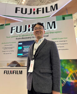
FUJIFILM Cellular Dynamics CEO Tom Hasegawa at his company’s booth at the American Society of Gene and Cell Therapy 2026 in Boston. [K. Davies]
Before long, the company set its sights on the U.S. market, and Hasegawa moved to New York to lead that effort, before returning to Tokyo in 2011 and assuming direction of the global marketing team. Three years later, he moved into business development and was put in charge of corporate planning and cultural affairs.
Hasegawa recalls reading about the induced pluripotent stem cells (iPSCs) by accident. In 2012, the Japanese scientist Shinya Yamanaka, PhD, famously won the Nobel Prize in Physiology or Medicine for the identification of factors crucial in the reprogramming of mature cells into pluripotent stem cells.
Following the acquisition of Cellular Dynamics, Fujifilm “asked me to be in charge” of this fledgling program in regenerative medicine, Hasegawa said. “This is a very well-known story in Japan because [Yamanaka] won the Nobel Prize and it is a very interesting technique. He’s a superstar! That’s why Fujifilm believes that the cell and gene therapy [CGT] field could be our next field of dreams.”
Thompson built a foundation in iPS cells in Cellular Dynamics and “ introduced lots of new [cell types] derived from iPS cells. Cellular Dynamics became a global leader for iPS-related products,” Hasegawa said. “Fujifilm’s concept was very interesting—we wanted to explore the synergy of imaging, analysis, and digital technologies to support the pharma industry. That’s what happened in 2015.”
Hasegawa says he has “a big dream” for the company’s growth. “One way is to deliver our iPS cell products, which include cardiomyocyte cells and neuron cells. We have 40 types of cells for toxicity and efficacy testing for pharma companies to do their R&D. We are a leading supplier of these iPS cells, which have high quality and also the same donor cell for multiple neurons to enable isogenic research.”
At one time, Hasegawa concedes, “we wanted to be a king of pharma, but we switched our strategy.” The company invested heavily in its services to support drug discovery through commercialization, including service lines across contract development and manufacturing (CDMO) and small-molecule drug development. In 2017, Fujifilm acquired reagent company Wako Pure Chemical, which has an HQ in Japan and a subsidiary in Richmond, VA.
Another strand to Fujifilm’s bow is as a provider of materials including reagent kits. “As you remember [in cinema], film is not necessarily the hero—cameras and cameramen are the heroes,” Hasegawa said. “We support [R&D], so that kind of mindset is very beneficial.”
Another thriving business is cell therapy utilizing iPSC technology. Hasegawa points to another subsidiary that provides cell therapy CDMO services in California. “We have an end-to-end service for cell therapy,” he said. In the cell therapy space, he is not looking for additional acquisitions, “because we shifted our strategy.” But for the supporting industry, “if there are good opportunities, we can of course have a discussion about it.”
Fujifilm says its $200-million investment in the new FCDI facility will “help secure America’s supply chain for biotech and regenerative medicine.” The new facility will feature state-of-the-art resources, including a Center of Excellence for genome editing and laboratories for cell culture manufacturing and process development. The space will quadruple capacity for R&D and manufacturing and enhance the company’s capabilities in drug discovery support and process development.
The company believes the investment will help Fujifilm keep pace with the rapidly expanding cell therapy market—and put a smile on the faces of patients and company executives alike.
Researchers from Rutgers University have shown that even limited exposure to modern medical care is associated with rapid changes in gut microbial diversity among remote Indigenous communities in the Amazon. The research, published in Cell Reports, studied the effects on Indigenous people in Venezuela where subsistence lifestyles have remained largely unchanged until the introduction of a World Health Organization–supported medical program for the treatment of the parasitic infectious disease onchocerciasis, also known as river blindness. After receiving medical care, the researchers discovered that gut microbial communities in the people began to shift toward patterns more commonly seen in industrialized populations after only a few medical visits and included measurable declines in microbial diversity.
“The study gives us a better idea of how sensitive human gut microbes are,” said senior author Maria G. Dominguez-Bello, PhD, a professor of microbiome and health at Rutgers. “It opens the door for future research on how we can restore our microbiota after using medicines like antibiotics, which can deplete organisms in our gut.”
Prior research has established that urbanization-related changes in diet, lifestyle, and environment can alter the gut microbiome, but those factors are often intertwined. In this research, the unique setting and population allowed the Rutgers team to isolate the effects of repeated medical exposure in populations with minimal prior contact with modern healthcare.
“Many factors contribute to reduced microbial diversity associated with Westernization, complicating efforts to identify early drivers of microbiome change,” the researchers noted, adding that the Amazonian villages provided a setting where “low-exposure villages show higher baseline gut microbiota diversity than the medium-exposure village, and microbiota diversity declines over time in association with repeated exposure, particularly in children.”
The study followed 335 participants across multiple villages, collecting fecal samples and body-site swabs before and during repeated medical visits between October 2015 and February 2016. The villages were categorized by prior exposure to outsiders and medicine, allowing comparisons between low-exposure communities and a medium-exposure village that had a longer history of modern medical services. Researchers collected samples from the gut, mouth, nose, and skin in conjunction with the WHO’s quarterly visits that delivered antiparasitic treatments and basic care.
“This study leverages a rare longitudinal dataset from remote Amazonian Indigenous communities to examine how the human microbiome shifts during the earliest stages of contact with external institutions, prior to major dietary or lifestyle urbanization,” the researchers wrote.
In the gut microbiome, data from the collected samples showed a decline in taxa commonly associated with fiber metabolism and an increased abundance of bacterial groups more commonly seen in industrialized populations. Functional gene profiles also shifted, with increased representation of genes linked to simple carbohydrate metabolism and antimicrobial resistance, and reduced representation of genes involved in fiber fermentation and other metabolic processes.
The study also found that microbial changes were most pronounced in children, pointing toward a heightened sensitivity of developing microbiomes to repeated medical exposure. In addition to gut changes, oral, nasal, and skin microbiomes also shifted: oral microbial diversity declined; nasal diversity increased after initial visits; and skin communities showed reductions in diversity and shifts in composition.
Previous research cited by the authors has linked industrialized lifestyles with reduced microbial diversity, altered microbial networks, and increased prevalence of genes associated with antimicrobial resistance. However, the current study provides a unique glimpse at the effects of the earliest stages of treatment by modern medicine before shifts in diet or infrastructure occur.
This new data highlights the need to find the balance between essential medical interventions and preservation of microbial diversity. While treatments such as those for river blindness remain critical for reducing infectious disease burden, the researchers suggest that repeated exposure to medical care may coincide with microbial restructuring that reduces diversity and alters functional capacity.
“Understanding and navigating this balance is essential not only for microbiome science but also for ethical, culturally informed approaches that respect both biological and social dimensions of wellbeing,” the researchers noted.
Future research will examine ways to protect microbial diversity during necessary medical interventions and expanding longitudinal studies that assess resilience and recovery of the microbiome after repeated exposure to medicines.
Background: Parkinson disease (PD) is a progressive neurodegenerative disorder that poses complex challenges for persons with PD, informal caregivers, and health care professionals. With growing interest in digital and predictive artificial intelligence (AI) tools for disease management, understanding the needs and digital readiness of these stakeholder groups is crucial. Objective: This work aims to (1) identify digital practices for PD management among persons with PD, at-risk individuals, caregivers, and health care professionals; (2) compare these practices across groups; (3) explore stakeholder desires for AI-based tools; and (4) assess alignments and gaps to inform tailored AI solutions. Methods: An anonymous cross-sectional online survey of an exploratory nature was distributed (from December 2024 to October 2025) in 5 languages and completed by 255 respondents. Descriptive statistics summarized responses to 41 questions, including stakeholder-specific items. tests were performed to examine stakeholder differences in desired AI features. Results: Interest in predictive AI was high across stakeholder groups. Symptom tracking was the most desired feature (selected by more than 76% of the respondents), and personalized treatment recommendations came second for both persons with PD and health care professionals; however, stakeholder priorities diverged in other areas. Health care professionals rated improving patient and informal caregiver engagement as significantly more important than persons with PD did, (n=205)=34.78, <.001, and Cramer V=0.41. Despite considerable interest, the reported use of digital tools was limited, as most persons with PD did not use symptom-tracking apps or wearables, nor were they currently monitoring their condition, although many expressed intentions to begin. Conclusions: While predictive AI tools were viewed positively across groups, there were significant gaps in stakeholder preferences, highlighting the importance of tailored, context-aware design. Early diagnosis was not prioritized by persons with PD or health care professionals, likely reflecting the complexity of diagnosing PD in the absence of disease-modifying therapies. Coupled with the emphasis placed on preventive lifestyle guidance by persons with PD and those at risk, this highlights the importance of actionability in AI-based monitoring and prediction. Such actionability may also enhance perceived relevance and uptake, given that reported interest in digital health tools and self-tracking exceeded actual use. These findings offer early-stage insight to guide the development of future AI-based solutions for PD.
RNA splicing, in which different coding RNA, or exons, are joined together after noncoding regions, or introns, are removed, allows for a large array of RNA transcript isoforms with distinct sequences, and functions in tissue- and cell-type-specific patterns. Conversely, transcript isoform alterations can sensitively reflect dynamic changes in cellular states. Aberrant splicing is closely associated with major diseases, such as cancer.
In a new study published in Nature Computational Science titled, “HELIX: a scalable model for predicting context-dependent regulation of RNA splicing and isoform usage,” researchers from the Chinese Academy of Sciences have developed an AI-driven framework that enables highly accurate prediction of RNA splicing and isoform usage by integrating genomic sequence features with tissue-specific RNA binding protein (RBP) expression profiles. The work offers valuable insights for splicing regulatory patterns, pathogenic variant interpretation, and precision medicine research.
Isoform usage is jointly regulated by multiple layers of control, including regulatory elements, such as splicing enhancers and silencers on exons and introns, and tissue microenvironments. Scientists have been challenged to accurately characterize and predict RNA splicing and isoform usage across tissues, cell types, and disease states.
The study’s AI framework, Hierarchical Explainable LSTM for Isoform eXpression (HELIX), overcomes the limitations of conventional approaches via a two-layer deep-learning architecture.
First, the framework integrates DNA sequence information with the expression profiles of 1,499 RBPs. Long short-term memory (LSTM) networks are then employed to effectively capture the complex dependencies and competitive relationships among multiple splice sites.
This design enables precise, reliable prediction of RNA splicing and transcript isoform usage. The model was trained and optimized on large-scale short- and long-read RNA-seq datasets covering 30 distinct human tissues, allowing accurate quantification of complex transcript structures and isoform usage. Results show that HELIX substantially outperforms existing mainstream methods in both splicing strength prediction and overall isoform usage prediction.
In disease-related studies, HELIX deciphered aberrant RNA splicing and transcript isoform alterations. Notably, the researchers identified widespread splicing dysregulation and abnormal isoform usage in tumor cells using large colorectal cancer cohorts.
The results reveal strong correlations among such alterations and genomic mutations, RBP dysregulation, and patient clinical profiles. Results support that splicing abnormalities can serve as key molecular signatures for tumor progression and guiding patient stratification.
The team also developed scHELIX, a single-cell RNA sequencing extension of HELIX. scHELIX supports high-resolution profiling of transcript isoform usage across different cell types and tumor subpopulations, which offer a refined view of intratumoral heterogeneity.
The findings reveal distinct RNA splicing and isoform usage patterns among tumor subclones, providing new clues for tumor evolution research and potential therapeutic target discovery.
Background: In the context of COVID-19, infection spread through human contact networks remains a major public health challenge. Beyond cumulative infections and deaths, it is necessary to understand which contacts matter most, and which population segments contribute most to transmission under different social conditions. In multilayer urban networks with community structure, routine contacts coexist with incidental encounters, and it remains unclear whether incidental encounters can alter epidemic burden and the main contributors to transmission when per-layer contact caps and routine-contact minima are unchanged (for the nonrandom layers). Objective: Under explicit daily-contact constraints, we examined (1) how changing overall contact opportunities affects epidemic speed and burden when incidental encounters are held fixed, and (2) whether increasing incidental encounters alone, per-layer contact caps, and routine-contact minima fixed (for the nonrandom layers), shifts the main contributors to transmission from a high-contact group to a medium-contact group, and the underlying network mechanism. Methods: We constructed a multilayer potential contact network for a synthetic urban population of 10,038 individuals, representing household, school, workplace, distance-driven activities, and incidental encounters as separate layers. Daily contact networks were sampled from the potential network each day, and transmission was simulated for 120 days using a Susceptible-Exposed-Infectious-Removed model with vaccination. Individuals were classified into high-contact and medium-contact groups based on baseline contact intensity, and group contribution combined each group’s share of infectious individuals and its per-infectious effective transmission yield. Contact-constraint parameters were calibrated using an online survey in Tokyo and Kanagawa (n=1089), and scenario comparisons and parameter sweeps were used to locate the transition point. Results: With incidental encounters held fixed, higher overall contact opportunities produced earlier and higher epidemic peaks and larger cumulative infections and deaths, whereas reduced opportunities slowed and prolonged spread. Holding overall contact opportunities and routine contacts fixed, increasing incidental encounters shifted the main contributors to transmission: higher-contact individuals accounted for more effective transmissions at low incidental contact, whereas medium-contact individuals accounted for more beyond a clear transition point. Network visualization and schematics suggest a bridge-allocation mechanism, where stronger incidental contact adds cross-community bridges that more often terminate at medium-contact individuals and carry infection into less-affected communities. Across R=30 replicate runs under fixed settings, the dominance flip was consistently observed, and the estimated threshold W∗ showed a narrow but nonzero distribution (reported as median and IQR). Conclusions: In multilayer urban contact networks with community structure, our results indicate that intensifying incidental encounters can change the main contributors to transmission even when overall contact opportunities and routine contacts are unchanged. We present an analysis framework under explicit daily-contact constraints to identify this contributor shift and its transition point, supporting comparisons of intervention priorities across social contact conditions.
<img src="https://jmir-production.s3.us-east-2.amazonaws.com/thumbs/1fbd573eb4e3a2afda02c2afd8220499" />
SAN FRANCISCO — While Tippi MacKenzie was a postdoctoral fellow in the early 2000s, she and her lab mates experimented with using the then-new technology of gene replacement therapy to try to treat inherited disorders in mice before they were born. Over and over it worked. They cured mice with hemophilia and mice with tyrosinemia. And the whole time, people kept telling her that gene therapy in human fetuses was just around the corner, just five years away.
It’s now been 25 years, and such a reality has yet to materialize. But after promising discussions with the Food and Drug Administration, MacKenzie is now closer than anyone’s ever been.
Her team has submitted an investigational new drug (IND) application to the agency seeking approval for a small trial that aims to treat five fetal patients with a rare lysosomal storage disorder. The agency told them they could bypass animal testing, because the safety profile of the vector they plan to use already has been so well characterized by other academics and companies developing gene therapies for kids and adults.
AI is increasingly being used to help determine breast cancer risk, with two studies showing its value for primary prevention and in women of diverse ancestry.
The first study, in Science Translational Medicine, revealed that AI could outperform standard clinical risk tools at identifying women of European ancestry who were at greatest risk of developing breast cancer within the next decade.
Because current AI-based models mostly predict breast cancer risk in the short term, Mikael Eriksson, PhD, from the Karolinska Institute, and fellow researchers created a model for invasive and in situ breast cancer that covered a 10-year period.
“Considering that a tumor can take five to 20 years to develop into a screen- or clinically detected cancer, a 10-year or lifetime risk projection time is reasonable,” they noted.
The researchers validated their image-derived AI-based 10-year risk model using digital mammograms from 8676 women in two cohorts from the U.S. and Sweden, of whom 1633 had breast cancer.
They compared their model with three other risk assessment tools: the Tyrer-Cuzick-v8, which uses personal and family history to determine the lifetime risk of breast cancer; the Breast Cancer Surveillance Consortium v3 tool, which estimates the risk of developing invasive risk cancer over five years; and the Mirai AI algorithm for predicting breast cancer risk.
The model calculated that the 10-year risk of breast cancer was 3.83% for the North-American group and 3.14% for the Scandinavian group, and it showed promise for predicting the risk of invasive tumors.
The AI model performed significantly better at predicting invasive breast cancer than the three comparator models both overall, in women aged 50 to 69 years and those with estrogen-receptor-positive breast cancers.
“An image-derived AI-based risk model developed for 10-year risk assessment for identifying individuals in mammographic screening who may benefit from primary prevention strategies identifies up to 40% of breast cancers to be at high risk at study entry in U.S. and Swedish validation case cohorts per clinical guidelines,” summarized Eriksson and co-workers.
The second study, in Science Advances, revealed how AI could be of value for women from more diverse backgrounds, and that it had value regardless of race and ethnicity.
The research was driven by the observation that conventional prediction models incorporating genetic and clinical factors including breast density underperform in women who do not have European ancestry.
Shu Jiang, PhD, from Washington University School of Medicine in St Louis, and co-workers set out to evaluate the generalizability of an AI-derived mammogram risk score (MRS), a summary of texture features which captures intrinsic breast-tissue characteristics that are the basis for cancer to develop.
To do this, they used data from two large North American cohorts that represented more than 226,000 racially diverse women undergoing routine breast screening with mammography.
This was then used to examine the generalizability of MRS with breast cancer risk across non-Hispanic white (NHW), non-Hispanic Black (NHB), East Asian, South Asian, and Indigenous women.
Jiang and team found that the MRS was a strong predictor of breast cancer risk independent of race or ethnicity, demonstrating its potential for broader clinical utility.
Across the two independent screening cohorts, the MRS increased with age in line with rising breast cancer risk. It was a strong and consistent predictor of breast cancer risk across race and ethnic groups as shown by the association study and comparison of distributions and demonstrated excellent calibration in all groups.
Its remained robust across full-field digital mammograms or synthetic two-dimensional digital breast tomosynthesis, although the limited numbers of images precluded an analysis of how performance varied across manufacturers.
“Overall, these results support that MRS is a powerful breast cancer risk predictor that does not depend on race or ethnicity, supporting its potential for broader clinical adoption and use in varied populations of women undergoing routine screening mammography to identify those at increased risk,” Jiang and coworkers concluded.
Scientists have developed a new biomarker test that can noninvasively detect kidney fibrosis from a urine sample. A study published today in Science Translational Medicine reports the test could accurately differentiate between mild and severe disease, something that is not yet possible with the diagnostic methods available today.
“Noninvasive and accurate diagnosis of kidney fibrosis remains unavailable in clinics, limiting effective patient management,” write the authors of the study, led by Zhou Jiaguo, PhD, professor of pharmacology at Sun Yat-sen University in China. “Although biopsy is considered the gold standard for assessing fibrotic progression, this invasive approach is subject to sampling error and interobserver variability, which may bias the diagnosis and is improper for repeated monitoring.”
Chronic kidney disease affects approximately 12% of the global population, with rates steadily rising as the world’s population ages. Characterized by the excessive formation of scar tissue, kidney fibrosis is a hallmark of chronic kidney disease that causes the proggressive impairment of kidney function, eventually leading to end-stage renal disease that requires dialysis or transplantation.
Diagnosing kidney fibrosis at early stages can be critical for preventing irreversible damage to the kidneys. However, current diagnostic methods present some major limitations. While biopsies are invasive and difficult to accurately interpret, blood biomarker tests cannot distinguish kidney fibrosis from other kidney conditions or from fibrosis in other organs.
To circumvent these limitations, Jiaguo’s team identified two biomarkers that are specific to kidney fibrosis and can accurately differentiate this diagnosis from other related conditions. These are the transglutaminase 2 (TG2) enzyme and its substrate, lysyl oxidase–derived allysine (LysAld), which are both upregulated during the formation of fibrotic kidney tissue.
The researchers developed a fluorescent reporter that binds to LysAld in the kidney and is then cleaved by TG2, turning on its fluorescence. Higher biomarker levels result in a stronger signal, providing an indication of how advanced fibrosis is in the patient’s kidneys.
In a small cohort of 35 participants, the molecular imaging test could distinguish patients with kidney fibrosis from healthy controls with 84% sensitivity and 94% specificity. In addition, the test could differentiate mild and severe cases of fibrosis, with results confirmed with histological imaging. In contrast, traditional clinical measurements such as glomerular filtration rate, serum creatinine, and blood urea nitrogen were not able to classify patients according to the severity of the condition.
Large-scale studies will be needed to establish this novel approach as a diagnostic and prognostic. Going forward, Jiaguo and colleagues are also interested in exploring using this test to monitor disease progression over time as a potential tool to evaluate treatment response and help clinicians make critical decisions to improve patient outcomes.
“This noninvasive activatable reporter holds translational potential for early identification of renal fibrosis and patient stratification of [chronic kidney disease] to improve clinical management and patient outcome,” write the researchers. “We do not envision the probe as a standalone replacement for current standards; synergistic combinations with other biomarkers could further enhance its clinical utility.”