In a new study published in Science titled, “Mitochondrial metabolism and signaling direct dendritic cell function in antitumor immunity,” researchers fromSt. Jude Children’s Research Hospital have uncovered a new metabolic mechanism for how tumors disable immune “gatekeeper” cells that initiate response in the presence of cancer. The results offer a new path to improve immunotherapy.
Dendritic cells alert and activate tumor-killing immune cells as a critical part of anticancer immune response. The authors found that tumors reduce dendritic cell function by minimizing mitochondrial fitness to prevent anticancer immune response. Correspondingly, boosting mitochondrial function in dendritic cells enhances antitumor immune activity and strengthens the efficacy of existing immunotherapies.
Within the nutrient-sparse tumor microenvironment, dendritic cells progressively lose mitochondrial activity, which drives cell dysfunction and weakens immune defenses against cancer. When dendritic cells with high mitochondrial activity were introduced into tumors in preclinical mouse models, results showed that immunogenic activity was restored while improving tumor control.
“We found that tumors reprogram mitochondrial metabolism in dendritic cells, reducing their ability to activate the immune system against cancer,” said Hongbo Chi, PhD, St. Jude Department of Immunology chair and corresponding author of the study. “By enhancing mitochondrial function, we could restore dendritic cell activity and rescue antitumor immunity.”
Immunotherapies for cancer, such as immune checkpoint blockade, have greatly improved care for many malignancies, but have not been successful in all cancers. To determine whether these findings could improve immunotherapy effectiveness in tumor-bearing mice, the authors evaluated the administered dendritic cells with high mitochondrial activity in combination with immune checkpoint blockade.
“We saw the most pronounced therapeutic effect in mice treated with the combination of dendritic cells that had high mitochondrial activity and immune checkpoint blockade,” said co-first author Zhiyuan You, PhD, researcher at St. Jude Department of Immunology. “Those combinations synergistically slowed or stopped tumor growth and extended survival far more than either treatment alone.”
To test long-term effects, the researchers exposed combination therapy treated mice to a new tumor after a few months. New tumor growth stopped for these mice, indicating durable, long-term immune memory.
To better understand the relationship between mitochondrial function and dendritic cells, the researchers examined metabolic pathways affected by the tumor microenvironment. They identified a signaling axis composed of two proteins, OPA1 and NRF1, that regulate communication between mitochondria and the nucleus. Expression was greatly downregulated in dendritic cells during tumor progression and acted as a metabolic switch to shut down dendritic cell immunogenic activity.
“We’re seeing a direct regulation of dendritic cells by the tumor microenvironment,” said co-first author Jiyeon Kim, PhD, researcher at St. Jude Department of Immunology. “We have characterized how that results in mitochondrial reprogramming of dendritic cells to benefit cancer, giving us new opportunities to reverse the process.”
The study’s mechanistic insights enable new directions to rewire dendritic cell function and enhance cancer treatments.
“These findings reinforce the central role of dendritic cells in cancer immunity,” Chi said. “By exploring their mitochondrial function in the tumor microenvironment, we have provided a proof-of-principle of how we may be able to improve the next generation of immunotherapies.”
UCLA scientists have developed a simple and cost-effective blood test that, in early studies in more than 1,000 people, showed promise in detecting multiple cancers, various liver conditions, and organ abnormalities simultaneously.
The new method, called MethylScan, works by analyzing cell-free DNA (cfDNA), tiny fragments of genetic material released into the blood when cells die. Because cells from every organ shed DNA into the bloodstream, cfDNA carries molecular signals that reflect what is happening throughout the body.
The researchers say MethylScan could represent a powerful and more affordable approach to early disease detection and comprehensive health monitoring. “Early detection is crucial,” said research lead Jasmine Zhou, PhD, a professor of pathology and laboratory medicine and investigator at the UCLA Health Jonsson Comprehensive Cancer Center. “Survival rates are far higher when cancers are caught before they spread. If you detect cancer at stage one, outcomes are dramatically better than at stage four.” Zhou is senior author of the team’s published paper in PNAS, titled “Toward the simultaneous detection of multiple diseases with a highly cost-effective cell-free DNA methylome test.”
“When cells die, they do not simply vanish; they leave behind molecular traces, including cell-free DNA (cfDNA) in the blood stream,” the authors wrote. “cfDNA is a mixture of DNA fragments released from various organs, offering valuable insights into the health of these organs.” Zhou added, “Every day, 50 to 70 billion cells in our body die. They don’t just disappear, their DNA goes into the bloodstream. That means we already have information from all our organs circulating in the blood.”
The idea of using blood to detect cancer, sometimes called a liquid biopsy, isn’t new. Some tests already look for mutations in tumor DNA to screen for certain cancers. But those tests often focus on a limited number of genetic changes and can be expensive, in part because they require deep sequencing to detect faint tumor signals.
Instead of searching for mutations, the UCLA team examined DNA methylation, chemical tags attached to DNA that help regulate gene activity. Methylation patterns differ by tissue type and can change when cells become cancerous or diseased. “Unlike an individual’s genome, which remains largely stable across tissues and over time (except for rare somatic mutations), the DNA methylome is tissue-specific and dynamically changes with the tissue’s disease status,” the team continued.
“DNA methylation reflects the health status of a tissue,” said co-corresponding author Wenyuan Li, PhD, a professor of pathology and laboratory medicine at UCLA and co-corresponding author of the study. “It’s a very informative signal.”
The challenge is that most cell-free DNA in the bloodstream doesn’t come from tumors or injured organs. About 80% to 90% originates from normal blood cells. That creates background noise, making it difficult and costly to detect the relatively rare fragments that might signal early cancer. “A major challenge in using the cfDNA methylome for disease detection is the high cost of sequencing,” the team stated. “In healthy individuals, about 85% of cfDNA originates from blood cells, creating substantial background noise that can obscure cfDNA from tumors or diseased organs.” And as the authors further pointed out, “Current cfDNA methylation assays primarily focus on single clinical indications by targeting specific genomic loci.”
For their newly reported research the team built on past work to develop a technique to remove much of the background DNA before sequencing. Using specialized enzymes, they selectively cut away unmethylated DNA fragments that largely come from blood cells. By designing a genome-wide hybridization panel, the captured DNA fragments are enriched for methylated DNA from solid organs, including those that are potentially diseased. “The MethylScan method is a targeted methylation assay that combines Methylation-Sensitive Restriction Enzymes (MSRE) digestion with a custom panel to enrich hypermethylated cfDNA from tissues beyond blood, enabling cost-effective detection of multiple diseases,” they wrote.
By removing the noise, the researchers say they can dramatically reduce the amount of sequencing needed, lowering costs while maintaining sensitivity. Achieving an effective sequencing depth of 300× per sample requires only 5 Gb of data, which would cost less than $20 if the price per gigabase is under $4.
To test the accuracy of MethylScan, the researchers analyzed blood samples from 1,061 people, including patients with liver, lung, ovarian and stomach cancers; individuals with liver diseases such as hepatitis B, hepatitis C, alcohol-related liver disease, and metabolic-associated liver disease; people with benign lung nodules; and healthy participants. Machine learning algorithms were then applied to analyze the complex methylation data.
For multi-cancer detection, the test achieved a high level of overall accuracy. At a specificity of 98%, meaning few false positives, it detected about 63% of cancers across all stages and roughly 55% of early-stage cancers. The test also performed well in liver cancer surveillance among high-risk individuals, including those with liver cirrhosis or HBV, detecting nearly 80% of cases at a specificity of just over 90%, meaning a less than 10% false positive rate.
Beyond simply detecting cancer, the methylation patterns helped identify where in the body a signal was coming from, known as the tissue of origin. “Being able to trace signals back to their source is important because a positive blood test needs to be followed by imaging or other diagnostic procedures directed at the right organ,” said Li.
MethylScan can work like a health radar for the body. By reading DNA signals in the blood, it can tell when specific organs, such as the liver or lungs, are under stress or damaged, even without knowing the disease in advance. The researchers also showed that the blood test could distinguish between different types of liver disease, including viral hepatitis and metabolic-associated liver disease. It correctly classified about 85% of patients, suggesting blood-based DNA testing could reduce the need for invasive liver biopsies.
Although larger prospective trials will be needed to confirm its performance in real-world screening, Zhou said the work represents an important step toward a single, affordable blood assay that can detect a broad spectrum of diseases earlier and more comprehensively than current methods allow.
“Because cfDNA in blood originates from multiple organs, and MethylScan captures a broad spectrum of robust hypermethylation markers, this assay has the potential to detect a variety of diseases, provided that appropriate training cohorts are available,” the investigators stated. “This versatile approach enables affordable, wide-ranging cfDNA tests that can identify various health conditions simultaneously, with the potential to transform early disease detection and health monitoring across diverse clinical settings.
Zhou added, “This study demonstrates that blood-based methylation profiling can deliver clinically meaningful information across multiple diseases. It’s an exciting advancement that brings us closer to realizing the dream of a single assay for universal disease detection.”
As a postdoctoral researcher at the University of Pennsylvania, Haijiao Liu, PhD, helped advance tumor-on-a-chip technology, a feat of bioengineering that mimics the microenvironment of malignant human tumors. Led by Dan Dongeun Huh, PhD, a Penn Engineering professor and trailblazer of organ-on-a-chip technology, Liu and his team explanted lung adenocarcinoma tumors onto the transparent chips to test their perfusion with chimeric antigen receptor (CAR) T cells. Their findings were published in October in Nature Biotechnology, with Liu as first author.
Now on paternity leave in Toronto, Liu spoke with Lindsey Leake about the implications of this work, the challenges inherent to tumor-on-a-chip studies, and his plans to launch a lab of his own this fall.
Q: Walk me through the creation of the tumor-on-a-chip. What went into its design?
Haijiao Liu: It’s essentially inspired by the need for alternative tumor models. This is speaking to the traditionally used animal tumor models and some existing in vitro tumor models, especially for the study of immunotherapies.
For example, when I started at Penn around 2018, Penn Medicine was pioneering this immunotherapy called CAR T-cell therapy, which is basically aiming to harness the patient’s own immune system, specifically the patient’s own T cells, to help fight the cancer. Penn Medicine was demonstrating huge clinical success using this CAR T therapy to treat blood cancers, such as leukemias and lymphomas. In a huge contrast to this, the solid cancer arena has seen a limited response from this new immunotherapy. So there’s this great need to study why this has not been successful, and that comes down to the consensus that the solid tumor has this really complex microenvironment.
In the category called tumor-on-a-chip, people try to control the cultural environment, the biochemical and biophysical environment of tumor cell cultures. We can use this for simple drug testing—see how the tumor growth will be affected or how effectively they can be killed. However, these existing tumor-on-chip or in vitro tumor models are still very simple. They don’t usually recreate or reproduce the complex structure of the human solid tumors that I described, like where they often include complex vessel networks.
I took the lead to address the need and the challenges of reproducing and then investigating, or probing, the dynamic interactions between those CAR T cells and human solid tumors entirely in vitro.
Q: How does the vascularization work on the chip?
Liu: It took several years to start, from the idea of building this more advanced tumor-on-a-chip technology toward proving it’s actually useful. I started by focusing on this one aspect, which is the CAR T-cell trafficking and their functions after they traffic to fight the tumors, and that will involve the recreation of the structural interface between the tumor and this complex vascular network that’s present in human tumors.
I was inspired by in vivo tumor transplantation, where traditionally, people take human tumors and then transplant them in a bulk, intact format into animal models. So my idea was, if we want to focus more on the human biology, if we want to engineer this entirely in vitro, how about we design a vascular bedding, like a miniature living model?
We basically took advantage of the self-assembly capability of human-sourced endothelial cells, combined with certain stromal fibroblasts, or stromal cells. With a bit of optimization, engineering, tweaking, then we can allow them to form capillary-like vascular networks in our engineered models.
Q: What are the advantages of recreating the tumor microenvironment in this way? That is, is the Petri dish becoming obsolete in cancer research?
Liu: The unique advantage of this way of engineering is to have a higher level of control over the structures of the tissue-tissue interface that we can build. For example, we can engineer different culture chambers. We can engineer different access windows with this model. That allows us to construct, step by step, the vascular bedding and then the tumor transplantation. Also, by forming these perfusable vessels—by the way, we can provide the infusion and flow of the CAR T cells, just like they are infused and flow in the patient—that gives us the leverage to reconstruct, probe, and then control these tissue functions in a highly precise manner.
Q: How did you and your colleagues at Penn explore CAR T-cell activity on the chip?
Liu: We first used different functional assays, like immunostaining and ELISA (enzyme-linked immunosorbent assay) assays, to characterize how the CAR T cells are doing and how they are interacting with the tumors in our engineered model. Then we disassembled this engineered tissue to extract all the cells for flow cytometry, to further characterize their functional phenotypes.
With the help of our collaborators and other people in the lab, I took advantage of this engineered model of vascular tumors interacting with CAR T cells for multi-omics analysis. For example, I was able to extract all the cells and send them for single-cell RNA seq[uencing]. We were able to look at the gene expressions of each individual cell from all the cell types that we included in this model. In this way, we have almost like a superpower to probe and read into how each cell—including the CAR Ts and tumors and the vessels—how they are responding and interacting with each other at the molecular gene levels. This is so powerful that it helped me to discover novel interactions between these parties and also new druggable targets.
The message is that through the development of this more advanced tumor-on-a-chip technology—combined with advanced multi-omics analytics and advanced computational analysis—we were able to provide this powerful in vitro technology to apply to accelerate the development of cell therapies, such as the CAR T immunotherapies for cancer, but also other complex diseases.
Q: What are the overall implications of this latest research?
Liu: With a growing understanding of human biology at the cellular and tissue levels, I think we’re seeing that our ability to engineer and design biological systems is also growing. More than ever, we have these advances in the ability to precisely construct, investigate, and then eventually control very complex tissue functions, and even organ functions. For example, our demonstrated tumor-on-a-chip technology is like presenting a miniature sandbox; we can literally see and predict the battlefield of CAR T therapy in cancer.
If we combine these advanced engineering technologies with emerging technologies in spatial multi-omics and the unprecedented productivity of the AI revolution, we will be able to accelerate the understanding of more complex human biology and extract more biological insights, and then apply that to accelerate the development of safer and more efficacious drugs and therapies, such as immunotherapies in cancer.
Q: What are the limitations of organ-on-chip technology that need to be overcome?
Liu: I think there are challenges on two fronts. The first limitation is the lack of complexity. We’re claiming that what we just published is a sufficiently complex system for us to deeply probe and understand the dynamics of CAR T tumor interactions. Still, if we’re speaking next level of translational power or potential, then we need to pursue a higher complexity that incorporates the missing but critical components from in vivo.
The other side of the coin is that if you make this engineered model more complex, you make it more challenging to reproduce or to scale up or to translate to other labs. But also, that points to an opportunity and growing room for translation, to standardize every single step, from the construction to the analysis of these engineered models, and to automate these processes as much as possible.
Q: What do you envision for your new lab?
Liu: I have a lot of things I want to do. I’m eager to establish my own team. The overarching and the unifying theme of the new lab will be to develop the next generation of in vitro complex tissue models, or I call it assembloid tissue models. Assembloid basically means there’s a stem cell-based, three-dimensional complex tissue model that intentionally incorporates different cell types, to emulate the critical tissue-tissue interactions that determine the tissue- and organ-level functions. I still need to make a big decision where the lab could be; it could be in Canada and it could also be in China.
Q: What impact might tumors-on-a-chip have on the future of precision medicine?
Liu: It’s attracting a lot of attention from biologists and clinicians who are heavily focused on using the traditional tissue models—animal models, for example, or the simple dish cultures—for their studies of interest. So the biggest impact I can foresee with our technology is that now it’s more mature. I can see it being gradually, and maybe quickly, adapted into more traditional biological labs, to help them dissect the complex biological questions they’re asking, or to accelerate the evaluation of the exciting new drugs or therapies they’re developing. Overall, I can see that accelerate this development pipeline of new drugs and therapies in precision medicine.
Lindsey Leake is an award-winning, independent health reporter based outside Washington, D.C. She spent 15 years as a staff journalist at outlets including Fortune, the USA TODAY Network and Sinclair Broadcast Group. She holds an MA in Science Writing from Johns Hopkins University, an MA in Journalism and Digital Storytelling from American University and a BA from Princeton University.
Researchers at Baylor College of Medicine have identified a mechanism driving progressive heart disease in people with myotonic dystrophy type 1 (DM1) that operates independently of genetic repeat expansion, providing new evidence of how cardiac damage develops and when it may be reversible. The study, published in the Journal of Clinical InvestigationInsight, shows that sustained expression of expanded CUG (CUGexp) repeat RNA drives cumulative cardiac injury through structural remodeling, including myocardial fibrosis, chamber dilation, and impaired contractility, while also disrupting electrical conduction pathways that regulate heart rhythm.
“Cardiac manifestations affect most DM1 patients,” said corresponding author Thomas A. Cooper, MD. “Cardiac problems are primarily electric conduction abnormalities, seen in up to 75% of adult DM1 cases, which can result in life-threatening arrhythmias accounting for 25% mortality and the second leading cause of death in DM1.”
DM1 is an autosomal dominant disorder and the most common cause of adult-onset muscular dystrophy. It is caused by an expanded CTG repeat in the DMPK gene, with affected individuals carrying between 50 and more than 4,000 repeats, compared to five to 37 in unaffected individuals. The expanded CTG repeat causes mutant transcripts that form expanded CUG (CUGexp) RNA foci and sequester muscleblind-like (MBNL) RNA-binding proteins. The result is a loss of function of MBNL.
DM1 affects multiple systems, including skeletal muscle, the central nervous system, the gastrointestinal tract, and the heart. Cardiac impairment is common in people with the disease and is often fatal. Electrical conduction abnormalities, including prolonged PR, QRS and QTc intervals, atrioventricular block and bundle branch block, occur in up to 75% of adults with DM1. Patients may also develop atrial fibrillation, tachycardia and, less commonly, ventricular arrhythmias. Structural changes such as left ventricular dysfunction, hypertrophy, dilation and fibrosis contribute to morbidity and mortality. These conditions typically worsen with age and are more severe in males.
The Baylor team used a transgenic mouse model engineered to express toxic CUG repeat RNA in the heart. Unlike human disease, the number of CTG repeats in this model remained stable over time, allowing researchers to isolate the effects of prolonged RNA toxicity without the confounding factor of repeat expansion. The researchers measured the cardiac function of the mice over a period of 14 months.
According to the researchers, the data showed that “sustained CUGexp RNA expression caused progressive cardiac enlargement, contractile dysfunction, conduction delay, myocardial fibrosis, and reduced survival, while MBNL-dependent splicing defects remained static, consistent with the stable repeat length.” These findings indicate that cardiac deterioration can occur even without increasing loss of MBNL function. This finding runs contrary to current thinking, which centers on the role of repeat expansion as the primary driver of disease progression.
In this research, the model mice developed enlarged hearts and electrical abnormalities early on. Over time, these changes progressed to cause weakened cardiac function, fibrosis and dilation of heart chambers.
The study also sought to find whether the cardiac damage could be reversed by halting expression of the toxic RNA. When RNA expression was stopped after a short duration, heart size, electrical function and structure largely returned to normal.
Yet when exposure to the toxic RNA was prolonged, the mice did not exhibit complete recovery. While the molecular defects such as abnormal RNA splicing were fully corrected, structural damage, including fibrosis and conduction abnormalities, persisted. This shows that early intervention could be one method to prevent irreversible changes such as fibrosis, which disrupts electrical signaling and increases the risk of arrhythmias.
Prior research had linked increasing CTG repeat length with DM1 severity and earlier onset, but the extent to which other mechanisms contribute to progression remained unclear. By isolating RNA toxicity, the Baylor researchers have provided evidence that chronic cellular stress, structural remodeling and potentially other RNA-mediated effects play roles in disease pathology independent of repeat expansion.
Future research will look to identify any additional mechanisms that may contribute to disease progression, and will include studying the long-term effects of RNA toxicity, changes in RNA-binding proteins, and potential links to premature cellular aging.
Studying sexually transmitted infections (STIs) has long been constrained by a fundamental problem: the available models fail to fully capture the complexity of the human body. Traditional cell cultures oversimplify biology, while animal models often do not accurately reflect human infection dynamics.
Now, scientists at the University of Maryland School of Medicine and collaborators have developed the first immune-capable “cervix-on-a-chip,” a microengineered system that recreates the human cervical environment with unprecedented realism. The work, published in Science Advances, could significantly accelerate the development of new treatments and prevention strategies for STIs.
A long-standing gap in STI research
STIs remain a major global health burden. According to the World Health Organization, nearly one million new infections occur every day worldwide, with chlamydia alone accounting for roughly 129 million cases annually. In the United States, chlamydia and gonorrhea together generate an estimated $1 billion in direct medical costs each year.
Beyond their prevalence, these infections can lead to serious complications, particularly in women, including infertility, pelvic inflammatory disease, and adverse pregnancy outcomes.
Despite this, researchers have struggled to study how infections develop and progress in the human cervix, a key site of infection, under realistic conditions.
“This new model will revolutionize how scientists study STIs,” said Jacques Ravel, PhD, co-lead author of the study. “By integrating engineering, microbiology, immunology, and microbiome science, we were able to build a model that more closely reflects human biology and the complexity of the cervical microenvironment.”
Recreating the cervix in the lab
The newly developed system belongs to a class of technologies known as organ-on-a-chip models, or microphysiological systems. These platforms are designed to mimic the structure and function of human tissues using living cells and controlled physical environments.
In this case, the researchers constructed a miniature model of the cervix using human cervical epithelial cells layered on a porous membrane, with supportive tissue cells beneath. Fluids flow across both sides of the membrane, replicating the dynamic conditions found in the body.
The model also incorporates immune cells and microbial communities, allowing scientists to study how these components interact during infection.
“A key goal was to develop a complex model system that is both practical and accessible,” said Jason Gleghorn, PhD, who led the model development. “The need for this model was particularly critical for studying the vaginal microbiome, which we know plays an important role in susceptibility to STIs.”
Capturing the role of the microbiome
One of the defining features of the cervix-on-a-chip is its ability to include different types of vaginal microbiomes, something that has been difficult to replicate in previous models.
The researchers tested the system using two of the most common STIs: chlamydia (Chlamydia trachomatis) and gonorrhea (Neisseria gonorrhoeae). They found that the outcome of infection depended strongly on the type of microbiome present.
In models dominated by Lactobacillus crispatus, a bacterial species commonly associated with vaginal health, infections were significantly limited. In contrast, when less protective microbiomes were introduced, infections became more severe.
“One of the most exciting findings was that just like in women, protective microbiomes dominated by Lactobacillus crispatus limited infection in the model,” Ravel said. “In contrast, when we introduced ‘nonoptimal’ microbiomes, infections worsened.”
These results reinforce growing evidence that the vaginal microbiome plays a central role in determining susceptibility to STIs.
Toward better treatments—and prevention
Beyond improving understanding, the new model provides a practical platform for testing potential therapies.
Because it closely mimics human biology, the cervix-on-a-chip can be used to evaluate new treatments under realistic conditions. This includes not only traditional antimicrobial drugs but also emerging approaches such as probiotics and live biotherapeutics designed to restore protective microbiomes.
“This model provides a powerful new tool to develop faster, more effective, and personalized treatments,” Ravel said. “For the first time, we can simulate what happens in the human body rather than relying solely on petri dish systems or inadequate animal models.”
A platform for broader applications
The implications of the technology extend beyond the infections tested in the study. The cervix-on-a-chip could be adapted to study a wide range of pathogens, as well as broader questions about reproductive health, inflammation, and host–microbe interactions.
The researchers emphasized accessibility in the model’s design, aiming to make it usable by scientists outside of specialized bioengineering labs. This could accelerate adoption and expand its impact across the field.
A step toward more human-relevant science
The development of immune-capable organ-on-a-chip systems represents a broader shift in biomedical research toward more human-relevant experimental models. By integrating multiple components of human biology—cells, tissues, immune responses, and microbiomes—these systems offer a more accurate view of disease processes.
In the context of STIs, where subtle interactions between host and microbes can determine outcomes, such realism is particularly valuable.
As researchers continue to refine these platforms, they may help bridge the gap between laboratory studies and real-world biology—ultimately enabling earlier, more precise, and more effective interventions.
For now, the cervix-on-a-chip marks a significant step forward, providing scientists with a tool that captures the complexity of the human cervix in a way that was previously out of reach.
Research led by Osaka International Cancer Institute in Japan shows that rates of therapy-related acute myeloid leukemia, linked to treatment for an earlier cancer, are going up as the overall number of cancer survivors increases.
As reported in the journal Cancer, the researchers showed the rates of treatment-related acute myeloid leukemia (AML) almost tripled between 1990 and 2020. The share of overall AML cases taken up by these patients has also increased from 4.4% to 8.2% over a similar time period.
With increasing numbers of cancer survivors in the U.S. and elsewhere, understanding the adverse effects of cancer treatment is becoming increasingly more important. Therapy‑related AML is a rare but serious complication of cytotoxic chemotherapy, and different chemotherapy drugs carry different levels of leukemia risk. Radiotherapy can also lead to this condition, but the risk is considered lower than that of chemotherapy.
“Several population‐based studies have analyzed the long‐term trend of therapy-related AML incidence, and the results are controversial,” write lead author Kenji Kishimoto, MD, PhD, a researcher at the Osaka International Cancer Institute, and colleagues.
“A national population‐based study did not demonstrate an increase in the incidence of therapy-related AML in Denmark between 2000 and 2013. In contrast, a significant increase in therapy-related AML incidence between 1997 and 2015 was identified in a Swedish nationwide study.”
As few such studies have been carried out in Asia, Kishimoto and colleagues analyzed how such rates have changed over time and whether the initial cancers leading to therapy-related AML have changed in Japan.
The researchers identified 9841 patients with AML in the Osaka Cancer Registry, 636 (6.5%) of whom had therapy-related AML. These patients were older when diagnosed than other AML patients at a median of 69 versus 66 years. There were also slightly more women with therapy-related AML than standard AML at 45% versus 40%. The time lag between first cancer and resultant AML was between two and 11 years (median five years).
In 1990, the incidence of therapy related AML was 0.13 per 100,000 people, but this had increased to 0.36 per 100,000 people by 2020. The share of all AML cases made up by therapy-related cases almost doubled from 4.4% in 1990 to 8.2% after 2010.
The most common earlier cancers before therapy-related AML were blood cancers (23%), breast cancer (15%), colorectal cancer (12%), and gastric cancer (9%). The mix shifted over time and gastric cancer became less common as the first cancer, while breast, head‑and‑neck, and lung cancers became more prominent.
“The study provides an important step towards better understanding how the nature of therapy-related AML is changing with the increasing number of cancer survivors,” said Kishimoto in a press release.
“Findings from this study lay the foundation for further studies to elucidate the mechanism of the change in therapy-related AML epidemiology,” add the authors.
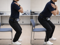
Background: Increasing life expectancy has increased focus on the health-related consequences of aging, such as sarcopenia and frailty. Given the prevalence of these conditions among older individuals and the frequent resulting long-term care needs, early detection and intervention are crucial. Objective: This study aimed to validate a novel smartphone-based system measuring acceleration during the sit-to-stand movement to detect sarcopenia and frailty. Methods: Participants were 587 individuals from the Otassha study cohort who underwent health assessments in 2023, of whom 569 (96.9%) completed 2 supervised sit-to-stand trials while holding a smartphone on the lower abdomen. Sarcopenia and frailty were diagnosed using the Asian Working Group for Sarcopenia 2019 criteria and the revised Japanese Cardiovascular Health Study criteria, respectively. Peak force, rising time (T1), and stabilization time (T2) were extracted from acceleration signals, and reproducibility was examined using the intraclass correlation coefficient (ICC(2,1)). Predictive models were developed using elastic net penalized logistic regression, and model performance was evaluated using 500 bootstrap resamples. Benchmark models using age and sex, walking speed, and grip strength were also constructed for comparison. Results: Sarcopenia and frailty were identified in 16.7% (95/569) and 9% (51/569) of the participants, respectively. Peak force demonstrated excellent reliability (ICC=0.863), whereas T1 and T2 showed lower reproducibility (ICC<0.30). For sarcopenia, the smartphone model achieved a bootstrap area under the receiver operating characteristic curve (AUC) of 0.800 and an optimism-corrected AUC of 0.781 (95% CI 0.733‐0.826), outperforming walking speed (0.663) and age and sex (0.656) and ranking second only to grip strength (0.845). For frailty, the smartphone model showed moderate discrimination, with an optimism-corrected AUC of 0.659 (95% CI 0.587‐0.736), exceeding age and sex (0.604), whereas walking speed remained the strongest predictor (0.751). Conclusions: Smartphone-derived sit-to-stand acceleration provides a practical and scalable approach for screening for sarcopenia and frailty in community-dwelling older adults. While traditional indicators such as grip strength and walking speed remain the most accurate predictors, smartphone-based measurements offer meaningful complementary information and may support large-scale functional screening and early detection initiatives in superaged societies.
Background: Men face a substantially higher risk of suicide. Effective suicide prevention strategies for men should specifically target gender-related risk factors, such as their lower likelihood of seeking professional help. Objective: This study investigates the use and impact of a suicide prevention website for men between March 1, 2023, and December 31, 2024. The Männer Stärken website is the first suicide prevention platform for men in Germany, with the primary aim of facilitating help-seeking behavior. The development of the platform was informed by interviews with men who had attempted suicide, as well as by existing evidence on effective communication strategies for engaging men at risk. Methods: This exploratory study combines quantitative web analytics and survey data with a qualitative analysis of open-ended responses from a feedback form. Using the web analytics tool Matomo, data were collected on the number of visits to the website and the subpages they accessed. In addition, 291 anonymous feedback forms were analyzed regarding visitors’ perceptions of the website’s helpfulness and its potential to support help-seeking behavior. A further component involved an online survey (n=40) examining whether a short suicide prevention film featured on the website could increase the intention to seek help. Results: During the study period, the website recorded 29,279 visits. A majority (n=291) of the respondents reported via the feedback form that they found the website helpful (n=201, 69.1%) and believed it could encourage help-seeking behavior (59.8%). In the evaluation of the short film, a significant increase in participants’ intentions to seek help was observed in situations involving suicidal ideation and personal difficulties and when considering professional support services. This effect was not observed with regard to informal sources of support, such as friends or family. Conclusions: The data suggest that the website is being used. Among those who completed the anonymous survey form (N=291), a majority reported that the website fulfills its primary aim of providing helpful pathways to support services. The evaluation of the short film further supports this conclusion. However, certain limitations must be acknowledged: since the data were collected in a field setting, the ability to draw firm conclusions about the characteristics or representativeness of the visitor sample is limited. In addition, the sample size for the short film evaluation was small. Nevertheless, the findings point to a clear need for gender-specific suicide prevention initiatives. They indicate promising directions for the development of effective, low-threshold measures, which merit further investigation in future research.
<img src="https://jmir-production.s3.us-east-2.amazonaws.com/thumbs/075608c52d64fbf781c284052cabc6f6" />
Posted on
<![CDATA[A relationship between inflammatory skin disease and depression revealed in new research.]]>
Extracting cytoplasmic material such as proteins, RNA, and mitochondria often relies on cell lysis using detergents or enzymes, which destroy the cells. Ultrasound and other sophisticated physical disruption methods need to be carefully tuned to avoid damaging biomolecules, potentially rendering them too time-consuming.
Delivering material into cells presents further challenges. Lipid-based carriers are limited to small molecules, viral vectors are costly, and microinjection techniques are difficult to scale. To date, no approach allows for controlled and efficient cytoplasmic transfer without compromising cell viability, according to researchers from Waseda University in Japan.
The team published a study “A Nanotube Injector for Cytoplasmic Transfer and Enhanced Mitochondrial Function” in Small Science that reports the development of a nanotube membrane-based injector—a platform that combines nanomaterials and fluid physics to directly transfer cytoplasmic contents between cell populations. The system consists of a thin gold membrane with vertically aligned nanotubes mounted on a glass tube. When this membrane is carefully pressed against cultured cells, the nanotubes penetrate the phospholipid bilayer of the living cells without causing significant damage. By adjusting the internal air pressure of the glass tube, the researchers can “suck up” cytoplasmic material from the source cells, hold it as the tube is repositioned over the target cell culture, and gently flush it into this new population using microliters of a buffer solution.
Credit: Waseda University
Through several experiments using fluorescent dyes and protein assays, the researchers say they confirmed that cytoplasmic contents could be extracted in a pressure-dependent manner. They also found that careful selection of nanotube diameter, nanotube density, and applied pressure was key to minimizing cellular damage. Notably, under optimized conditions, cell viability hovered around 95%, with a cytoplasmic transfer efficiency of well over 90%, note the scientists.
To further test the capabilities of their platform, the team investigated whether it could transfer intact mitochondria. To this end, they labeled mitochondria in donor cells with a fluorescent tag and observed them in the recipient cells via confocal microscopy. They found that dozens of mitochondria could be reliably delivered per cell.
Most importantly, according to Takeo Miyake, PhD, team leader, these mitochondria remained functional, as evidenced by markedly higher levels of adenosine triphosphate (ATP) produced in recipient cells compared to controls.
“This technology establishes a new paradigm for cell manipulation—transforming cells not by genetic modification but by reconstructing intracellular composition itself,” explains Miyake, adding that such controlled cytoplasmic engineering, enabled by the proposed nanotube injector, could support the development of next-generation cell therapies, improved disease models, and more precise drug screening platforms.
“Directly transferring healthy mitochondria or cytoplasmic components into target cells is particularly relevant for regenerative medicine, where therapeutic cells often suffer from reduced metabolic activity or functional heterogeneity after isolation and expansion,” highlights Miyake, “By restoring or augmenting mitochondrial function without genetic modification, the technology offers a new strategy to improve cell quality prior to transplantation.”
Overall, this innovative system paves the way for a new level of control in cell biology research, as well as bioengineering and biomedical applications, points out the research team.