

Inside interoception: The hidden sense of how you feel inside

MIT Technology Review Explains: Let our writers untangle the complex, messy world of science and technology to help you understand what’s coming next. You can read more from the series here.

Your brain lives in the dark space of your skull. Yet it knows when the wind lifts the hairs on your skin, when your heart is racing, when your gut tightens with fear.

It’s also, right now, predicting what you’ll read next as your eyes move across this page. It’s picking up signals that help it make sense of what’s happening around you and prepare you to act if you need to stay safe. You aren’t usually aware that your brain is doing all that.

Our senses take in information at a staggering rate—roughly 11 million bits flood in every second from our skin, eyes, ears, and more. That’s nearly three paperback novels’ worth of data every second. Only a sliver reaches our conscious awareness.  Researchers estimate that our conscious minds can process roughly 10 to 60 bits of information per second, about the rate at which you’re reading this sentence. That’s a ratio of about one conscious bit to hundreds of thousands of unconscious bits.

And that’s a mercy. As Moriah Thomason, a neuroscientist at NYU Langone, says, “Thank goodness we’re built like this. There’s a layer of what we have access to in conscious awareness. And then we have a right-under-the-surface amount. There is only a certain amount we are meant to ‘hold in mind’ in order to function successfully.” 

What you are aware of: Your stomach growling when you’re hungry. Your palms sweating before you speak in public. The breath you just took, if you pay attention to it. Even your heartbeat, which some people can sense from the inside without feeling their pulse in their wrist.

Scientists have a word for how we sense ourselves from the inside: interoception. 

The term was coined in 1906 by the British neurophysiologist Charles Sherrington. For most of the 20th century it remained largely confined to textbooks. Today, thanks to a 2021 Nobel Prize and new tools that can map the interoceptive system across the body, the study of this facility is suddenly quite hot. As researchers decode how signals move between body and brain, a clearer picture is starting to take shape—with implications for how we understand and treat conditions from obesity to chronic pain to anxiety.

The field began to take off in the 1990s. In 1994, the neurologist Antonio Damasio published a book with a pointed title: Descartes’ Error. He challenged the historical separation of thinking and feeling, arguing that our ability to choose and act is driven by feelings, and those feelings in turn are shaped by the body’s signals, such as your gut clenching or your skin going clammy. When we lose that connection between feeling and thinking, as one of Damasio’s patients did after surgery to treat a brain tumor, we may still be able to reason with perfect logic about the pros and cons of traveling on a Tuesday or a Wednesday. But without the emotional signals that help us predict what a choice will feel like, our reason spins and circles, and we cannot decide.

A contemporary of Damasio’s, the neuroscientist Bud Craig, spent his career asking one question: How do you feel? He charted how the brain builds an inner map of the body and updates it in real time every moment you are alive.

Think of the captain’s bridge on the USS Enterprise, where a live map displays the status of the ship’s critical systems: oxygen levels, energy availability, hull integrity, shield strength. Another set of indicators senses things outside the ship: asteroid belts, enemy ships, radiation, life signs, and spatial anomalies not yet understood.

Your brain, only about the size of your two fists pressed together, creates a map like this for your entire body, along with a map of the outside world, from data streaming in through your five senses. Together, they feed into your brain’s working model of you in the world, now and across time—where you are, who you are, your expectations for what’s about to happen (based on everything you know), and what all that means for you.

When someone asks “How are you doing?” we consult our maps and report back on our status. We might say we’re happy, depleted, anxious, or energetic. These feelings are always a braid of emotional and physical sensations. They’re what your interoceptive navigational system serves up to your awareness when you sense yourself from the inside.

As we grow up, we learn to interpret what these sensations mean—interpretations that, in turn, can alter our physiology, emotions, and behavior. Research by the psychologist Alia Crum shows that people who embrace a “stress is enhancing” mindset produce more growth hormones than people who have a “stress is debilitating” mindset. They also experience more positive emotions and greater cognitive flexibility.

Language also matters. We learn words for the textures of our feelings—words that then shape how we feel and act. People low in emotional “granularity”—as the psychologist Marc Brackett calls the ability to distinguish between closely related feelings—react more impulsively under stress and are less able to find meaning in difficult experiences. But mindsets and emotional intelligence are malleable. We can learn that “anxious” is different from “terrified,” and we can even reframe how we interpret our body’s sensations. Instead of thinking of the butterflies in our bellies as annoying, we can welcome them as our body’s way of preparing us for a peak performance.

Scientists have long understood that the interoceptive information informing these lived experiences travels via two major systems: nerves and humors (blood and lymph). Now they’re actively studying a third system—the “interstitium,” a network of fluid-filled spaces woven throughout the body’s connective fascia that may also play a role in communication.

But until recently, scientific understanding of this interoceptive system looked like a high-level schematic that left out vital details—how information travels from the outside environment in, how it moves from your body to your brain, and how it is integrated and interpreted within your brain. Researchers are now racing to explore what the neuroscientist Catherine Tallon-Baudry calls this “new continent of awareness.”

The wandering highway

One of the most active areas of research centers on the vagus nerve, the main component of the parasympathetic nervous system and an information highway carrying news from your organs up to your brain and back down to your body. The vagus has become a celebrity nerve, ubiquitous in wellness podcasts and trauma therapy. “Tone your vagus nerve.” “Activate your parasympathetic system.” The language suggests a single thing you can target, like a muscle. The reality, as Steve Liberles at Harvard Medical School is discovering, is far more interesting.

Liberles has spent most of his career mapping what he calls “the great wide unknown” of one of our largest and longest nerves. He speaks the way he works—methodically, without overselling. But the questions driving him are huge. How do we sense our body’s inner state? What information flows through which channels? And how does the brain decide what to do with it?

“When I’m nervous giving a talk in front of a thousand people,” he says, “my heart might race. I might get butterflies in my stomach. I might get goosebumps on my skin.” We all know what he’s talking about.

“It’s bizarre,” he muses. “Your brain has to send a signal to the gut, and then the gut back to the brain, to tell you you’re nervous?” He pauses. “This just shows there is this intimate connectivity between the brain and the body that’s real.”

The vagus is often called the calming nerve, because it controls “rest and digest” functions that quiet our body after the sympathetic nervous system revs us up with “fight or flight” impulses to handle danger or stress. 

But it is also doing something else: It’s listening to us inside. Anatomists have known for over a century that roughly 80% of its fibers carry information upward, from body to brain. Think of it as a two-lane highway with far more traffic headed north. What scientists are just beginning to understand in detail is what those signals are saying. 

Liberles is decoding the vagus with molecular precision and finding that its messaging system is unexpectedly diverse. So far, his research has uncovered dozens of types of vagus nerve cells, each wired to a specific organ. Team Red relays information about the heart; Team Blue, the gut.

Within those teams, each courier has a unique job that’s different from those all its teammates perform. Liberles found 10 types in the lungs alone. Until then, only one lung reflex had ever been identified, in 1868. One nerve courier carries information about breathing rate; another the stretch of your lungs; yet another information about airway threats, like food going down the wrong pipe.

“It’s super exciting to think about what each of these neurons is doing,” he told me in a conversation last fall, a flash of intensity breaking through the calm. “Where does it go in the body? What is it sensing? What is it controlling?”

The doors of the cell

Liberles is mapping the vagus information highways. But highways need on-ramps for signals to enter. For years, one of neurobiology’s biggest mysteries was the molecular on-ramp for our sense of touch.

Somewhere, something in our bodies was converting physical force into an electrical signal that the nervous system could understand. But no one knew how. 

Solving that mystery required a scientist willing to trust a hunch when the data couldn’t show the way. 

Ardem Patapoutian grew up in Lebanon and fled the country’s civil war at 18, landing in Los Angeles, where he delivered pizzas and wrote horoscopes for a local newspaper before falling in love with science at UCLA.

In the 1990s, as a postdoc at the University of California, San Francisco, he became fascinated with our sense of touch—the last of the five major senses not yet understood at the molecular level. The lung stretch signal that Liberles’s vagus neurons carry to the brain? No one had ever figured out how that signal began.

“How do you feel the embrace of a loved one? How do your fingers distinguish one texture of hair from another?” Patapoutian invites us to wonder in his 2021 Nobel Prize lecture. The problem: Most cellular communication works through chemistry. But mechanical force offers no molecule to bind. How does the body translate physical pressure into the electrochemical language that neurons speak?

Scientists knew that the answer had to be an ion channel—a protein gate embedded in cell membranes that opens to let electrically charged particles into the cell. But tracking down the one responsible for touch turned out to be absurdly difficult. Ion channels are a hundred thousandth the size of a cell, invisible to ordinary microscopes. Worse, they don’t resemble each other. You can’t recognize one by its shape or its sequence of amino acids. Even with one right in front of you, nothing would tell you it was there.

At Scripps, where he works now, Patapoutian decided to try an unusual approach. He’d try to find cells that showed sensitivity to touch and destroy their internal genetic blueprint one gene at a time—hunting for the move that would make the cell go numb. It was tedious, expensive, and possibly a dead end. “A lot of people made fun of us,” he says.

Two years in, Patapoutian’s collaborator Bertrand Coste had burned through half his postdoctoral appointment with no results. Patapoutian said: Another 30 genes, and then we decide whether to continue.

What kept them going, Patapoutian told me, was informed intuition. “As you gain more experience, you have this sense of what’s going to work, what’s not going to work. Sometimes the data cannot answer the question of when to stop or when to continue. There has to be another process. If you start trusting it, it gives you an avenue to continue.”

Coste knocked out candidate gene 72. Flatline. The cell had gone numb.

They’d found it—the mechanism behind something you feel every day.

They named the protein they identified PIEZO, from the Greek piezi, meaning pressure. There are two variations, PIEZO1 and PIEZO2, each responsible for sensing different kinds of pressure in the body. They’re elegant in their design—over 2,500 amino acids folded into a three-bladed propeller-shaped gate embedded in cell membranes. When pressure stretches the membrane, the gate opens and electrically charged ions flood through, translating physical pressure into an electrical signal that the brain can understand—all within milliseconds.

Patapoutian calls scientific discovery a dream that survives reality. He won the Nobel Prize in medicine in 2021 for his discovery of PIEZO, sharing the award with David Julius of UCSF for his work on how cells sense temperature. Now researchers are finding PIEZO proteins everywhere—skin, organs, blood vessels, and even red blood cells, where they help the cells squeeze through narrow capillaries. They’re how your brain knows where your hand is in space without looking at it, a sense called proprioception. They’re in plants too, enabling roots to sense pressure as they push down into the earth.

PIEZO was just the beginning. With a $14.5 million grant from the US National Institutes of Health, Patapoutian and his collaborators are now mapping the body’s entire interoceptive system—as many internal senses as he can find, he says.8

Patapoutian has translated his discovery into a unique form of public outreach. At scientific conferences, he sometimes rolls up his sleeve mid-lecture to reveal half his arm covered in ink—a gigantic PIEZO protein in exquisite anatomical detail, its blades spreading across his biceps. Then he flexes. The tattoo flexes with him, the structure bending exactly as the real protein does when pressure opens the gate.

“At a pub or a party,” he explains, smiling, “how else would I demonstrate this beautiful structure?”

Orchestrating the field

Steve Liberles is mapping a major interoception highway. Ardem Patapoutian discovered the gates of touch. Meanwhile, Wen Chen at the National Institutes of Health is pulling the field together, putting neuroscientists, immunologists, physiologists, and clinicians into the same room. The demand, she says, has been enormous.

She tested her pitch at a dinner party with NIH colleagues a few years ago. You’re hungry right now—that’s interoception. You’re thirsty—that’s interoception. Heads nodded as she pointed around the table.

“We can’t have just the brain or just the body,” she told me. “We need to look at the whole person.”

In 2018 she organized a symposium on interoception where Liberles was one of the invitees, along with researchers and practitioners of meditation and yoga. “It was not their thing,” she says, laughing as she recalls how uncomfortable some of the researchers looked. But the practitioners were excited to finally meet scientists who were studying the inner mechanisms of what they did.

That was followed by a series of NIH workshops on interoception that spanned topics from basic science to clinical practice. Patapoutian was the keynote speaker for the first one. 

The NIH began funding scientists to chart the neural circuits of interoception and bringing them together to talk about their findings. Partway through one of these meetings, the equipment failed for an hour. More than 1,000 people stayed online, waiting for it to come back.

“We were shocked at the turnout,” she says. “There was much bigger interest than we could have imagined.”

Chen is now building infrastructure to match the demand: a formal community, funding mechanisms, a venue where cardiologists and neuroscientists and clinicians can all find each other. And she’s redefining the field as she goes; interoception is not a one-way signal from body to brain but a continuous two-way communication system, each direction shaping the other in real time.10   

Liberles’s nervousness on stage is that two-way loop in action. Signals from his racing heart and belly butterflies travel up to the brain, which weaves them into an interpretation: This is anxiety, and this is what to do to handle it. His actions produce fresh signals that the brain reads in light of its ongoing predictions about what will happen next. In the body-brain communication loop, each player constantly updates the other.

I asked Wen what her work on interoception might mean for another inner sense: intuition. “People talk about ‘gut feelings,’” I said. “How does that relate to interoception?”

 “Intuition might be the bridge where interoception moves from unconscious processing to conscious awareness,” she answered. “If that’s true, then intuition is not magic. It’s physiology.”

But it depends on how we read the signals. Intuition is like pain. It tells you something, but it’s not always clear what. “Perhaps we can treat intuition as a source of data,” she says. “Meaningful, but probably not complete.”

“Maybe we can be grounded in both—in feeling and fact.”

Which raises a more personal question: What do you do with the signals your body is sending?

One avenue for exploration is therapeutic intervention—both pharmacological and neural stimulation. Vagal nerve stimulation has treated epilepsy and depression for four decades, but as Liberles puts it, it’s like pressing all the keys on the piano to hit one note. Weight-loss drugs like Ozempic act in part through vagal pathways but can cause nausea as a side effect, because the targeting isn’t precise enough. Map the body’s circuits with enough accuracy and you might hit the note you actually want.

Another area of active research is psychological and behavioral—teaching people how to detect and even shape interoceptive signals. Low interoceptive awareness is linked to mental-health disorders and stress-related physical conditions.11 But like emotional intelligence, it’s not fixed. Researchers are finding that people can boost their body awareness by, for example, learning to detect their heartbeats from the inside—now a common measure of interoceptive awareness.12 Other interventions focus on body-based therapies and conscious activation of the parasympathetic “rest and digest” system to improve emotional and physical well-being. The placebo effect is another example of the mind acting on the body through expectation alone.

The signals we once dismissed as vague feelings—when your gut tightens before you know why, when your body says yes or no before your mind catches up—those are real. How we interpret them and whether we act on them is another frontier.

It’s clear that gut feelings play a role in scientific research, especially when the path forward looks foggy. Patapoutian’s informed intuition kept him and his colleagues going long enough to find PIEZO, a reminder that major discoveries often start with a hunch that is later tested against evidence. Chen puts it well: Maybe we can be grounded in both feeling and fact.

Katherine W. Isaacs is a writer and senior lecturer at the MIT Sloan School of Management. Her teaching and research focus on the intersection of psychology, technology, and innovation. Originally trained as a biologist and later as a social psychologist, she is currently working on a book called Gut Feel, about intuition, interoception, and embodied decision-making.
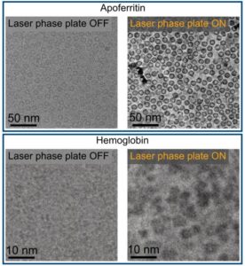
Laser‑Driven Phase Contrast Enhances Cryo‑EM Resolution of Small Proteins

You know when you are at the eye doctor getting an updated prescription, and suddenly the world snaps into sharper focus? Physicists at the University of California (UC), Berkeley, have now done something similar for electron microscopy. By introducing phase contrast into a cryo‑electron microscope, they have delivered dramatically sharper images of some of biology’s smallest and most elusive proteins.

The advance comes from a new laser phase plate (LPP), described in the paper “Laser phase plate improves structure determination of small proteins by cryo‑EM,” which was published recently in Science. Led by physicist Holger Mueller, PhD, of UC Berkeley and Lawrence Berkeley National Laboratory, the team demonstrated that a laser‑driven phase plate can overcome one of cryo‑EM’s most persistent limitations: poor contrast for small proteins.

Cryo-EM images of two proteins, apoferritin and hemoglobin, taken without and with a laser phase plate. The images are analyzed in a computer to produce detailed 3D structures of the proteins. [Holger Müller, Jessie Zhang/UC Berkeley]

Cryo‑EM has transformed structural biology over the past decade, earning a Nobel Prize in 2017 for enabling high‑resolution structures without crystallization. But despite its impact, the technique still struggles with proteins below ~70 kilodaltons—a size range that includes about 90% of the human proteome. “Because of signal-to-noise limitations, the majority of human and animal proteins are too small to be analyzed by these methods [cryo-EM and cryoelectron tomography]. The increase in signal-to-noise ratio provided by this laser phase plate is expected to overcome these important limitations.”

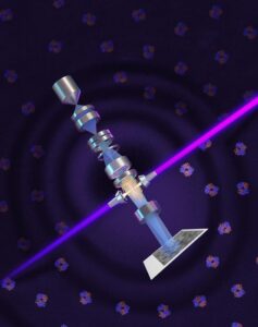
The new LPP begins to address that problem. The LPP uses an intense, continuous‑wave laser to shift the phase of the electron beam itself. This produces true phase contrast without dimming or destabilizing the beam. Mueller described the laser focus as “75 kilowatts focused to a few microns… That’s more powerful than what you use for welding. It has more power than a military laser. It builds up the brightest continuous laser focus ever.”

Installed in a custom Thermo Fisher Titan Krios, the LPP immediately improved the clarity and resolvability of small proteins, including hemoglobin, which sits at the lower limit of what today’s cryo‑EM instruments can handle. As the authors wrote in the abstract: “Here, we show that the laser phase plate (LPP)… enhances the resolution in single-particle reconstruction of small proteins by improving specimen-motion correction, recovery of information from the early frames, as well as particle visualization, 3D classification, and alignment.”

phase plate cover Cryo-EM
A laser (purple) is powerfully amplified by highly polished mirrors and focused on the electron beam (blue) to shift its phase and increase the cryo-EM microscope’s contrast, allowing biologists to image smaller proteins and the crowded structures inside cells. [Sayo Studio]

These improvements were achieved using standard defocus ranges and reconstruction workflows. “For the most challenging cases—small particles, bad specimens—the laser produces a very considerable advantage,” Mueller said.


The impact extends beyond single‑particle analysis. Cryo‑electron tomography (cryo‑ET), which assembles multiple angular views of a molecule or protein into a three-dimensional image, stands to benefit even more. “With cryo-ET, we’re looking at small, very complicated cellular material that’s incredibly crowded inside the cell,” said Bridget Carragher, PhD, founding technical director of imaging at Biohub. “It’s like a forest of trees, and you’re trying to find one leaf on one tree in there. Cryo-ET needs a dramatic step forward in contrast, so we can start to see what’s going on inside the cell. That’s what the laser phase plate promises to give us.”

Biohub is developing a dual‑laser version of the system, designed to reduce component wear and minimize aberrations. Meanwhile, Mueller’s team is pushing toward imaging proteins as small as 17 kilodaltons, a threshold that would open access to vast regions of the human proteome previously invisible to cryo‑EM.

“This technology is a step function change for biology,” said Stephani Otte, PhD, Biohub’s vice president of imaging science. “What was once invisible will become visible—and that changes everything about how we understand disease.”

“The bottom line is, if you have a large protein and a really good sample—a fresh one or one frozen without bubbles, for example—you may not need the phase plate to get a single, high-quality image. But for a small protein and a bad sample, laser-on is best,” Mueller said. “This could fill an enormous gap in our knowledge of protein structures that can’t be crystallized or are too small for today’s cryo-EM. And it will be revolutionary for cryo-ET.”

The post Laser‑Driven Phase Contrast Enhances Cryo‑EM Resolution of Small Proteins appeared first on GEN – Genetic Engineering and Biotechnology News.

Pioneering Innovation in Transplant Diagnostics

Sara Barrington is CEO of Verici Dx, a precision diagnostics company redefining how transplant clinicians understand and predict graft health. By combining multi-omic analysis with proprietary artificial intelligence (AI) models, Verici Dx delivers predictive, actionable intelligence that helps clinicians optimize therapy, inform biopsy decisions, and stratify risk for complex transplant patients.

In this edition of Asked & Answered, Barrington speaks with Inside Precision Medicine Editor in Chief Damian Doherty about pioneering innovation in transplant diagnostics to better reflect biological and patient complexity, and ultimately, improve health outcomes.


Q: Tell me about Verici Dx, its founding, mission, and products.

Sara Barrington: Verici Dx was founded with a very clear purpose: to fundamentally improve how transplant clinicians are informed and manage graft health. Transplant medicine requires high-stakes, time-sensitive decision-making to promote longevity of the graft, minimize the risk of complications, and improve the patient’s quality of life.

Sara Barrington
Sara Barrington

Kidney transplantation has long struggled with one fundamental challenge: accurately identifying rejection early and distinguishing it from other causes of injury and graft dysfunction. Clinicians often rely on conventional blood markers such as serum creatinine or, more recently, donor-derived cell-free DNA. While these tools are useful for signaling that “something is wrong,” they are injury markers and lack a level of precision important in rejection biology.

Creatinine levels, for example, increase only after functional damage has already occurred and cannot differentiate rejection from non-immune causes of injury such as ischemic reperfusion injury, drug toxicity, or viral infections like BK virus–associated nephropathy. Biomarkers can generate false positives, triggering biopsies or treatment escalation that may not be necessary and can expose patients to avoidable risk.

This lack of specificity has made rejection difficult to manage proactively. By the time rejection is clearly identified, tissue injury may already be established. At the same time, clinicians are understandably cautious about intensifying immunosuppression without clear evidence, given the associated risks of infection, malignancy, and toxicity.

Our mission is to close this information gap by delivering precision diagnostics that reflect the true complexity of transplant biology and translate that complexity into insights clinicians can proactively and confidently apply to patient care.

We operate at the intersection of multi-omic science, advanced analytics, and clinical reality. We know that transplant rejection and graft injury are not single-signal events, but are multifactorial, dynamic biological processes. We design our diagnostics to capture that complex biology in a way that mirrors how clinicians think about their patients, rather than forcing clinical decision-making to fit the limitations of older technologies.

Our lead product, Tutivia™, is a blood-based test that assesses the immune and broader, relevant biological status of kidney transplant patients to identify the risk of all forms of acute rejection, including T cell (TCMR) and antibody-mediated rejection (ABMR). Tutivia combines transcriptomics with proprietary AI models to address this precise unmet need: biologically accurate insight into what is actually happening within the graft.

The test delivers a risk score that enables more precise immunosuppression management by distinguishing rejection from other causes of graft injury, such as BK virus–associated nephropathy and ischemia-reperfusion injury, which can confound conventional blood tests like serum creatinine and donor-derived cell-free DNA.

By focusing on the immune and other pathways associated with rejection, Tutivia moves beyond damage markers and toward a direct assessment of rejection biology itself. That shift, from injury detection to biological interpretation, is what makes the test such a valuable tool for transplant clinicians.

Thermo Fisher Scientific recently launched its One Lambda™ Pre-Transplant Risk Assessment (PTRA) assay, using pre-transplant prognostic technology licensed from Verici Dx. The assay provides a risk score for early acute rejection based on a patient’s unique gene profile, helping clinicians better balance the risk of rejection against the potential side effects of over-immunosuppression. Using a 29-gene mRNA signature, the PTRA assay stratifies patients into high- and low-risk categories, with clinical validation showing high-risk patients are seven times more likely to experience early acute rejection.


Q: Tutivia is built on RNA sequencing. Why is precision transcriptomic analysis well-suited for identifying rejection biology?

Barrington: The need for reliable biomarkers is crucial for individualizing therapy that offers the potential to extend allograft survival.

Early research underpinning the science leading to the development of Tutivia was led by principal investigators at The Mount Sinai Hospital in New York, one of the world’s leading transplant research centers, combining decades of clinical expertise with rigorous science. The underlying science reflects the fact that rejection in kidney transplant patients is not driven by a single molecule or pathway, it results from coordinated immune and other biologic pathways across multiple cell types, signaling cascades, and regulatory mechanisms. Injury-driven biomarkers capture only a narrow slice of this biology and can be influenced by non-rejection factors, limiting their specificity and reliability.

Precision transcriptomic analysis, on the other hand, measures gene expression patterns across thousands of genes simultaneously. In developing specific tests, this analysis is the basis for gene selection using unsupervised deep learning mathematical modeling, allowing the most impactful and reproducible gene expression signals and interactions to be included in the final signature.

Tutivia uses a peripheral blood next-generation sequencing assay to evaluate a 17-gene mRNA signature in combination with a proprietary artificial intelligence algorithm to categorize kidney transplant patients as at low risk or high risk of acute rejection.

This provides a comprehensive view of immune activation, regulation, and injury responses within the transplanted organ, allowing the identification of signature changes that are truly characteristic of rejection biology.

In short, RNA sequencing lets us ask better biological questions: not just whether the kidney is injured, but whether immune-mediated rejection is driving that injury. That distinction is critical for precision medicine in transplantation.


Q: What does earlier and more precise identification of rejection biology mean for patient management and outcomes?

Barrington: Earlier identification of rejection biology can create an opportunity to intervene before irreversible damage occurs. When clinicians have confidence in what the biology is telling them, they can make timely, targeted decisions that may preserve graft function.

This precision also supports more individualized care. Rather than applying broad treatment strategies, clinicians can tailor immunosuppression based on biological risk, potentially reducing over-treatment and its associated complications.

For patients, this can translate into fewer unnecessary biopsies, fewer adverse events related to immunosuppression, and a greater likelihood of long-term graft survival. Ultimately, better information leads to better decisions, and better decisions lead to better outcomes.

Our vision is that precision diagnostics like Tutivia become a standard component of transplant management, providing earlier insight into rejection biology, supporting more informed clinical decisions, and helping clinicians intervene at the right time for the right patient.


Q: AI is central to your platform. How does Verici Dx apply AI differently from others in diagnostics?

Barrington: We apply AI with rigor and restraint. We use advanced machine learning and mathematical modeling to analyze highly complex biological data, but always within a disciplined scientific framework. Our models are developed using unsupervised approaches and validated extensively on inclusive patient populations. Once validated, they are locked and reproducible; they are not evolving over time. That distinction matters because clinicians are making life-altering decisions based on these results, and trust, consistency, and validation are essential.


Q: Validation and standards are recurring themes for Verici Dx. How was Tutivia validated?

Barrington: Tutivia was validated through a rigorous clinical study that included a broad and diverse transplant patient population, rather than narrowly curated cohorts. We deliberately chose a more difficult validation path because clinical reality is complex, and diagnostics must perform under those conditions. While this approach requires more time and effort, it ensures the test is reliable, reproducible, and clinically relevant across the full spectrum of patients that clinicians see every day.


Q: What’s in the future for the company?

Barrington: Verici Dx is focused on deepening the clinical adoption of Tutivia as well as expanding the studies demonstrating the utility of an earlier and more precise biomarker in improving patient outcomes. Long-term outcomes for transplant patients involve more biological pathways, and significant complexity is involved in managing care. We are focused on developing and validating tests using both transcriptomic and proteomic precision tools to assist clinicians and their patients in this journey.


Damian Doherty has been in media and publishing for over 30 years, beginning at News Corporation. Damian has managed, edited, and launched life science titles in drug discovery and precision medicine. He was features editor of Drug Discovery World and founded the Precision Medicine Leaders Summit and the Journal of Precision Medicine. He edited AIMed magazine before launching Photo51Media, a platform for illuminating untold, compelling stories in precision healthcare. Damian joined Mary Ann Liebert in 2021 to help steer the new rebrand and relaunch of Clinical OMICS to Inside Precision Medicine.

The post Pioneering Innovation in Transplant Diagnostics appeared first on Inside Precision Medicine.

Stopping Neurodegeneration in Place with Vaccines

Developing truly disease-changing treatments for Alzheimer’s disease and other neurodegenerative conditions has proved challenging, with many failed trials over the last few decades. The approval of Eisai/Biogen’s monoclonal antibody lecanemab in 2023, the first such treatment to have a positive, albeit modest, impact on symptoms of Alzheimer’s disease, was therefore received with enthusiasm by many.

Critics of lecanemab and Eli Lilly’s donanemab, approved a year later for the same indication, argue that the small benefit gained from the drugs does not outweigh the economic costs, possible side effects, and burden of regular intravenous infusions.

Using a vaccine-style approach, where the treatment prompts the body to generate its own antibodies, has the potential to solve these problems. Several companies are developing active immunotherapies to target Alzheimer’s and Parkinson’s disease, as well as other neurological conditions.

Andrea Pfiefer
Andrea Pfiefer, PhD
CEO and Co-founder
AC Immune

“What we’re trying to do is link the antigen to a carrier and bring it into a form which mimics the pathology,” explained Andrea Pfeifer, PhD, CEO and co-founder of Swiss company AC Immune, a leading biotech taking the active immunotherapy route to target Alzheimer’s and Parkinson’s disease.

“We inject it into the immune system, and what it recognizes is the misfolded protein. So, because of that, the immune system only makes antibodies against this pathological protein.”

Although none have yet reached the market, the active vaccine-style approach potentially has a number of advantages over passive treatment with monoclonal antibodies. Importantly, fewer rounds of treatment are required. The exact dosing is yet to be determined, but it would certainly be less frequent than the regular infusions of lecanemab or donanemab that are currently prescribed. This would help reduce costs and treatment burdens for patients and their families. There is also likely to be less risk of amyloid-related imaging abnormalities (ARIA) due to the relatively slower onset of antibody generation by the body.

“If you have to take the patient every two to four weeks to get a two-hour infusion in a hospital, and then you have to wait and do imaging, it’s really burdensome,” said Pfeifer.

“After a certain while, they just don’t want to go. … They say, ‘Sorry, we believe your science, we believe everything, but we don’t want this.”

To date, most vaccine trials have enrolled people with at least some degree of Alzheimer’s or Parkinson’s disease, as preventive vaccine trials need to be large and long in duration. Theoretically, developing a preventive vaccine is a feasible approach, as many neurodegenerative diseases typically have a slow onset before noticeable symptoms appear. However, reliable biomarkers that can accurately predict disease onset have been in short supply.

Alzinova’s ALZ-101
ALZ-101 – Alzinova’s disease-modifying vaccine candidate for Alzheimer’s disease

This is changing, though. Last year, two blood tests that measure phosphorylated tau and amyloid ratios were approved by the U.S. Food and Drug Administration (FDA) for Alzheimer’s diagnosis. The biomarker field is less developed for Parkinson’s and other neurodegenerative diseases, such as amyotrophic lateral sclerosis (ALS), but things are slowly improving.

Promising mid-stage results from front-runners like AC Immune and Alzinova suggest that this pathway has merit, but whether they can succeed in larger registrational trials going forward is unclear.

Tord Labuda
Tord Labuda, PhD
CEO, Alzinova

“It remains to be seen how our immune system reacts. I think that when we look at the titers we see from the vaccine, they’re similar to what we get with passive immunizations. So I think there are a lot of things that point in the right direction,” said Tord Labuda, PhD, CEO of Swedish biotech Alzinova, which has an amyloid beta vaccine in development.

“Personally, I don’t think that the vaccine or passive immunization is the real challenge. The real challenge is to have the right target.”

Taking a more active approach to immunotherapy

While passive immunotherapies like lecanemab and donanemab are groundbreaking in that they are the first disease-modifying treatments for Alzheimer’s to be approved by the FDA and they slow cognitive decline by around 25%–35% over two years, these drugs can cause significant side effects such as ARIA.

This can cause brain swelling and bleeding in some people. Individuals at highest risk for some neurodegenerative conditions, such as carriers of the APOE4 gene variant, are prevented from accessing these therapies at all, as they have a higher-than-average risk of experiencing ARIA-like side effects.

Treating neurodegenerative diseases like Alzheimer’s and Parkinson’s disease when symptoms commence is problematic, as currently, there is no known method of regaining neuronal function once it has been lost.

Classic pathology studies suggest that, by the time the typical motor symptoms of Parkinson’s appear, around 50%–70% of dopaminergic neurons are already lost from the brain. Similarly, in early clinical Alzheimer’s disease, where those affected have mild dementia, hippocampal volume in the brain seen on imaging is already up to 25% lower than that of age-matched controls. This means, however good the treatment is, the patients will never regain complete function.

“There’s no way we can restore these neurons,” said Roman Kniazev, CEO of U.S.-biotech Nuravax, which is developing several different Alzheimer’s vaccines. “They are gone forever. So that is why the best strategy, and this is our motto in our company, is to not let the pathology kill the neurons.”

Researchers trying to develop new therapies for Alzheimer’s and Parkinson’s are increasingly moving towards an early or even preventive approach. The idea of a vaccine-like approach to targeting Alzheimer’s is not new. Animal work in the late 1990s showed that vaccinating against amyloid‑beta could clear plaques and improve cognition in transgenic mouse models. This led to the development of Elan/Wyeth’s amyloid‑beta vaccine, AN1792.

It entered Phase I/II trials for mild‑to‑moderate Alzheimer’s and successfully induced anti‑amyloid beta antibodies in some patients. But the study had to be stopped in 2002 because around six percent of the participants developed meningoencephalitis, linked to T cell–mediated inflammation in the brain. Despite strong amyloid plaque clearance, there was little impact on symptoms in the clinical trial participants.

Several other candidates, designed to avoid the T-cell activation seen with AN1792, have been unsuccessfully trialed over the last two decades. Although largely safe and antibody-producing, most of these programs were discontinued due to a lack of efficacy.

The field has persevered despite this, with improvements in technology, safety, biomarkers, and clinical trial design having led to a new generation of vaccine candidates that, while not yet approved, are showing good results in Phase I and II trials.

Neil Warma
Neil Warma
CEO, ProMIS Neurosciences

“I think what we’ve learned over the past five or 10 years from other vaccine approaches is that the next generation are those that come with a very precise approach to generating antibodies against the toxic species of a given protein for that specific neurodegenerative disease,” said Neil Warma, CEO of ProMIS Neurosciences, a U.S.-based company taking an antibody and a vaccine-based approach to treating Alzheimer’s and other neurodegenerative diseases.

“Those are really the things that make the vaccine approach much more interesting now. The sophistication of the biomarkers, the data we’ve learned from past and current products in the market, and then this ability to design and create antibodies that are highly specific to a targeted pathogenic form of the protein.”

AC Immune has three active immunotherapy candidates in Phase II: an anti-amyloid beta therapy, ACI-24, that is being developed with Takeda; an anti-tau therapy, ACF 35, being developed with Johnson & Johnson; and an anti-alpha synuclein candidate targeting Parkinson’s disease.

Staining of neurons in the brain
Staining of neurons in the brain from Neil Cashman’s lab-1.

The company reported good results for its Parkinson’s Phase II study at the end of last year. The interim study results showed a 100% response rate and a good safety profile in people with early Parkinson’s disease. Alpha-synuclein and neurofilament light levels in the blood, as well as scores from standard movement tests, suggested that disease stabilization had occurred.

If confirmed by results from the second part of the study, this would be the first time that disease modification, rather than symptom management, has shown promise for Parkinson’s disease. It would also confirm alpha synuclein as a pathogenic contributor to the condition.

“The statistical variability was very, very small, which was a surprise to us. Every single marker, preclinical biomarker, clinical imaging, went into the same direction,” said Pfeifer. “For me, what was particularly rewarding was that there was a connection between the titers, so the antibody response in the people versus the reduction of this pathology … the alpha-synuclein versus the imaging, which showed that the neurons can be protected.”

Better targeting to improve safety and efficacy

A common theme among vaccine developers in the neurodegeneration space is precision targeting. There is a strong consensus that a lack of specificity when picking targets is likely why many trials failed to show efficacy and had significant side effect issues in the past.

Different companies or research groups back slightly different protein targets, but the experts all emphasize the importance of avoiding binding to inert plaque in the brain in Alzheimer’s disease, and of targeting toxic, misfolded proteins that are disease-specific.

“We provide the body with active immunotherapy, and then the body induces the antibodies. But what is important is that these antibodies are really specific for the pathological form,” said Pfeifer.

AC Immune is not the only company with a keen focus on tightening up targeting in this area. Alzinova is specifically targeting toxic amyloid beta oligomers with its lead candidate ALZ-101, a therapeutic Alzheimer’s vaccine.

It received a recent FDA Fast Track designation for ALZ-101 after good safety and efficacy data were reported last year from its completed Phase Ib clinical trial.

“They have shown in many in vitro and in vivo models that … when you remove these toxic oligomers using antibodies towards them, you can basically neutralize the toxicity in these extracts towards the neurons,” explained Labuda.

He added that many of the “vaccines, as well as the monoclonal antibodies, are going for the N-terminal part of the protein. … By doing that, you will target the monomers, all the fibrils, but most importantly, also the plaques. Very little will be left to bind to something else that might be more important for the disease. I think this is what we see with the current treatments on the market. There’s a lot of off-target effects … and that’s why we have these huge challenges.”

ProMIS is also targeting amyloid beta oligomers in Alzheimer’s using both a monoclonal antibody and vaccine approach. It has developed a special method with the help of artificial intelligence to develop antibodies, taking both conformational shape and protein sequence into account.

“These three-dimensional shapes don’t exist on monomers, and they’re buried in plaque. We’ve tried and tried and tried to get these antibodies to bind monomers, to bind plaque, and they really don’t, which is good,” said Warma.

“We’ve done side-by-side testing with other antibodies to see if ours is truly differentiated. … In many different studies before we got to the clinic, PMN-310, our therapeutic antibody, was the only one that bound oligomers and avoided monomers and plaque. All the others cross-reacted with everything.”

ProMIS is testing its PMN-310 antibody before moving on to the vaccine approach. “If we can come with that one-two punch to say we’ve got a drug now that can treat patients with Alzheimer’s, we’ve got the ability to detect the onset of disease pathology in Alzheimer’s, and we have a vaccine that prevents you from developing that disease, I mean, that would be a pretty powerful combination,” noted Warma.

Nuravax is aiming to address two key shortcomings of first‑generation Alzheimer’s vaccines, such as AN1792: the risk of problematic T cell-driven inflammation and uneven immune responses in older adults. Its MultiTEP‑based candidates (AV‑1959R, AV‑1980R, and Duvax) are engineered to elicit a strong, antibody‑dominant response against amyloid‑beta and tau while minimizing activation of potentially autoreactive T cells and maintaining effectiveness.

“The platform which we developed makes the vaccine highly immunogenic, and this high immunogenic feature is essential for diseases in the brain,” said Kniazev.

Kiran Bhaskar
Kiran Bhaskar, PhD
Professor
University of New Mexico

Kiran Bhaskar, PhD, is a professor and group leader at the University of New Mexico. He has worked on Alzheimer’s disease for many years and is also a scientific co-founder of TheraVac Biologics. He and his colleagues are developing an anti-tau Alzheimer’s vaccine that is about to start human trials.

They are also aiming to reduce risks associated with immune reactions to vaccine adjuvants, which can contribute to ARIA, and have created a vaccine that does not need an adjuvant.

“We use a strategy called a virus-like particle,” he explained. “You don’t need to expect any side effects because of adjuvants. In this way, we trick the immune system into thinking that there is a viral attack on the body. It immediately starts an immune response against the virus-like particle and also anything sticking to the surface of those virus-like particles, which in this case is pathological or phosphorylated tau proteins.”

Overall, in the trials carried out so far in the active immune therapy space in Alzheimer’s disease, rates of ARIA have been very low, which is another selling point for the vaccine approach. “We don’t expect to see ARIA in active immunotherapy because we are using the host’s own immune response system to generate antibodies. … So that way there’ll be less unanticipated immune response,” explained Bhaskar.

syringe and vaccine bottle
Credit: Ake Ngiamsanguan / iStock / Getty Images Plus

Overcoming challenges on the road to the clinic

There is no doubt that active immunotherapies or vaccines to target Alzheimer’s and Parkinson’s disease are more advanced than they have ever been before, but they are still a long way from a mainstream rollout.

One reason the conversation has shifted is that the approval and broader use of the first anti-amyloid antibodies, for all their modest effect sizes and ARIA issues, shows that lowering the right protein species can slow deterioration. Using a more vaccine-like approach, where people make their own antibodies, does have the potential to make targeting these conditions safer, cheaper, and more accessible for patients.

But basic clinical questions remain unanswered. For example, how long vaccine-induced protection will last, how often boosters will be needed, and whether early stabilization of biomarkers and motor or cognitive scores—such as AC Immune’s interim Parkinson’s data or Alzinova’s Phase Ib Alzheimer’s study results—will translate into true preserved function a decade later.

Biomarkers are a big potential stumbling block for the development of preventive vaccines. The position is better for Alzheimer’s disease; the FDA’s approval of two blood-based Alzheimer’s biomarkers now gives developers a way to find people with silent pathologies without relying solely on positron emission tomography imaging. But there is still a lot more to do on this front, particularly in diseases like Parkinson’s, ALS, or other neurological or neurodegenerative diseases where less is known.

surgical glove pointing at a brain scan image
Credit: Martin Philip / iStock / Getty Images Plus

“We need better and safer treatments, but it’s also linked to having the right biomarkers, because if you have a risk factor, it doesn’t mean necessarily that you have the disease,” said Pfeifer. “These biomarkers are still not very well established. … The goal is absolutely to go to preclinical, and preclinical will require these biomarkers. If you ask me what is needed most in Parkinson’s right now, [it] is definitely to have better biomarkers.”

A second stumbling block for companies that want to develop vaccines that can effectively immunize people against neurodegenerative disease is that clinical trials of vaccines in populations of people with no symptoms have historically been large, expensive, and time-consuming—something out of reach of most biotechs without significant outside investment.

ProMIS and others are hoping that the efficacy of their therapeutic antibodies will boost their vaccine pipeline in the future. “If we show that it works in Alzheimer’s, then a similar approach should work in these other diseases,” said Warma. “Since it’s an almost identical process for vaccines, it also shows proof of concept for this whole wave of vaccines coming behind it.”

For now, active immunotherapies promise something that is more modest than true disease prevention but still crucially important. Namely, cheaper, less burdensome, and potentially safer ways to target the same disease biology as today’s monoclonal antibodies, ideally years earlier in the process.

The post Stopping Neurodegeneration in Place with Vaccines appeared first on Inside Precision Medicine.

Remote Monitoring Benefits Unclear After Hospitalization for Serious Infection

Remotely monitoring patients after they have been released from the hospital for serious infections does not reduce their risk of readmission, particularly for older adults, a randomized trial suggests.

The findings, released simultaneously in JAMA Network Open and at the Critical Care Reviews annual meeting in Belfast, emphasize the importance of studying potentially beneficial care models in real-world scenarios.

Remote monitoring involves the use of resources such as smartphone-based questionnaires, internet-connected scales, or blood pressure monitoring to allow patients to leave hospital.

These enable clinicians to monitor health from afar, while acting on alerts to ensure timely care.

While patients might prefer this type of care, the trial suggested that remote monitoring should be re-evaluated and individually tailored after acute care for sepsis and lower respiratory tract infections.

It did not increase the days patients spent at home and did not support U.S. federal agency efforts to reduce hospital readmissions by reimbursing remote monitoring.

“Health systems, insurers and policymakers all want to reduce hospital readmissions, and most patients prefer to recover safely at home,” explained lead researcher Sachin Yende, PhD, from the University of Pittsburgh.

“Remote monitoring has been held up as a solution, is reimbursed by the Centers of Medicare & Medicaid and its use has grown. But, aside from a few conditions, there’s a dearth of high-quality data to show it reduces readmissions.”

In an attempt to source this information, Yende and team conducted a complex trial in 1286 adults, with a median age of 63 years, who were recovering at home after hospitalization for sepsis or lower respiratory tract infections at 19 institutions. Of these patients, 32.6% received intensive care.

Participants were randomly assigned to one of four remote monitoring strategies or usual care.

Usual care generally involved a phone call from a nurse after discharge followed by continuing management from a primary care physician. The remote monitoring interventions consisted of short or long health questionnaire with standard or enhanced clinical response teams.

The standard response involved nurses who responded to alerts highlighted in the questionnaires together with coordinated care involving the patient’s primary and specialty clinicians.

Enhanced care included certified registered nurse practitioners with palliative care expertise who could coordinate some of the care response and also access social workers.

The primary endpoint was days at home at 90 days after discharge.

The trial showed that remote monitoring programs with high- or low-intensity questionnaires combined with a nurse response team or a nurse practitioner-led response team did not increase the time spent at home.

At least one readmission occurred among 37.8% of patients in the control group, receiving usual care.

This compared with 39.7% in patients receiving the shorter questionnaire and standard response team, 44.2% for those receiving the long questionnaire with standard clinical response, 37.3% receiving the shorter questionnaire but enhanced response team, and 36.3% for those receiving the long questionnaire with enhanced clinical response.

These differences did not reach statistical significance.

However, among patients aged 65 years and older, the remote monitoring programs did reduced time spent at home and increased readmission rates compared with usual care.

In this older age group, both the standard and enhanced response groups had fewer home days compared with usual care, with corresponding cumulative odds ratios of 0.56 and 0.67.

“These findings support reevaluating and tailoring remote monitoring in post–acute care following sepsis and lower respiratory tract infection to support further alignment with patients’ needs and desire for personalized monitoring,” the researchers concluded.

The post Remote Monitoring Benefits Unclear After Hospitalization for Serious Infection appeared first on Inside Precision Medicine.

Potential Cocaine Addiction Targets Identified Through Genetic Mapping in Rats

Scientists at the University of California San Diego have reported the results of a genome-wide association study in rats that identified key biological drivers of cocaine addiction. Using a genetically diverse group of nearly 900 rats to map genetic markers associated with compulsive drug use, the researchers uncovered a potential new therapeutic target that resides in the liver rather than in the brain.

Current research in this field often focuses on the brain, but the UC San Diego team’s findings suggest that how the body metabolizes cocaine may be just as critical in determining whether somebody develops an addiction.

“Finding a liver-based enzyme that shapes cocaine-taking behavior was a real ‘aha’ moment for us,” said Olivier George, PhD, a professor of psychiatry at UC San Diego School of Medicine. The George lab led the addiction behavioral studies that provided the foundation for the research. “It reminds us that addiction isn’t only in the brain. It’s a complex puzzle involving how the entire body processes the drug.”

George is co-corresponding author of the team’s published paper in Nature Communications, titled “Genome-wide association study of cocaine self-administration behavior in Heterogeneous Stock rats.”

Cocaine use disorder (CUD) has a strong genetic component, the authors noted. “Twin studies estimate the heritability of cocaine dependence to be as high as 70%, a finding supported by recent comprehensive reviews,” they wrote.  GWAS have also uncovered a significant heritable component, the team continued, with single nucleotide polymorphism (SN)-based heritability estimated at 27-30%. However, scientists have struggled to pinpoint the specific genes that make certain individuals more vulnerable to addiction.

“The paucity of significant and replicated associations for CUD limits our understanding of this disorder, hampering our ability to identify novel pharmacological targets,” the investigators added. Co-corresponding author Abraham A. Palmer, PhD, professor of psychiatry at UC San Diego School of Medicine, who led the project’s intensive genetic modeling and analysis, further commented, “Identifying those genes in an important goal, because drugs could then be developed to target those genes, shifting genetically susceptible people to become more like genetically resistant people.”

To investigate further, the team carried out a GWAS in nearly 900 outbred Heterogeneous Stock (HS) rats—a model system capable of mimicking the vast genetic diversity found in human populations. By using HS rats the team was able to capture the critical differences between individuals who are genetically susceptible to addiction and those who are naturally more resistant. “Prior work has established the phenotypic diversity of HS rats across a broad range of addiction-relevant behaviors, including cocaine self-administration,” the researchers commented.

“The extended access model allowed us to characterize escalating intake, increased motivation to take the drug, and compulsive-like behavior despite negative consequences.” In addition to the GWAS results the researchers carried out a range of secondary analysis strategies to uncover what they describe as novel genetic drivers of cocaine self-administration behaviors.

Analyzing millions of genetic markers in each animal, the team discovered six major genetic regions linked to addiction-like behaviors, such as the escalation of drug intake and the time elapsed between doses. The researchers identified in the rats a specific group of carboxylesterase genes that are orthologous to the human CES1 gene, which are responsible for creating the enzyme that metabolizes cocaine. The study found that variations in these genes are closely linked to how frequently and compulsively rats self-administered the drug.

The findings also replicated a known genetic link found in humans (Trak2), providing a vital translational bridge between animal research and human medicine. This replication strengthens the argument that the biological pathways identified in the lab could eventually lead to real-world therapies. “Genes associated with CUD in humans remain limited, however our GWAS identified one gene (Trak2) that has also been identified by human GWAS of CUD, and the novel identification of Ces1 offers a fresh avenue for future studies,” they stated.

The collective findings suggest that by targeting the enzymes that metabolize cocaine with medicines, scientists might be able to alter how the drug affects the body, potentially reducing its addictive impact. In their paper they concluded “Our results replicate previous loci associated with CUD in humans and provide several novel biological insights including the potential of pharmacological strategies targeting carboxylesterases.”

Palmer said, “This work showcases the power of long-term, team-science collaboration that pairs experts in rodent behavior with quantitative geneticists. A decade of coordinated effort across multiple cohorts and federal partners made possible a discovery that no single lab could achieve alone.”

First author Montana Kay Lara, PhD, a postdoctoral researcher at UC San Diego School of Medicine, who helped bridge the gap between the study’s behavioral and genetic components, said, “Seeing the Ces1 signal validate a hypothesis that has been circulating for decades is incredibly exciting. It gives us a concrete target to test whether changing how cocaine is metabolized can blunt the drive toward compulsive use.”

The research team is now moving into the next phase of the project, which involves investigating exactly how these genetic mutations change function of the enzyme. They also hope to use the study’s extensive Preclinical Addiction Biobanks—collections of blood, urine, brain and other tissue samples—to identify biological markers that could one day help predict an individual’s risk of developing a substance use disorder.

The researchers hope that by leveraging this resource, they and other scientists working in this space will be able to translate genetic discoveries into diagnostic tools and new treatments that can help stabilize individuals struggling with addiction.

The post Potential Cocaine Addiction Targets Identified Through Genetic Mapping in Rats appeared first on GEN – Genetic Engineering and Biotechnology News.

Lipid-Protein Biomarker Makes Clinical Debut with Early Ovarian Cancer Detection

For Anna Jeter, the story behind AOA Dx’s promising ovarian cancer diagnostic begins with a misconception. Despite serving more than half the population, women’s health continues to be viewed by many investors as a niche category—an assumption Jeter believes has contributed to decades of underinvestment in some of medicine’s most pressing unmet needs. 

“Whenever you speak to investors about women’s health, there’s this perception that it’s somehow a smaller market,” Jeter, co-founder and chief regulatory officer of AOA Dx, told Inside Precision Medicine. “Women represent 51% of the population, but historically women’s health has been treated as a specialty category rather than a major healthcare market.” 

That perception has shaped innovation patterns across healthcare, particularly in diagnostics. Despite advances in genomics, liquid biopsy, and AI in oncology, ovarian cancer detection has stagnated. The biomarker Cancer Antigen 125 (CA-125), introduced in 1987, remains the primary blood-based tool used to evaluate women presenting with symptoms suggestive of ovarian malignancy. 

“There have been very few advancements in ovarian cancer,” Jeter said. “The standard of care today is still largely based on technology that became available almost 40 years ago.” 

Early detection could have massive, life-changing consequences, as ovarian cancer is often highly treatable when diagnosed early. More than 90% of women survive when the disease is identified at stage 1, yet nearly 80% of cases are diagnosed only after the cancer has progressed to advanced stages. 

After more than 15 years of developing diagnostics companies centered around women’s health, Jeter, Oriana Papin-Zoghbi, and Alex Fischer decided to take action and co-founded AOA Dx. Through an early partnership with McGill University, the team began investigating whether emerging advances in multi-omics could uncover biological signals that existing ovarian cancer diagnostics were missing. 

Anna Jeter AOA Dx
Anna Jeter, co-founder and chief regulatory officer of AOA Dx

AOA Dx recently announced that its investigational blood test, AKRIVIS GD, achieved 92% sensitivity for Stage I and II ovarian cancer detection in symptomatic women—nearly double the sensitivity historically associated with CA-125 alone. 

Yet Jeter argues the most important aspect of the findings is not the performance metric itself, but what enabled it: the successful translation of lipid biomarkers into a clinical oncology assay. “We are now looking at lipids as very specific biomarkers, which have made a major impact on our ability to detect ovarian cancer significantly earlier,” she said. “And we believe this approach can be translated into other cancers in the future.” 

In fact, it may even go beyond that, as the most notable multi-omic approaches using lipidomics and proteins, such as recent work for stratifying Alzheimer’s disease subtypes, are still in early research phases. 

Blood-based tests beyond DNA 

The announcement, which was recently made at the 2026 American Society of Clinical Oncology (ASCO) annual meeting, reflects a growing shift within cancer diagnostics away from single-analyte approaches and toward integrated biological modeling. Over the past decade, liquid biopsy innovation has largely centered on circulating tumor DNA (ctDNA), driven by advances in next-generation sequencing and the promise of minimally invasive genomic profiling. However, ctDNA performance has varied substantially across tumor types and disease stages, particularly in malignancies characterized by low tumor shedding during early progression. 

Jeter contends that ovarian cancer exemplifies these limitations. “Certain cancers shed ctDNA very effectively into circulation, and in those cases the technology performs very well,” Jeter said. “But in other cancers, either the shedding is insufficient for the sensitivity required at early stages, or the specificity profile becomes challenging. That has led to a broader realization across the field that a single biological layer cannot fully capture disease progression.” 

Increasingly, oncology researchers are converging around multi-omic frameworks that integrate orthogonal biological signals, including proteomic, metabolic, genomic, and transcriptomic features. Within that context, lipid metabolism has emerged as an area of renewed interest. 

Lipid signaling, long associated primarily with cardiovascular disease and metabolic disorders, now plays a deeply intertwined role in oncogenesis. Alterations in lipid metabolism contribute to membrane remodeling, inflammatory signaling, tumor proliferation, immune modulation, and metastatic adaptation. Tumors frequently undergo profound metabolic reprogramming early in disease development, creating detectable shifts in circulating lipid and metabolite profiles before conventional biomarkers become clinically informative. 

“What we have learned is that lipid metabolism appears to be highly active in the earliest stages of disease,” Jeter said. “That biology has been described in the literature for some time. The challenge has been translating those discoveries into a clinical-grade diagnostic assay.” 

That translation problem is nontrivial. Lipids present substantial analytical complexity due to structural diversity, isomeric overlap, and variability in sample handling and quantitation. Discovery-level lipidomics frequently produces promising signals that prove difficult to standardize under regulated clinical laboratory conditions. AOA Dx’s central claim is that it has successfully navigated that transition. 

The company’s platform integrates targeted lipidomic and metabolomic profiling using high-resolution mass spectrometry with protein biomarker analysis via immunoassays. Machine-learning models are then applied to derive composite signatures associated with ovarian malignancy. 

New clinical biomarker class 

According to the company, the assay was developed through biomarker discovery efforts involving more than 2,200 patient samples across multiple cohorts and demographic populations. 

Importantly, the ASCO dataset focused specifically on the intended-use population: symptomatic women presenting in routine clinical settings with nonspecific but concerning symptoms such as abdominal pain, bloating, urinary changes, or altered bowel habits. 

That population reflects a longstanding clinical challenge in gynecologic oncology. Although the majority of ovarian cancer patients report symptoms beginning relatively early in disease progression, those symptoms are often diffuse and overlap extensively with benign gastrointestinal or genitourinary conditions. As a result, diagnostic delays remain common, with patients frequently cycling through multiple evaluations before referral to gynecologic oncology. 

“About 85–95% of women begin experiencing symptoms as early as stage 1,” Jeter noted. “But the symptoms are nonspecific enough that providers often do not initially suspect ovarian cancer.” 

As a result, nearly 80% of ovarian cancers are still diagnosed at advanced stages, despite the dramatic survival differential associated with early intervention. When detected at stage 1 or 2, five-year survival rates exceed 90%. Once metastatic dissemination occurs, however, outcomes deteriorate sharply. 

The company’s results suggest that multi-omic integration may provide clinically meaningful improvements over historical CA-125 performance, particularly in early-stage disease where existing diagnostic sensitivity has remained inadequate. According to AOA Dx, AKRIVIS GD achieved nearly double the early-stage sensitivity historically associated with CA-125 alone. 

The broader implication is not simply improved biomarker performance but a reframing of how cancer detection may evolve over the next decade. Rather than relying on single molecular features, next-generation diagnostics are increasingly attempting to model disease as a systems-level biological phenomenon. 

“There’s a growing recognition that different biological pathways become informative at different stages of disease progression,” Jeter explained. “Some protein markers rise later. Some genomic signals are difficult to detect early. By integrating lipidomic, metabolomic, and proteomic information simultaneously, we’re able to observe the disease from multiple biological angles.” 

The company describes this infrastructure as a broader discovery engine known as GlycoLocate, which is designed to interrogate thousands of lipid and metabolite signatures across multiple disease states. Ovarian cancer represents the company’s first targeted clinical application, but executives indicate the platform is already being expanded into additional oncology indications, particularly within women’s health. 

The women’s health investment gap 

AOA Dx’s emergence also reflects a broader maturation within women’s health biotechnology, a sector that historically struggled to attract sustained institutional investment despite its substantial market opportunity. 

Jeter has spoken openly about what she views as a longstanding disconnect between investor perception and commercial reality in women’s health innovation. Earlier this year, she presented an investment analysis at the 2026 JP Morgan Healthcare Conference examining historical returns within the category. 

“Women’s health has traditionally been viewed as a niche market, despite women representing more than half the population,” she said. “Part of the issue is that many successful companies serving women have not historically been classified as women’s health investments, so the return profile of the sector has been systematically underestimated.” 

That dynamic may now be shifting as women’s health increasingly intersects with other high-growth sectors, including artificial intelligence, precision medicine, molecular diagnostics, and computational biology. AOA Dx itself sits squarely at that convergence point, combining AI-driven biomarker modeling with high-complexity laboratory diagnostics and oncology applications. 

The company is now advancing AKRIVIS GD toward validation studies and an early-access launch program. Equally important will be demonstrating clinical utility and securing reimbursement pathways, both of which remain major barriers for emerging diagnostics platforms. “It’s one thing to develop a test,” Jeter said. “It’s another thing entirely to ensure that patients can actually access it through reimbursement and coverage.” 

To support commercialization efforts, AOA Dx has expanded its leadership team with executives experienced in diagnostic platform deployment and LC-MS assay translation, including chief product officer, Chris Roberts, and senior director of biomarker and analytical development, Cory Bystrom, PhD. 

Whether lipidomics ultimately becomes a foundational pillar of oncology diagnostics remains uncertain. However, the field appears to be entering a transitional period in which metabolic biology is moving from peripheral exploratory science toward regulated clinical application. 

For AOA Dx, the ambition extends beyond ovarian cancer itself. The company sees lipid-based multi-omics as an entirely new diagnostic layer capable of complementing—and in some settings potentially surpassing—the limitations of genomics-only approaches. “What I often say,” Jeter reflected, “is that the situation feels very similar to where next-generation sequencing was 10 or 15 years ago.” 

If the company’s early data continues to hold in larger validation cohorts, AOA Dx may not simply be introducing another ovarian cancer assay. It may help to define the next biological frontier of liquid biopsy diagnostics. 

The post Lipid-Protein Biomarker Makes Clinical Debut with Early Ovarian Cancer Detection appeared first on Inside Precision Medicine.

Inside soccer’s data renaissance

Imagine tuning in to the opening kickoff of a World Cup match and seeing a player intentionally send the ball all the way down the pitch and right out of bounds on the opponent’s end. Casual fans might scratch their heads. Where’s the logic in surrendering possession seconds into a game? If you were Jesse Davis, though, you’d know that this play could be a prime setup to score. 

Davis is a professor of computer science at KU Leuven in Belgium and head of its Sports Analytics Lab, which has been at the vanguard of a data awakening in soccer since its inception more than a decade ago. Though the research group brings machine-­learning models to bear on a variety of sports—including basketball, volleyball, and field hockey—nowhere is its impact felt more than on the soccer pitch. 

Davis and his team of researchers employ advanced data analytics to reveal a range of (beg your pardon) game-changing findings that are shifting pro clubs’ decision-making. “His lab is the most influential sports analytics lab in soccer,” says Hugo Rios-Neto, data recruitment lead for Royal Sporting Club Anderlecht in Belgium. They’ve helped teams better evaluate their rosters, conceived ways to assess how efficient (or not) strategies are, and developed algorithms that uncover hidden tactical patterns.

Like, for instance, the value of kicking the ball out of bounds close to the goal and letting your opponent throw it back into play—a move that’s been popping up in some of the world’s top leagues over the last few years.

To make the statistical argument for this seemingly counterproductive move, Davis’s group built a training data set composed of more than 1.4 million passes and some 60,000 throw-ins—partly from the 2022 World Cup. They used tree ensemble models (essentially a mashup of decision trees) to simulate the tactic. The conclusion, which the researchers presented in a 2024 paper under the apt title “Boot it”: When the ball is in the middle third of the pitch, kicking it out of bounds on your opponents’ side of the field can put you within 10 actions (think passes and dribbles) of a goal. That can be a big deal in a game that has 1,500 or more actions per match and very little scoring. The idea, Davis explains, is that you’re setting yourself up to recover the ball in an advantageous situation.

Beyond providing discrete game-day insights, Davis also occupies a unique niche in the world of sports analytics, where many clubs now hire their own internal data teams to maintain a competitive edge. He makes most of his research freely available via open-source analytics tools, but the academic life also affords him the freedom to tackle more complex problems—like standardizing in-game data, a project that will make it easier to parse game footage and come up with winning strategies. 


Davis, 45, grew up in Wisconsin and spent his childhood enraptured by basketball and (American) football. Soccer was largely a nonentity to him until college, when the 2002 World Cup—in which Brazil famously swept the tournament—reeled him in. But the notion of going on to dissect the sport never crossed his mind. His doctoral studies in computer science at the University of Wisconsin–Madison had him working with radiologists to analyze mammography reports. 

In October 2010, he joined KU Leuven as a computer science professor looking at the intersection of AI and health care, with a focus on monitoring athletic performance. His research team studied, for instance, combining things like heart rate with other metrics to determine whether someone was overtraining. They also dove into the biomechanics of running.

The tactical and technical aspects of sports, and soccer specifically, became the subject of Davis’s professorial work when he hired Jan Van Haaren, an engineering student focused on artificial intelligence and a self-described soccer fanatic. He wondered if data analysis could be used to study things like passing, shooting, and ball progression—metrics the game was only just beginning to digitally crunch at the time. 

Davis realized that machine learning and other artificial-intelligence tools lent themselves well to the complexity, fluidity, and speed of soccer.

You need not be well versed in the moneyball-ization of pro sports to see that it’s relatively easy to apply deep statistical work to baseball or basketball. You can isolate actions like jump shots and assign value to ones taken close or far away. Soon a basketball coach realizes that a player who can’t make a layup, but shoots roughly as well from the three-point line as on mid-range jumpers, might as well go for the shot that gets more points. 

Soccer, by comparison, seemed like a poor candidate for that kind of analysis. “The vast, vast majority of actions really don’t lead to the outcome of a goal or even a shot,” says Rios-Neto. “So it’s hard to elaborate or derive a winning strategy from the data.”

But Van Haaren’s love of the sport, and Davis’s love of sports in general, inspired them to try. Over time, Davis realized that machine learning and other artificial-intelligence tools lent themselves well to the complexity, fluidity, and speed of soccer. In 2014, he officially stood up the Sports Analytics Lab. 

With a stable of about 10 students and postdocs at any one time, the lab began laying what Van Haaren calls the “intellectual foundations of how the game is analyzed today.” The researchers picked apart in-game actions, and suddenly they were valuing ball possession, penalty-kick strategy (aim for the center), and the merits of long shots on goal (take them). “One of the trends that’s been in soccer over the last five to 10 years is that the number of long shots has dramatically increased,” says Davis. “What the data let you do is really quantify what the probabilities of those things are.”


In the years since Davis and his team started untangling individual soccer tactics, their ideas have started to permeate clubs across Europe, like Belgium’s Club Brugge KV, as well as national soccer organizations in the US and Belgium. “The work coming out of the lab is genuinely useful,” Rios-Neto says, “and clubs apply it for a range of purposes.” 

Van Haaren, who’s now the director of football intelligence at Club Brugge, is one of many in-house analysts adapting the lab’s work to the pro game. “Our collaboration with the lab is centered on translating [the team’s] football philosophy into measurable, data-driven outputs,” he says. When a club wants to assess, say, how well a center-back is moving the ball down the field, it aims to tally how many times the ball ended up in the part of the pitch closest to the opposing team’s goal. It does this by combining event data, which records actions on the ball, with tracking data, which records player movement. This shows how well players fulfill their roles, which is useful in development and also when scouting for new recruits. 

Davis’s lab, meanwhile, is continuing to ask questions that apply to the game writ large. To determine if there’s an advantage to taking more long shots, for instance, postdoc Maaike Van Royand colleagues modeled the behavior of English Premier League teams using a Markov decision process—a computational framework in which some actions are under a person’s control while others are random. (That duality is particularly useful for soccer, where movement can feel anything but linear.) The results, presented in 2021 at the MIT Sloan Sports Analytics Conference, showed that Chelsea could gain 1.6 more goals per season by shooting from distance 20% more often.

Despite those kinds of insights from Davis’s lab and similar research groups that have sprung up over the last decade at institutions like MIT and Carnegie Mellon, soccer somewhat lags behind many other pro sports when it comes to collecting the data that analysts need. All teams employ people to watch video and use software to annotate specific in-game tactics—the details of which may make sense only to the most devoted fans. It’s a mostly manual process, one that can take up to six hours per game. “It’s a complete nightmare as a data analyst to work with,” says Davis.

So while the lab plays on, Davis has also joined up with researchers from other institutions in an effort to standardize data across all matches. The group is experimenting with transformers, the neural network architecture that underpins large language models like ChatGPT. If you can bring that to the world of soccer, a human game annotator could tag a tactic—a three-on-two breakaway, say—a few times, and that could train the model on the concept so it could tag subsequent instances on its own. “There’s been a lot of progress,” Davis says. “But it still remains quite hard.”

If we’re keeping score, though, the lab’s work has already made the analytics process easier thanks to open-source tools it’s put out there—some of which clock thousands of downloads a month. One is a framework called VAEP, a model that assesses the effects of all actions on the ball. Another is an xG (expected goals) model, which looks at the quality of a scoring chance. Still another is a package to synchronize event data with tracking data. “Lots of people in industry use our code in their daily workflows,” Davis says.

For him, the practical application of having their code out there is important, but the real (ahem) kick is watching theory become practice. As he says, “I’m really motivated to solve problems that arise in real settings and see my work have an impact.” 

Andrew Zaleski is a contributing writer at Washingtonian magazine. 

GSK to Acquire Nuvalent for $10.6B, Boosting Cancer Pipeline with Precision NSCLC Treatments

GlaxoSmithKline (GSK) has agreed to acquire Nuvalent for $10.6 billion, the companies said, in a deal designed to strengthen the buyer’s cancer pipeline with Nuvalent’s precision oncology treatments—including three non-small cell lung cancer (NSCLC) therapies, of which two are under FDA review with decisions expected later this year.

Boston-based Nuvalent’s pipeline is headed by the ROS1 inhibitor zidesamtinib (NVL-520) and the ALK inhibitor eladalkib (NVL-655), which according to the company represent potential best-in-class, next-generation, highly selective treatments for NSCLC. Both are brain penetrant. The FDA has set target decision dates of September 18 for zidesamtinib and November 27 for neladalkib, both of which have been granted the agency’s Breakthrough Therapy and Orphan Drug designations.

Zidesamtinib is designed to treat NSCLC tumors driven by ROS1 that have developed resistance to currently available ROS1 inhibitors, including tumors with the prevalent G2032R “solvent front” resistance mutation. Zidesamtinib is selective in order to minimize CNS adverse events related to off-target inhibition of the tropomyosin receptor kinase (TRK) family, and potentially drive durable responses for patients with ROS1-mutant variants, Nuvalent says.

Eladalkib was created to address treating tumors driven by ALK that have developed resistance to first-, second-, and third-generation ALK inhibitors, including tumors with both single or compound treatment-emergent ALK mutations such as those involving the G1202R “solvent front” mutation. Eladalkib is also designed to avoiding TRK family inhibition and to treat brain metastases.

The third NSCLC asset of Nuvalent, NVL-330, is a HER2 inhibitor now under study in Phase I trials for HER2-altered NSCLC. In addition, Nuvalent’s pipeline includes an unspecified number of preclinical programs focused on “addressing the limitations of existing therapies for clinically proven kinase targets in oncology,” the company states on its website.

“Today’s acquisition is a multi-product deal, consistent with our approach to acquire assets that have clinically proven targets and meaningfully address an efficacy and/or tolerability gap,” GSK CEO Luke Miels said in a statement. “The two lead products are potential best-in-class assets that could launch this year if approved by the FDA and offer significant new treatment options to patients with two forms of non-small cell lung cancer.”

GSK investors were less enthusiastic as its shares on the London Stock Exchange on Monday dipped 0.5% to 1,903.50 pence. However, Nuvalent shares jumped 39% on Nasdaq to $123.25.

The $10.6 billion Nuvalent acquisition is the third largest merger-and-acquisition (M&A) deal announced this year, behind the €10.7 billion ($12.355 billion) cash buyout offer for Italian-based Recordati being pursued by CVC Capital Partners and Groupe Bruxelles Lambert, which aim to take the company private; and Sun Pharmaceutical Industries’ planned $11.75 billion purchase of Organon, the women’s health drug developer spun out of Merck & Co., in a deal expected to close in early 2027.

Immediate sales opportunities

The Nuvalent candidates, GSK added, present immediate new sales growth opportunities, improving profit contributions from 2027, and a platform in lung cancer for rapid expansion with GSK’s Ris-Rez, a B7-H3 targeted antibody-drug conjugate (ADC) now in Phase III clinical development.

In a presentation to investors after announcing a series of business updates on May 27, Nuvalent projected an ROS1+ NSCLC treatment could generate ~$1.4 billion to $2.1 billion in peak year sales, with about 40% of those sales (from ~$570 million to $855M million) expected to come from the U.S.—multiples above the ~$150 million in peak year sales attained in 2019 by Xalkori® (crizotinib), marketed by Pfizer and Merck KGaA.

An ALK+ NSCLC treatment would potentially be even more lucrative, Nuvalent said last month, with projected worldwide peak year sales ranging from ~$3.4 billion to $5 billion, of which the U.S. would account for 40% of sales, or between ~$1.35 billion and $2 billion—well above the $519 million in peak sales attained in 2023 by Alecensa® (alectinib), marketed by Genentech, a member of the Roche Group and created by Roche-owned Chugai Pharmaceutical.

“Since our founding, we have leveraged our deep expertise in chemistry and structure-based drug design to develop a portfolio of novel, potentially best-in-class kinase inhibitors. Our close collaboration with leading physician-scientists and patient advocates has driven remarkable enrolment, accelerating development and building confidence in the clinical profile of these drugs,” Nuvalent CEO James Porter, PhD, stated. “We’re excited that GSK has recognized the significant value these programs can offer patients and shares our vision for practice-changing innovation.”

Positive pivotal data

In announcing the acquisition, GSK cited positive pivotal data Nuvalent presented at the IASLC 2025 World Conference on Lung Cancer and the 2026 ASCO Annual Meeting. Data at both conferences showed potential best-in-class profiles for zidesamtinib and neladalkib, with both treatments designed to deliver longer effective treatment with better quality of life than current therapies, through high target-selectivity, durable treatment response, improved tolerability, enhanced blood-brain barrier penetration for tumor spread, and broader coverage of ALK and ROS1 mutations.

ROS1- and ALK-altered NSCLC primarily affect non-smoking adults aged 40-50, GSK and Nuvalent said—a patient population the companies described as uniquely defined and engaged.

GSK said it will commence a tender offer to acquire all of Nuvalent’s outstanding shares of Class A and Class B common stock at a purchase price of $124 per share in cash within 10 business days. The expected purchase price represents a 40% premium to the last closing price and a 26% premium to the 30 calendar day volume-weighted average price.

Net of cash acquired, GSK estimated its aggregate investment in Nuvalent to be $9.4 billion.

GSK said the acquisition will not change its 2026 full-year guidance range of 7-9% core operating profit and core EPS growth. The acquisition is expected to contribute to revenue growth from 2027, be incremental to GSK’s existing ambition for sales of >£40 billion (>$53.56 billion) by 2031, and strengthen the company’s core operating profit through the two-year period of loss of exclusivity for its aging blockbuster dolutegravir (2028-2030).

Dolutegravir is an HIV-1 integrase strand transfer inhibitor (INSTI) marketed as the monotherapy Tivicay® by Viiv Healthcare, in which GSK holds a 78.3% majority stake (and Shionogi, the remaining 21.7% after Pfizer cashed out its 11.7% stake, receiving $1.88 billion). Dolutegravir is also included in Viiv’s fixed-dose HIV combination therapies Dovato (dolutegravir and lamivudine) and Juluca (dolutegravir and rilpivirine).

Adding to core profit, EPS

GSK said it expected to add to its core operating profit in 2027 and core earnings per share (EPS) in 2029 by acquiring Nuvalent, even after accounting for cost-cutting synergies and “reprioritization,” which it defines as the shifting of personnel, capital, and other resources away from lower-yield, early-stage research or legacy programs toward higher-value clinical assets and corporate activities. Nuvalent reported 228 full-time employees, of which 144 are engaged in R&D, in its Form 10-K annual report for 2025, filed February 26.

Should the transaction close in Q3 2026 as expected, GSK said it expects low single-digit percentage dilution to core EPS this year through 2028.

The company said it will fund the Nuvalent acquisition primarily from new and existing debt facilities plus cash, with no impact expected to its credit rating. GSK ended Q1 with £3.442 billion ($4.608 billion) in cash and cash equivalents, up 1.3% from £3.397 billion ($4.548 billion) at the end of 2025.

The transaction is subject to customary closing conditions, including the tender of a majority of Nuvalent’s outstanding shares of Class A common stock in the tender offer and the expiration or termination of the applicable waiting period under the Hart-Scott-Rodino Act in the U.S. Soon after the closing of the tender offer, GSK expects to acquire any remaining shares of Nuvalent through a second-step merger under Delaware law at the same price per share.

GSK said it will account for the transaction as a business combination and assume Nuvalent’s existing revenue-sharing arrangements of low-single-digit royalties payable to Royalty Pharma and Deerfield. Royalty Pharma in December acquired for up to $315 million a pre-existing royalty interest in zidesamtinib and neladalkib from an undisclosed third party. Deerfield is Nuvalent’s largest shareholder.

“GSK’s proven track record, infrastructure, and expertise will support the successful commercialization of zidesamtinib and neladalkib, as well as accelerate advancement of our broader discovery pipeline,” Porter added.

The post GSK to Acquire Nuvalent for $10.6B, Boosting Cancer Pipeline with Precision NSCLC Treatments appeared first on GEN – Genetic Engineering and Biotechnology News.